Loading
PDBj
MenuPDBj@FacebookPDBj@X(formerly Twitter)PDBj@BlueSkyPDBj@YouTubewwPDB FoundationwwPDB
RCSB PDBPDBeBMRBAdv. SearchSearch help
Search by PDB author
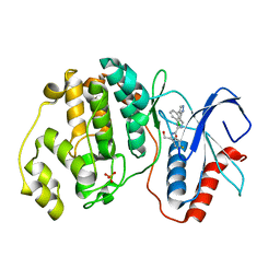
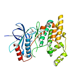
3G90
DownloadVisualize
BU of 3g90 by Molmil
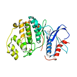
JNK-3 bound to (Z)-5-fluoro-1-((6-fluoro-4H-benzo[d][1,3]dioxin-8-yl)methyl)-3-(hydroxyimino)indolin-2-one
Descriptor: (3E)-5-fluoro-1-[(6-fluoro-4H-1,3-benzodioxin-8-yl)methyl]-1H-indole-2,3-dione 3-oxime, Mitogen-activated protein kinase 10
Authors:Xie, X, Jacobs, M.D.
Deposit date:2009-02-12
Release date:2009-02-24
Last modified:2024-02-21
Method:X-RAY DIFFRACTION (2.4 Å)
Cite:Structure-based design and parallel synthesis of N-benzyl isatin oximes as JNK3 MAP kinase inhibitors.
Bioorg.Med.Chem.Lett., 19, 2009
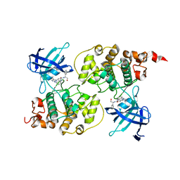
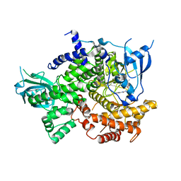
7KKE
DownloadVisualize
BU of 7kke by Molmil
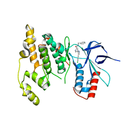
Phosphoinositide 3-Kinase gamma bound to a thiazole inhibitor
Descriptor: N-[2-(3,3-dimethylbutoxy)ethyl]-N'-{4-methyl-5-[(pyridin-4-yl)ethynyl]-1,3-thiazol-2-yl}urea, Phosphatidylinositol 4,5-bisphosphate 3-kinase catalytic subunit gamma isoform
Authors:Jacobs, M.D, Griffith, J.P.
Deposit date:2020-10-27
Release date:2021-03-24
Last modified:2023-10-18
Method:X-RAY DIFFRACTION (2.81 Å)
Cite:Discovery of a Novel Series of Potent and Selective Alkynylthiazole-Derived PI3K gamma Inhibitors.
Acs Med.Chem.Lett., 12, 2021
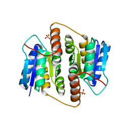
5E95
DownloadVisualize
BU of 5e95 by Molmil
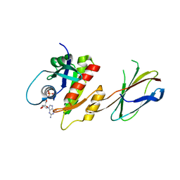
Crystal Structure of Mb(NS1)/H-Ras Complex
Descriptor: GTPase HRas, GUANOSINE-5'-DIPHOSPHATE, MAGNESIUM ION, ...
Authors:Eguchi, R.R, Sha, F, Gupta, A, Koide, A, Koide, S.
Deposit date:2015-10-14
Release date:2016-11-02
Last modified:2023-09-27
Method:X-RAY DIFFRACTION (1.402 Å)
Cite:Inhibition of RAS function through targeting an allosteric regulatory site.
Nat. Chem. Biol., 13, 2017
2OJG
DownloadVisualize
BU of 2ojg by Molmil
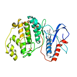
Crystal structure of ERK2 in complex with N,N-dimethyl-4-(4-phenyl-1H-pyrazol-3-yl)-1H-pyrrole-2-carboxamide
Descriptor: Mitogen-activated protein kinase 1, N,N-DIMETHYL-4-(4-PHENYL-1H-PYRAZOL-3-YL)-1H-PYRROLE-2-CARBOXAMIDE, SULFATE ION
Authors:Xie, X, Jacobs, M.D.
Deposit date:2007-01-12
Release date:2007-02-06
Last modified:2023-12-27
Method:X-RAY DIFFRACTION (2 Å)
Cite:Flipped Out: Structure-Guided Design of Selective Pyrazolylpyrrole ERK Inhibitors.
J.Med.Chem., 50, 2007
2OJI
DownloadVisualize
BU of 2oji by Molmil
Crystal structure of ERK2 in complex with N-benzyl-4-(4-(3-chlorophenyl)-1H-pyrazol-3-yl)-1H-pyrrole-2-carboxamide
Descriptor: Mitogen-activated protein kinase 1, N-BENZYL-4-[4-(3-CHLOROPHENYL)-1H-PYRAZOL-3-YL]-1H-PYRROLE-2-CARBOXAMIDE, SULFATE ION
Authors:Xie, X, Jacobs, M.D.
Deposit date:2007-01-12
Release date:2007-02-06
Last modified:2023-12-27
Method:X-RAY DIFFRACTION (2.6 Å)
Cite:Flipped Out: Structure-Guided Design of Selective Pyrazolylpyrrole ERK Inhibitors.
J.Med.Chem., 50, 2007
2OK1
DownloadVisualize
BU of 2ok1 by Molmil
Crystal structure of JNK3 bound to N-benzyl-4-(4-(3-chlorophenyl)-1H-pyrazol-3-yl)-1H-pyrrole-2-carboxamide
Descriptor: Mitogen-activated protein kinase 10, N-BENZYL-4-[4-(3-CHLOROPHENYL)-1H-PYRAZOL-3-YL]-1H-PYRROLE-2-CARBOXAMIDE
Authors:Xie, X, Jacobs, M.D.
Deposit date:2007-01-15
Release date:2007-02-06
Last modified:2023-12-27
Method:X-RAY DIFFRACTION (2.4 Å)
Cite:Flipped Out: Structure-Guided Design of Selective Pyrazolylpyrrole ERK Inhibitors.
J.Med.Chem., 50, 2007
2OJJ
DownloadVisualize
BU of 2ojj by Molmil
Crystal structure of ERK2 in complex with (S)-N-(1-(3-chloro-4-fluorophenyl)-2-hydroxyethyl)-4-(4-(3-chlorophenyl)-1H-pyrazol-3-yl)-1H-pyrrole-2-carboxamide
Descriptor: (S)-N-(1-(3-CHLORO-4-FLUOROPHENYL)-2-HYDROXYETHYL)-4-(4-(3-CHLOROPHENYL)-1H-PYRAZOL-3-YL)-1H-PYRROLE-2-CARBOXAMIDE, Mitogen-activated protein kinase 1, SULFATE ION
Authors:Xie, X, Jacobs, M.D.
Deposit date:2007-01-12
Release date:2007-02-06
Last modified:2023-12-27
Method:X-RAY DIFFRACTION (2.4 Å)
Cite:Flipped Out: Structure-Guided Design of Selective Pyrazolylpyrrole ERK Inhibitors.
J.Med.Chem., 50, 2007
3I4B
DownloadVisualize
BU of 3i4b by Molmil
Crystal structure of GSK3b in complex with a pyrimidylpyrrole inhibitor
Descriptor: Glycogen synthase kinase-3 beta, N-[(1S)-2-hydroxy-1-phenylethyl]-4-[5-methyl-2-(phenylamino)pyrimidin-4-yl]-1H-pyrrole-2-carboxamide
Authors:Ter Haar, E.
Deposit date:2009-07-01
Release date:2010-01-12
Last modified:2024-04-03
Method:X-RAY DIFFRACTION (2.3 Å)
Cite:Structure-guided design of potent and selective pyrimidylpyrrole inhibitors of extracellular signal-regulated kinase (ERK) using conformational control.
J.Med.Chem., 52, 2009
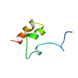
1MGX
DownloadVisualize
BU of 1mgx by Molmil
COAGULATION FACTOR, MG(II), NMR, 7 STRUCTURES (BACKBONE ATOMS ONLY)
Descriptor: COAGULATION FACTOR IX
Authors:Freedman, S.J, Furie, B.C, Furie, B, Baleja, J.D.
Deposit date:1995-06-21
Release date:1996-11-08
Last modified:2017-11-29
Method:SOLUTION NMR
Cite:Identification of the phospholipid binding site in the vitamin K-dependent blood coagulation protein factor IX.
J.Biol.Chem., 271, 1996
3CUK
DownloadVisualize
BU of 3cuk by Molmil
Crystal structure of human D-amino acid oxidase: bound to an inhibitor
Descriptor: 4H-furo[3,2-b]pyrrole-5-carboxylic acid, D-amino-acid oxidase, FLAVIN-ADENINE DINUCLEOTIDE
Authors:Prasad, S, Munshi, S.
Deposit date:2008-04-16
Release date:2008-07-22
Last modified:2023-08-30
Method:X-RAY DIFFRACTION (2.49 Å)
Cite:The discovery of fused pyrrole carboxylic acids as novel, potent D-amino acid oxidase (DAO) inhibitors.
Bioorg.Med.Chem.Lett., 18, 2008
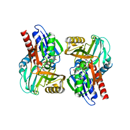
2BES
DownloadVisualize
BU of 2bes by Molmil
Structure of Mycobacterium tuberculosis Ribose-5-Phosphate Isomerase, RpiB, Rv2465c, in complex with 4-phospho-D-erythronohydroxamic acid.
Descriptor: 4-PHOSPHO-D-ERYTHRONOHYDROXAMIC ACID, CARBOHYDRATE-PHOSPHATE ISOMERASE
Authors:Roos, A.K, Ericsson, D.J, Mowbray, S.L.
Deposit date:2004-11-30
Release date:2004-12-03
Last modified:2023-12-13
Method:X-RAY DIFFRACTION (2.1 Å)
Cite:Competitive Inhibitors of Mycobacterium Tuberculosis Ribose-5-Phosphate Isomerase B Reveal New Information About the Reaction Mechanism.
J.Biol.Chem., 280, 2005
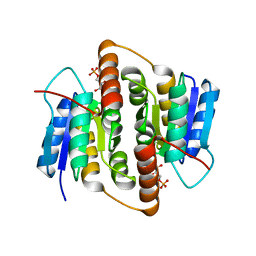
2BET
DownloadVisualize
BU of 2bet by Molmil
Structure of Mycobacterium tuberculosis Ribose-5-Phosphate Isomerase, RpiB, Rv2465c, in complex with 4-phospho-D-erythronate.
Descriptor: 4-PHOSPHO-D-ERYTHRONATE, CARBOHYDRATE-PHOSPHATE ISOMERASE
Authors:Roos, A.K, Ericsson, D.J, Mowbray, S.L.
Deposit date:2004-11-30
Release date:2004-12-03
Last modified:2023-12-13
Method:X-RAY DIFFRACTION (2.2 Å)
Cite:Competitive Inhibitors of Mycobacterium Tuberculosis Ribose-5-Phosphate Isomerase B Reveal New Information About the Reaction Mechanism
J.Biol.Chem., 280, 2005
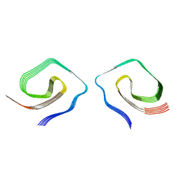
7UN5
DownloadVisualize
BU of 7un5 by Molmil
Structure of Type II Prion filaments from Gerstmann-Straussler-Scheinker disease
Descriptor: Major prion protein
Authors:Ozcan, K.A, Hoq, M.R, Bharath, S.R, Jiang, W.
Deposit date:2022-04-08
Release date:2022-07-27
Last modified:2024-03-27
Method:ELECTRON MICROSCOPY (3.13 Å)
Cite:Cryo-EM structures of prion protein filaments from Gerstmann-Straussler-Scheinker disease.
Acta Neuropathol, 144, 2022
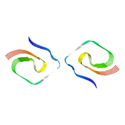
7UMQ
DownloadVisualize
BU of 7umq by Molmil
Structure of Type I Prion filaments from Gerstmann-Straussler-Scheinker disease
Descriptor: Major prion protein
Authors:Ozcan, K.A, Hoq, M.R, Bharath, S.R, Jiang, W.
Deposit date:2022-04-07
Release date:2022-07-27
Last modified:2024-03-27
Method:ELECTRON MICROSCOPY (3.29 Å)
Cite:Cryo-EM structures of prion protein filaments from Gerstmann-Straussler-Scheinker disease.
Acta Neuropathol, 144, 2022
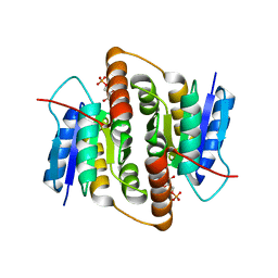
1USL
DownloadVisualize
BU of 1usl by Molmil
Structure Of Mycobacterium tuberculosis Ribose-5-Phosphate Isomerase, RpiB, Rv2465c, Complexed With Phosphate.
Descriptor: PHOSPHATE ION, RIBOSE 5-PHOSPHATE ISOMERASE B
Authors:Roos, A.K, Andersson, C.E, Unge, T, Jones, T.A, Mowbray, S.L.
Deposit date:2003-11-25
Release date:2004-01-02
Last modified:2023-12-13
Method:X-RAY DIFFRACTION (1.88 Å)
Cite:Mycobacterium Tuberculosis Ribose-5-Phosphate Isomerase Has a Known Fold, But a Novel Active Site
J.Mol.Biol., 335, 2004
<12


227344

PDB entries from 2024-11-13

PDB statisticsPDBj update infoContact PDBjnumon