Loading
PDBj
MenuPDBj@FacebookPDBj@TwitterPDBj@YouTubewwPDB FoundationwwPDB
RCSB PDBPDBeBMRBAdv. SearchSearch help
Search by PDB author
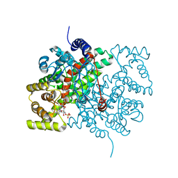
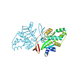
1CTS
DownloadVisualize
BU of 1cts by Molmil
CRYSTALLOGRAPHIC REFINEMENT AND ATOMIC MODELS OF TWO DIFFERENT FORMS OF CITRATE SYNTHASE AT 2.7 AND 1.7 ANGSTROMS RESOLUTION
Descriptor: CITRATE SYNTHASE, CITRIC ACID
Authors:Remington, S, Wiegand, G, Huber, R.
Deposit date:1984-01-27
Release date:1984-07-20
Last modified:2024-02-07
Method:X-RAY DIFFRACTION (2.7 Å)
Cite:Crystallographic refinement and atomic models of two different forms of citrate synthase at 2.7 and 1.7 A resolution.
J.Mol.Biol., 158, 1982
6JHU
DownloadVisualize
BU of 6jhu by Molmil
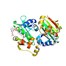
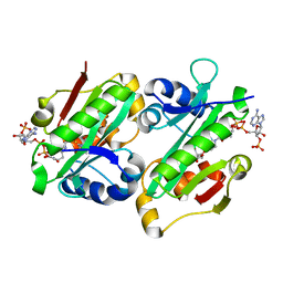
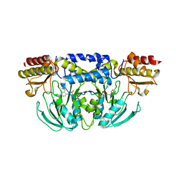
Crystal Structure Of Biotin Protein Ligase From Leishmania Major in complex with Biotinyl-5-AMP
Descriptor: BIOTINYL-5-AMP, Biotin/lipoate protein ligase-like protein, SULFATE ION
Authors:Rajak, M, Patel, A, Sundd, M.
Deposit date:2019-02-19
Release date:2020-04-08
Last modified:2023-11-22
Method:X-RAY DIFFRACTION (1.97 Å)
Cite:Leishmania major biotin protein ligase forms a unique cross-handshake dimer
Acta Crystallogr.,Sect.D, 77, 2021
5YO2
DownloadVisualize
BU of 5yo2 by Molmil
The crystal structure of Rv2747 from Mycobacterium tuberculosis in complex with Acetyl CoA and L-Arginine
Descriptor: ACETYL COENZYME *A, ARGININE, Amino-acid acetyltransferase
Authors:Singh, E, Tiruttani Subhramanyam, U.K, Pal, R.K, Srinivasan, A, Gourinath, S, Das, U.
Deposit date:2017-10-26
Release date:2018-11-07
Last modified:2024-03-27
Method:X-RAY DIFFRACTION (2.997 Å)
Cite:Structural insights into the substrate binding mechanism of novel ArgA from Mycobacterium tuberculosis
Int. J. Biol. Macromol., 125, 2019
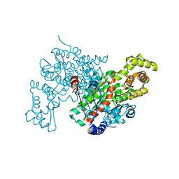
3CSC
DownloadVisualize
BU of 3csc by Molmil
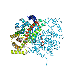
STRUCTURE OF TERNARY COMPLEXES OF CITRATE SYNTHASE WITH D-AND L-MALATE: MECHANISTIC IMPLICATIONS
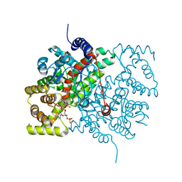
Descriptor: (2S)-2-hydroxybutanedioic acid, ACETYL COENZYME *A, CITRATE SYNTHASE
Authors:Karpusas, M, Holland, D, Remington, S.J.
Deposit date:1990-05-07
Release date:1991-04-15
Last modified:2024-02-21
Method:X-RAY DIFFRACTION (1.9 Å)
Cite:1.9-A structures of ternary complexes of citrate synthase with D- and L-malate: mechanistic implications.
Biochemistry, 30, 1991
5ZAU
DownloadVisualize
BU of 5zau by Molmil
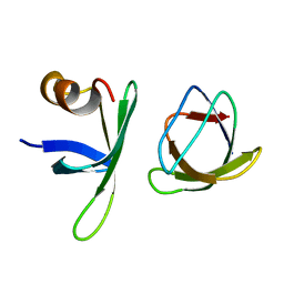
Complex of the human FYN SH3 and monobody binder
Descriptor: Monobody Binder, Tyrosine-protein kinase Fyn
Authors:Reddy, P.P, Gulyani, A, Das, R.
Deposit date:2018-02-09
Release date:2019-09-11
Last modified:2024-05-15
Method:SOLUTION NMR
Cite:A Fyn biosensor reveals pulsatile, spatially localized kinase activity and signaling crosstalk in live mammalian cells.
Elife, 9, 2020
3CTS
DownloadVisualize
BU of 3cts by Molmil
CRYSTALLOGRAPHIC REFINEMENT AND ATOMIC MODELS OF TWO DIFFERENT FORMS OF CITRATE SYNTHASE AT 2.7 AND 1.7 ANGSTROMS RESOLUTION
Descriptor: CITRATE SYNTHASE, CITRIC ACID, COENZYME A
Authors:Remington, S, Wiegand, G, Huber, R.
Deposit date:1984-01-27
Release date:1984-07-20
Last modified:2024-02-21
Method:X-RAY DIFFRACTION (1.7 Å)
Cite:Crystallographic refinement and atomic models of two different forms of citrate synthase at 2.7 and 1.7 A resolution.
J.Mol.Biol., 158, 1982
6ADD
DownloadVisualize
BU of 6add by Molmil
The crystal structure of Rv2747 from Mycobacterium tuberculosis in complex with CoA and NLQ
Descriptor: Amino-acid acetyltransferase, COENZYME A, N~2~-ACETYL-L-GLUTAMINE
Authors:Das, U, Singh, E, Pal, R.K, Tiruttani Subhramanyam, U.K, Gourinath, S, Srinivasan, A.
Deposit date:2018-07-31
Release date:2018-12-26
Last modified:2023-11-22
Method:X-RAY DIFFRACTION (2.301 Å)
Cite:Structural insights into the substrate binding mechanism of novel ArgA from Mycobacterium tuberculosis
Int. J. Biol. Macromol., 125, 2019
5YD2
DownloadVisualize
BU of 5yd2 by Molmil
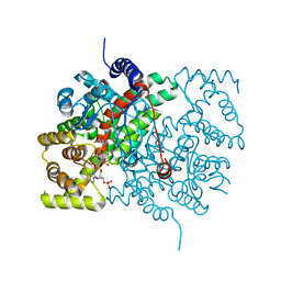
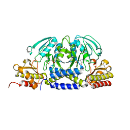
Crystal Structure of Delta 4 mutant of EhPSAT (Phosphoserine aminotransferase of Entamoeba histolytica)
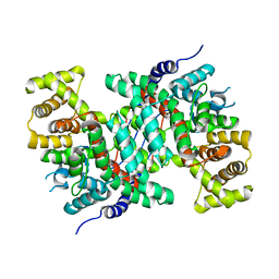
Descriptor: CHLORIDE ION, PYRIDOXAL-5'-PHOSPHATE, Phosphoserine aminotransferase
Authors:Singh, R.K, Gourinath, S.
Deposit date:2017-09-10
Release date:2018-10-31
Last modified:2023-11-22
Method:X-RAY DIFFRACTION (2.35 Å)
Cite:N-terminal residues are crucial for quaternary structure and active site conformation for the phosphoserine aminotransferase from enteric human parasite E. histolytica.
Int.J.Biol.Macromol., 132, 2019
5YII
DownloadVisualize
BU of 5yii by Molmil
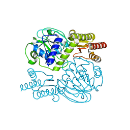
Crystal Structure of 45 amino acid deleted from N-terminal of Phosphoserine Aminotransferase (PSAT) of Entamoeba histolytica
Descriptor: Phosphoserine aminotransferase, THIOCYANATE ION
Authors:Singh, R.K, Gourinath, S.
Deposit date:2017-10-04
Release date:2018-10-24
Last modified:2023-11-22
Method:X-RAY DIFFRACTION (1.8 Å)
Cite:N-terminal residues are crucial for quaternary structure and active site conformation for the phosphoserine aminotransferase from enteric human parasite E. histolytica.
Int.J.Biol.Macromol., 132, 2019
3UAS
DownloadVisualize
BU of 3uas by Molmil
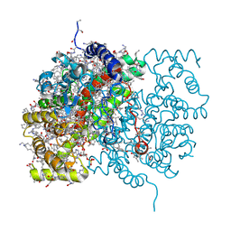
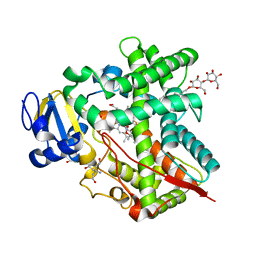
Cytochrome P450 2B4 covalently bound to the mechanism-based inactivator 9-ethynylphenanthrene
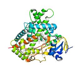
Descriptor: 5-CYCLOHEXYL-1-PENTYL-BETA-D-MALTOSIDE, Cytochrome P450 2B4, PROTOPORPHYRIN IX CONTAINING FE, ...
Authors:Gay, S.C, Zhang, H, Shah, M.B, Stout, C.D, Halpert, J.R, Hollenberg, P.F.
Deposit date:2011-10-21
Release date:2013-01-09
Last modified:2023-09-13
Method:X-RAY DIFFRACTION (2.939 Å)
Cite:Potent Mechanism-Based Inactivation of Cytochrome P450 2B4 by 9-Ethynylphenanthrene: Implications for Allosteric Modulation of Cytochrome P450 Catalysis.
Biochemistry, 52, 2013
8TDO
DownloadVisualize
BU of 8tdo by Molmil
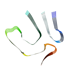
Cryo-EM structure of cardiac amyloid fibril from a variant ATTR I84S amyloidosis patient-3, variant-type morphology
Descriptor: Transthyretin
Authors:Nguyen, B.A, Singh, V, Saelices, L.
Deposit date:2023-07-04
Release date:2023-12-13
Last modified:2024-03-13
Method:ELECTRON MICROSCOPY (3.1 Å)
Cite:Structural polymorphism of amyloid fibrils in ATTR amyloidosis revealed by cryo-electron microscopy.
Nat Commun, 15, 2024
8TDN
DownloadVisualize
BU of 8tdn by Molmil
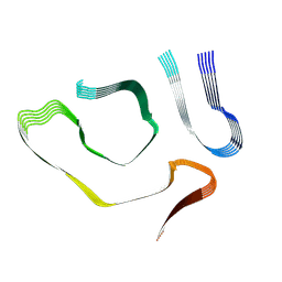
Cryo-EM structure of cardiac amyloid fibril from a variant ATTR I84S amyloidosis patient-3, wild-type morphology
Descriptor: Transthyretin
Authors:Nguyen, B.A, Singh, V, Saelices, L.
Deposit date:2023-07-04
Release date:2023-12-13
Last modified:2024-03-13
Method:ELECTRON MICROSCOPY (3.1 Å)
Cite:Structural polymorphism of amyloid fibrils in ATTR amyloidosis revealed by cryo-electron microscopy.
Nat Commun, 15, 2024
8TGO
DownloadVisualize
BU of 8tgo by Molmil
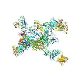
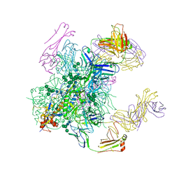
Crystal structure of the BG505 triple tandem trimer gp140 HIV-1 Env in complex with PGT124 and 35O22
Descriptor: 2-acetamido-2-deoxy-beta-D-glucopyranose, 2-acetamido-2-deoxy-beta-D-glucopyranose-(1-4)-2-acetamido-2-deoxy-beta-D-glucopyranose, 35O22 scFv, ...
Authors:Xian, Y, Yuan, M, Wilson, I.A.
Deposit date:2023-07-12
Release date:2024-04-17
Last modified:2024-10-30
Method:X-RAY DIFFRACTION (5.75 Å)
Cite:Triple tandem trimer immunogens for HIV-1 and influenza nucleic acid-based vaccines.
Npj Vaccines, 9, 2024
5YB0
DownloadVisualize
BU of 5yb0 by Molmil
Crystal Structure of Wild Type Phosphoserine aminotransferase (PSAT) from E. histolytica
Descriptor: CHLORIDE ION, PYRIDOXAL-5'-PHOSPHATE, Phosphoserine aminotransferase
Authors:Singh, R.K, Gourinath, S.
Deposit date:2017-09-02
Release date:2018-10-31
Last modified:2024-05-08
Method:X-RAY DIFFRACTION (2.94 Å)
Cite:N-terminal residues are crucial for quaternary structure and active site conformation for the phosphoserine aminotransferase from enteric human parasite E. histolytica.
Int.J.Biol.Macromol., 132, 2019
3TK3
DownloadVisualize
BU of 3tk3 by Molmil
Cytochrome P450 2B4 mutant L437A in complex with 4-(4-chlorophenyl)imidazole
Descriptor: 4-(4-CHLOROPHENYL)IMIDAZOLE, Cytochrome P450 2B4, PROTOPORPHYRIN IX CONTAINING FE
Authors:Gay, S.C, Jang, H.H, Wilderman, P.R, Zhang, Q, Stout, C.D, Halpert, J.R.
Deposit date:2011-08-25
Release date:2011-11-16
Last modified:2023-09-13
Method:X-RAY DIFFRACTION (2.8001 Å)
Cite:Investigation by site-directed mutagenesis of the role of cytochrome P450 2B4 non-active-site residues in protein-ligand interactions based on crystal structures of the ligand-bound enzyme.
Febs J., 279, 2012
5CTS
DownloadVisualize
BU of 5cts by Molmil
PROPOSED MECHANISM FOR THE CONDENSATION REACTION OF CITRATE SYNTHASE. 1.9-ANGSTROMS STRUCTURE OF THE TERNARY COMPLEX WITH OXALOACETATE AND CARBOXYMETHYL COENZYME A
Descriptor: CARBOXYMETHYL COENZYME *A, CITRATE SYNTHASE, OXALOACETATE ION
Authors:Karpusas, M, Branchaud, B, Remington, S.J.
Deposit date:1989-11-16
Release date:1990-10-15
Last modified:2024-03-06
Method:X-RAY DIFFRACTION (1.9 Å)
Cite:Proposed mechanism for the condensation reaction of citrate synthase: 1.9-A structure of the ternary complex with oxaloacetate and carboxymethyl coenzyme A.
Biochemistry, 29, 1990
5CSC
DownloadVisualize
BU of 5csc by Molmil
STRUCTURE OF AN OPEN FORM OF CHICKEN HEART CITRATE SYNTHASE AT 2.8 ANGSTROMS RESOLUTION
Descriptor: CITRATE SYNTHASE
Authors:Liao, D.-I, Karpusas, M, Remington, S.J.
Deposit date:1990-05-07
Release date:1991-04-15
Last modified:2024-03-06
Method:X-RAY DIFFRACTION (2.8 Å)
Cite:Crystal structure of an open conformation of citrate synthase from chicken heart at 2.8-A resolution
Biochemistry, 30, 1991
6AVN
DownloadVisualize
BU of 6avn by Molmil
Crystal structure of unbound anti-HIV antibody Fab PGV19 at 2.5 A
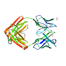
Descriptor: GLYCEROL, PENTAETHYLENE GLYCOL, PGV19 Fab heavy chain, ...
Authors:Sarkar, A, Wilson, I.A.
Deposit date:2017-09-03
Release date:2018-06-27
Last modified:2024-10-09
Method:X-RAY DIFFRACTION (2.5 Å)
Cite:Structure of a cleavage-independent HIV Env recapitulates the glycoprotein architecture of the native cleaved trimer.
Nat Commun, 9, 2018
6B0N
DownloadVisualize
BU of 6b0n by Molmil
Crystal structure of the cleavage-independent prefusion HIV Env glycoprotein trimer of the clade A BG505 isolate (NFL construct) in complex with Fabs PGT122 and PGV19 at 3.39 A
Descriptor: 2-acetamido-2-deoxy-beta-D-glucopyranose, 2-acetamido-2-deoxy-beta-D-glucopyranose-(1-4)-2-acetamido-2-deoxy-beta-D-glucopyranose, Envelope glycoprotein gp140, ...
Authors:Sarkar, A, Irimia, A, Wilson, I.A.
Deposit date:2017-09-14
Release date:2018-05-30
Last modified:2023-10-04
Method:X-RAY DIFFRACTION (3.4 Å)
Cite:Structure of a cleavage-independent HIV Env recapitulates the glycoprotein architecture of the native cleaved trimer.
Nat Commun, 9, 2018
1OUY
DownloadVisualize
BU of 1ouy by Molmil
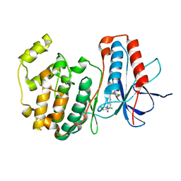
The structure of p38 alpha in complex with a dihydropyrido-pyrimidine inhibitor
Descriptor: 1-(2,6-DICHLOROPHENYL)-6-[(2,4-DIFLUOROPHENYL)SULFANYL]-7-(1,2,3,6-TETRAHYDRO-4-PYRIDINYL)-3,4-DIHYDROPYRIDO[3,2-D]PYRIMIDIN-2(1H)-ONE, Mitogen-activated protein kinase 14
Authors:Fitzgerald, C.E, Patel, S.B, Becker, J.W, Cameron, P.M, Zaller, D, Pikounis, V.B, O'Keefe, S.J, Scapin, G.
Deposit date:2003-03-25
Release date:2003-09-02
Last modified:2023-08-16
Method:X-RAY DIFFRACTION (2.5 Å)
Cite:Structural basis for p38alpha MAP kinase quinazolinone and pyridol-pyrimidine inhibitor specificity
Nat.Struct.Biol., 10, 2003
4LGX
DownloadVisualize
BU of 4lgx by Molmil
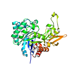
Structure of Chitinase D from Serratia proteamaculans revealed an unusually constrained substrate binding site
Descriptor: ACETATE ION, GLYCEROL, Glycoside hydrolase family 18
Authors:Madhuprakash, J, Singh, A, Kumar, S, Sinha, M, Kaur, P, Sharma, S, Podile, A.R, Singh, T.P.
Deposit date:2013-06-30
Release date:2013-10-02
Last modified:2023-11-08
Method:X-RAY DIFFRACTION (1.49 Å)
Cite:Inverse relationship between chitobiase and transglycosylation activities of chitinase-D from Serratia proteamaculans revealed by mutational and biophysical analyses.
Sci Rep, 5, 2015
2CSC
DownloadVisualize
BU of 2csc by Molmil
Structure of ternary complexes of CITRATE SYNTHASE WITH D-AND L-MALATE: mechanistic implications
Descriptor: CARBOXYMETHYL COENZYME *A, CITRATE SYNTHASE, D-MALATE
Authors:Karpusas, M, Holland, D, Remington, S.J.
Deposit date:1990-05-07
Release date:1991-04-15
Last modified:2024-02-14
Method:X-RAY DIFFRACTION (1.7 Å)
Cite:1.9-A structures of ternary complexes of citrate synthase with D- and L-malate: mechanistic implications.
Biochemistry, 30, 1991
2CTS
DownloadVisualize
BU of 2cts by Molmil
CRYSTALLOGRAPHIC REFINEMENT AND ATOMIC MODELS OF TWO DIFFERENT FORMS OF CITRATE SYNTHASE AT 2.7 AND 1.7 ANGSTROMS RESOLUTION
Descriptor: CITRATE SYNTHASE, CITRIC ACID, COENZYME A
Authors:Remington, S, Wiegand, G, Huber, R.
Deposit date:1984-01-27
Release date:1984-07-20
Last modified:2024-02-14
Method:X-RAY DIFFRACTION (2 Å)
Cite:Crystallographic refinement and atomic models of two different forms of citrate synthase at 2.7 and 1.7 A resolution.
J.Mol.Biol., 158, 1982
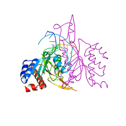
1ERI
DownloadVisualize
BU of 1eri by Molmil
X-RAY STRUCTURE OF THE DNA-ECO RI ENDONUCLEASE-DNA RECOGNITION COMPLEX: THE RECOGNITION NETWORK AND THE INTEGRATION OF RECOGNITION AND CLEAVAGE
Descriptor: DNA (5'-D(*TP*CP*GP*CP*GP*AP*AP*TP*TP*CP*GP*CP*G)-3'), PROTEIN (ECO RI ENDONUCLEASE (E.C.3.1.21.4))
Authors:Kim, Y, Grable, J.C, Love, R, Greene, P.J, Rosenberg, J.M.
Deposit date:1994-05-18
Release date:1995-02-07
Last modified:2024-02-07
Method:X-RAY DIFFRACTION (2.5 Å)
Cite:Refinement of Eco RI endonuclease crystal structure: a revised protein chain tracing.
Science, 249, 1990
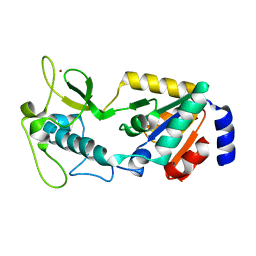
8GBL
DownloadVisualize
BU of 8gbl by Molmil
Structure of Apo Human SIRT5
Descriptor: NAD-dependent protein deacylase sirtuin-5, mitochondrial, ZINC ION
Authors:Petrunak, E.M, Stuckey, J.A.
Deposit date:2023-02-26
Release date:2024-06-05
Last modified:2024-06-19
Method:X-RAY DIFFRACTION (2.24 Å)
Cite:Human SIRT5 variants with reduced stability and activity do not cause neuropathology in mice.
Iscience, 27, 2024

226707

PDB entries from 2024-10-30

PDB statisticsPDBj update infoContact PDBjnumon