Loading
PDBj
MenuPDBj@FacebookPDBj@TwitterPDBj@YouTubewwPDB FoundationwwPDB
RCSB PDBPDBeBMRBAdv. SearchSearch help
Search by PDB author
6D4L
DownloadVisualize
BU of 6d4l by Molmil
Joint X-ray/neutron structure of DNA oligonucleotide d(GTGGCCAC)2 with 2'-SeCH3 modification on Cyt5
Descriptor: DNA (5'-D(*GP*TP*GP*GP*(CSL)P*CP*AP*C)-3'), MAGNESIUM ION
Authors:Kovalevsky, A, Huang, Z, Vandavasi, V.G.
Deposit date:2018-04-18
Release date:2018-10-17
Last modified:2023-10-04
Method:NEUTRON DIFFRACTION (1.56 Å), X-RAY DIFFRACTION
Cite:Temperature-Induced Replacement of Phosphate Proton with Metal Ion Captured in Neutron Structures of A-DNA.
Structure, 26, 2018
3KCL
DownloadVisualize
BU of 3kcl by Molmil
Room temperature neutron structure of D-Xylose Isomerase in complex with two Cd2+ cations and d12-D-alpha-glucose in the ring form (refined jointly with X-ray structure 3KBM)
Descriptor: CADMIUM ION, Xylose isomerase, alpha-D-glucopyranose
Authors:Kovalevsky, A.Y, Langan, P.
Deposit date:2009-10-21
Release date:2010-06-16
Last modified:2024-04-03
Method:NEUTRON DIFFRACTION (2 Å), X-RAY DIFFRACTION
Cite:Metal ion roles and the movement of hydrogen during reaction catalyzed by D-xylose isomerase: a joint x-ray and neutron diffraction study.
Structure, 18, 2010
3KBJ
DownloadVisualize
BU of 3kbj by Molmil
Room temperature X-ray structure of apo-D-Xylose Isomerase
Descriptor: Xylose isomerase
Authors:Kovalevsky, A.Y, Hanson, L, Langan, P.
Deposit date:2009-10-20
Release date:2010-09-29
Last modified:2024-02-21
Method:X-RAY DIFFRACTION (2 Å)
Cite:Identification of the Elusive Hydronium Ion Exchanging Roles with a Proton in an Enzyme at Lower pH Values.
Angew.Chem.Int.Ed.Engl., 50, 2011
3KCO
DownloadVisualize
BU of 3kco by Molmil
Room temperature neutron structure of D-Xylose Isomerase in complex with two Ni2+ cations and d12-D-glucose in the linear form (refined jointly with X-ray structure 3KBN)
Descriptor: D-glucose, NICKEL (II) ION, Xylose isomerase
Authors:Kovalevsky, A.Y, Langan, P.
Deposit date:2009-10-21
Release date:2010-06-16
Last modified:2024-04-03
Method:NEUTRON DIFFRACTION (1.8 Å), X-RAY DIFFRACTION
Cite:Metal ion roles and the movement of hydrogen during reaction catalyzed by D-xylose isomerase: a joint x-ray and neutron diffraction study.
Structure, 18, 2010
3KBN
DownloadVisualize
BU of 3kbn by Molmil
Room temperature structure of D-Xylose Isomerase in complex with 2Ni(2+) co-factors and d12-D-glucose in the linear form
Descriptor: D-glucose, NICKEL (II) ION, Xylose isomerase
Authors:Kovalevsky, A.Y, Hanson, L, Langan, P.
Deposit date:2009-10-20
Release date:2010-06-16
Last modified:2023-09-06
Method:X-RAY DIFFRACTION (1.53 Å)
Cite:Metal ion roles and the movement of hydrogen during reaction catalyzed by D-xylose isomerase: a joint x-ray and neutron diffraction study.
Structure, 18, 2010
3KYU
DownloadVisualize
BU of 3kyu by Molmil
X-ray crystal structure determination of fully perdeuterated rubredoxin at 100K
Descriptor: FE (III) ION, Rubredoxin
Authors:Gardberg, A.S.
Deposit date:2009-12-07
Release date:2010-04-28
Last modified:2023-09-06
Method:X-RAY DIFFRACTION (1.1 Å)
Cite:Unambiguous determination of H-atom positions: comparing results from neutron and high-resolution X-ray crystallography.
Acta Crystallogr.,Sect.D, 66, 2010
3KYV
DownloadVisualize
BU of 3kyv by Molmil
Denovo X-ray crystal structure determination of H-labeled perdeuterated rubredoxin at 100K
Descriptor: FE (III) ION, Rubredoxin
Authors:Gardberg, A.S.
Deposit date:2009-12-07
Release date:2010-04-28
Last modified:2024-02-21
Method:X-RAY DIFFRACTION (1.1 Å)
Cite:Unambiguous determination of H-atom positions: comparing results from neutron and high-resolution X-ray crystallography.
Acta Crystallogr.,Sect.D, 66, 2010
3KYW
DownloadVisualize
BU of 3kyw by Molmil
Xray crystal structure determination of H-labeled perdeuterated rubredoxin at 295K
Descriptor: FE (III) ION, Rubredoxin
Authors:Gardberg, A.S.
Deposit date:2009-12-07
Release date:2010-04-28
Last modified:2023-09-06
Method:X-RAY DIFFRACTION (1.1 Å)
Cite:Unambiguous determination of H-atom positions: comparing results from neutron and high-resolution X-ray crystallography.
Acta Crystallogr.,Sect.D, 66, 2010
3KYX
DownloadVisualize
BU of 3kyx by Molmil
Joint Xray/neutron crystal structure determination of fully perdeuterated rubredoxin at 295K
Descriptor: FE (III) ION, Rubredoxin
Authors:Gardberg, A.S, Meilleur, F.
Deposit date:2009-12-07
Release date:2010-04-28
Last modified:2023-09-06
Method:NEUTRON DIFFRACTION (1.675 Å), X-RAY DIFFRACTION
Cite:Unambiguous determination of H-atom positions: comparing results from neutron and high-resolution X-ray crystallography.
Acta Crystallogr.,Sect.D, 66, 2010
6FJI
DownloadVisualize
BU of 6fji by Molmil
Joint neutron and x-ray crystal structure of human carbonic anhydrase IX mimic (apo).
Descriptor: Carbonic anhydrase 2, ZINC ION
Authors:Fisher, S.Z.
Deposit date:2018-01-22
Release date:2019-02-06
Last modified:2024-05-01
Method:NEUTRON DIFFRACTION (1.6 Å), X-RAY DIFFRACTION
Cite:Using neutron crystallography to elucidate the basis of selective inhibition of carbonic anhydrase by saccharin and a derivative.
J. Struct. Biol., 205, 2019
6FJJ
DownloadVisualize
BU of 6fjj by Molmil
Joint neutron and x-ray crystal structure of human carbonic anhydrase IX mimic (saccharin).
Descriptor: 1,2-BENZISOTHIAZOL-3(2H)-ONE 1,1-DIOXIDE, Carbonic anhydrase 2, ZINC ION
Authors:Fisher, S.Z.
Deposit date:2018-01-22
Release date:2019-02-06
Last modified:2024-05-01
Method:NEUTRON DIFFRACTION (1.5 Å), X-RAY DIFFRACTION
Cite:Using neutron crystallography to elucidate the basis of selective inhibition of carbonic anhydrase by saccharin and a derivative.
J. Struct. Biol., 205, 2019
6D54
DownloadVisualize
BU of 6d54 by Molmil
Low Temperature joint X-ray/neutron structure of DNA oligonucleotide d(GTGGCCAC)2 with 2'-SeCH3 modification on Cyt5
Descriptor: DNA (5'-D(*GP*TP*GP*GP*(CSL)P*CP*AP*C)-3'), MAGNESIUM ION
Authors:Kovalevsky, A, Huang, Z, Vandavasi, V.G.
Deposit date:2018-04-19
Release date:2018-10-17
Last modified:2023-10-04
Method:NEUTRON DIFFRACTION (1.65 Å), X-RAY DIFFRACTION
Cite:Temperature-Induced Replacement of Phosphate Proton with Metal Ion Captured in Neutron Structures of A-DNA.
Structure, 26, 2018
6GCY
DownloadVisualize
BU of 6gcy by Molmil
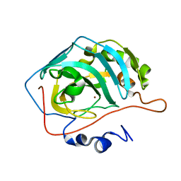
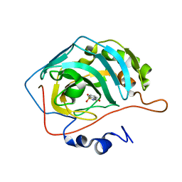
Joint neutron and x-ray crystal structure of human carbonic anhydrase IX mimic (saccharin-sugar conjugate complex)
Descriptor: Carbonic anhydrase 2, ZINC ION, [1-(1,1-dioxido-3-oxo-2,3-dihydro-1,2-benzothiazol-6-yl)-1H-1,2,3-triazol-4-yl]methyl alpha-L-idopyranoside
Authors:Fisher, S.Z, Koruza, K.
Deposit date:2018-04-20
Release date:2019-02-06
Last modified:2024-05-01
Method:NEUTRON DIFFRACTION (1.3 Å), X-RAY DIFFRACTION
Cite:Using neutron crystallography to elucidate the basis of selective inhibition of carbonic anhydrase by saccharin and a derivative.
J. Struct. Biol., 205, 2019
3KBS
DownloadVisualize
BU of 3kbs by Molmil
Room Temperature X-ray structure of D-Xylose Isomerase in complex with 2Cd(2+) co-factors
Descriptor: CADMIUM ION, Xylose isomerase
Authors:Kovalevsky, A.Y, Hanson, L, Langan, P.
Deposit date:2009-10-20
Release date:2010-06-16
Last modified:2024-02-21
Method:X-RAY DIFFRACTION (1.8 Å)
Cite:Metal ion roles and the movement of hydrogen during reaction catalyzed by D-xylose isomerase: a joint x-ray and neutron diffraction study.
Structure, 18, 2010
3KBM
DownloadVisualize
BU of 3kbm by Molmil
Room Temperature X-ray structure of D-Xylose Isomerase complexed with 2Cd(2+) co-factors and d12-D-alpha-glucose in the cyclic form
Descriptor: CADMIUM ION, Xylose isomerase, alpha-D-glucopyranose
Authors:Kovalevsky, A.Y, Hanson, L, Langan, P.
Deposit date:2009-10-20
Release date:2010-06-16
Last modified:2023-09-06
Method:X-RAY DIFFRACTION (2 Å)
Cite:Metal ion roles and the movement of hydrogen during reaction catalyzed by D-xylose isomerase: a joint x-ray and neutron diffraction study.
Structure, 18, 2010
3KBV
DownloadVisualize
BU of 3kbv by Molmil
Room temperature structure of D-Xylose Isomerase in complex with 2Ni(2+) co-factors
Descriptor: NICKEL (II) ION, Xylose isomerase
Authors:Kovalevsky, A.Y, Hanson, L, Langan, P.
Deposit date:2009-10-20
Release date:2010-06-16
Last modified:2024-02-21
Method:X-RAY DIFFRACTION (1.8 Å)
Cite:Metal ion roles and the movement of hydrogen during reaction catalyzed by D-xylose isomerase: a joint x-ray and neutron diffraction study.
Structure, 18, 2010
3KCJ
DownloadVisualize
BU of 3kcj by Molmil
Room temperature neutron structure of apo-D-Xylose Isomerase (refined jointly with X-ray structure 3KBJ)
Descriptor: Xylose isomerase
Authors:Kovalevsky, A.Y, Langan, P.
Deposit date:2009-10-21
Release date:2010-09-29
Last modified:2024-04-03
Method:NEUTRON DIFFRACTION (1.8 Å), X-RAY DIFFRACTION
Cite:Identification of the Elusive Hydronium Ion Exchanging Roles with a Proton in an Enzyme at Lower pH Values
Angew.Chem.Int.Ed.Engl., 50, 2011
3KBW
DownloadVisualize
BU of 3kbw by Molmil
Room temperature X-ray mixed-metal structure of D-Xylose Isomerase in complex with Ni(2+) and Mg(2+) co-factors
Descriptor: MAGNESIUM ION, NICKEL (II) ION, Xylose isomerase
Authors:Kovalevsky, A.Y, Hanson, L, Langan, P.
Deposit date:2009-10-20
Release date:2010-06-16
Last modified:2024-02-21
Method:X-RAY DIFFRACTION (1.6 Å)
Cite:Metal ion roles and the movement of hydrogen during reaction catalyzed by D-xylose isomerase: a joint x-ray and neutron diffraction study.
Structure, 18, 2010
6E21
DownloadVisualize
BU of 6e21 by Molmil
Joint X-ray/neutron structure of PKAc with products Sr2-ADP and phosphorylated peptide SP20
Descriptor: ADENOSINE-5'-DIPHOSPHATE, STRONTIUM ION, cAMP-dependent protein kinase catalytic subunit alpha, ...
Authors:Kovalevsky, A, Gerlits, O.O, Taylor, S.
Deposit date:2018-07-10
Release date:2019-04-03
Last modified:2023-10-25
Method:NEUTRON DIFFRACTION (2 Å), X-RAY DIFFRACTION
Cite:Zooming in on protons: Neutron structure of protein kinase A trapped in a product complex.
Sci Adv, 5, 2019
6GTL
DownloadVisualize
BU of 6gtl by Molmil
Achromobacter cycloclastes copper nitrite reductase at pH 6.0
Descriptor: COPPER (II) ION, Copper-containing nitrite reductase, MALONATE ION, ...
Authors:Halsted, T.P, Eady, R.R, Antonyuk, S.V, Hasnain, S.S.
Deposit date:2018-06-18
Release date:2019-07-03
Last modified:2024-05-15
Method:X-RAY DIFFRACTION (1.5 Å)
Cite:Catalytically important damage-free structures of a copper nitrite reductase obtained by femtosecond X-ray laser and room-temperature neutron crystallography.
Iucrj, 6, 2019
3KYY
DownloadVisualize
BU of 3kyy by Molmil
Joint Xray/neutron crystal structure determination of H-labeled perdeuterated rubredoxin at 295K
Descriptor: FE (III) ION, Rubredoxin
Authors:Gardberg, A.S.
Deposit date:2009-12-07
Release date:2010-04-28
Last modified:2023-09-13
Method:NEUTRON DIFFRACTION (1.1 Å), X-RAY DIFFRACTION
Cite:Unambiguous determination of H-atom positions: comparing results from neutron and high-resolution X-ray crystallography.
Acta Crystallogr.,Sect.D, 66, 2010
4JEC
DownloadVisualize
BU of 4jec by Molmil
Joint neutron and X-ray structure of per-deuterated HIV-1 protease in complex with clinical inhibitor amprenavir
Descriptor: CHLORIDE ION, HIV-1 protease, {3-[(4-AMINO-BENZENESULFONYL)-ISOBUTYL-AMINO]-1-BENZYL-2-HYDROXY-PROPYL}-CARBAMIC ACID TETRAHYDRO-FURAN-3-YL ESTER
Authors:Kovalevsky, A.Y, Weber, I.T, Langan, P.
Deposit date:2013-02-26
Release date:2013-07-24
Last modified:2024-02-28
Method:NEUTRON DIFFRACTION (2.01 Å), X-RAY DIFFRACTION
Cite:Joint X-ray/Neutron Crystallographic Study of HIV-1 Protease with Clinical Inhibitor Amprenavir: Insights for Drug Design.
J.Med.Chem., 56, 2013
4DVO
DownloadVisualize
BU of 4dvo by Molmil
Room-temperature joint X-ray/neutron structure of D-xylose isomerase in complex with 2Ni2+ and per-deuterated D-sorbitol at pH 5.9
Descriptor: NICKEL (II) ION, Xylose isomerase, sorbitol
Authors:Kovalevsky, A.Y, Hanson, L, Langan, P.
Deposit date:2012-02-23
Release date:2012-08-29
Last modified:2024-02-28
Method:NEUTRON DIFFRACTION (2 Å), X-RAY DIFFRACTION
Cite:Inhibition of D-xylose isomerase by polyols: atomic details by joint X-ray/neutron crystallography.
Acta Crystallogr.,Sect.D, 68, 2012
4DUO
DownloadVisualize
BU of 4duo by Molmil
Room-temperature X-ray structure of D-Xylose Isomerase in complex with 2Mg2+ ions and xylitol at pH 7.7
Descriptor: MAGNESIUM ION, Xylitol, Xylose isomerase
Authors:Kovalevsky, A.Y, Hanson, L, Langan, P.
Deposit date:2012-02-22
Release date:2012-08-29
Last modified:2024-02-28
Method:X-RAY DIFFRACTION (2 Å)
Cite:Inhibition of D-xylose isomerase by polyols: atomic details by joint X-ray/neutron crystallography.
Acta Crystallogr.,Sect.D, 68, 2012
5T8H
DownloadVisualize
BU of 5t8h by Molmil
Joint X-ray/neutron structure of HIV-1 protease triple mutant (V32I,I47V,V82I) with amprenavir at pH 6.0
Descriptor: Protease, {3-[(4-AMINO-BENZENESULFONYL)-ISOBUTYL-AMINO]-1-BENZYL-2-HYDROXY-PROPYL}-CARBAMIC ACID TETRAHYDRO-FURAN-3-YL ESTER
Authors:Kovalevsky, A.Y, Gerlits, O.O.
Deposit date:2016-09-07
Release date:2017-03-01
Last modified:2024-04-03
Method:NEUTRON DIFFRACTION (1.85 Å), X-RAY DIFFRACTION
Cite:Room Temperature Neutron Crystallography of Drug Resistant HIV-1 Protease Uncovers Limitations of X-ray Structural Analysis at 100 K.
J. Med. Chem., 60, 2017

226262

數據於2024-10-16公開中

PDB statisticsPDBj update infoContact PDBjnumon