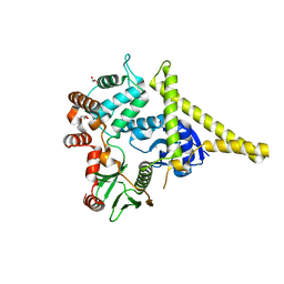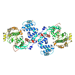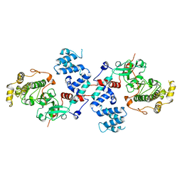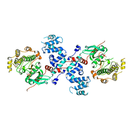
Loading
PDBj
MenuPDBj@FacebookPDBj@TwitterPDBj@YouTubewwPDB FoundationwwPDB
RCSB PDBPDBeBMRBAdv. SearchSearch help
Search by PDB author
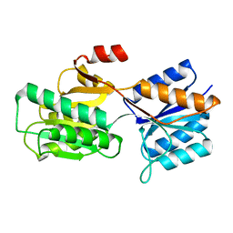
7ABP
DownloadVisualize
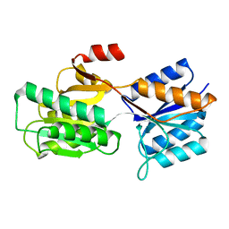
BU of 7abp by Molmil
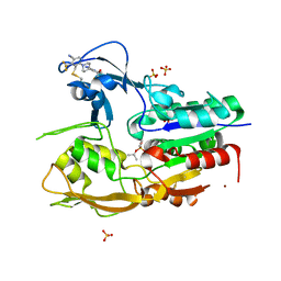
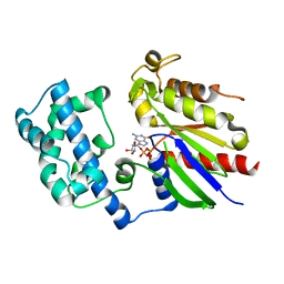
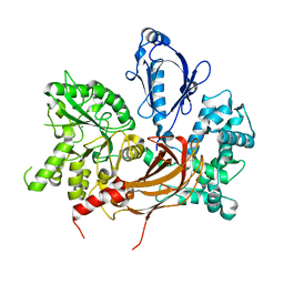
SUGAR-BINDING AND CRYSTALLOGRAPHIC STUDIES OF AN ARABINOSE-BINDING PROTEIN MUTANT (MET108LEU) WHICH EXHIBITS ENHANCED AFFINITY AND ALTERED SPECIFICITY
Descriptor: L-ARABINOSE-BINDING PROTEIN, alpha-D-fucopyranose, beta-D-fucopyranose
Authors:Vermersch, P.S, Tesmer, J.J.G, Quiocho, F.A.
Deposit date:1991-04-25
Release date:1991-10-15
Last modified:2024-03-06
Method:X-RAY DIFFRACTION (1.67 Å)
Cite:Sugar-binding and crystallographic studies of an arabinose-binding protein mutant (Met108Leu) that exhibits enhanced affinity and altered specificity.
Biochemistry, 30, 1991
9ABP
DownloadVisualize
BU of 9abp by Molmil
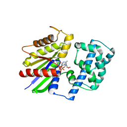
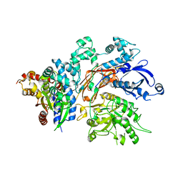
A PRO TO GLY MUTATION IN THE HINGE OF THE ARABINOSE-BINDING PROTEIN ENHANCES BINDING AND ALTERS SPECIFICITY: SUGAR-BINDING AND CRYSTALLOGRAPHIC STUDIES
Descriptor: L-ARABINOSE-BINDING PROTEIN, alpha-D-galactopyranose, beta-D-galactopyranose
Authors:Vermersch, P.S, Tesmer, J.J.G, Quiocho, F.A.
Deposit date:1991-11-15
Release date:1992-01-15
Last modified:2024-02-14
Method:X-RAY DIFFRACTION (1.97 Å)
Cite:A Pro to Gly mutation in the hinge of the arabinose-binding protein enhances binding and alters specificity. Sugar-binding and crystallographic studies.
J.Biol.Chem., 265, 1990
2RGN
DownloadVisualize
BU of 2rgn by Molmil
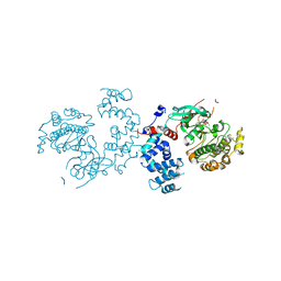
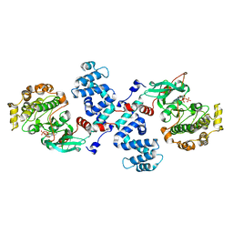
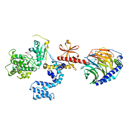
Crystal Structure of p63RhoGEF complex with Galpha-q and RhoA
Descriptor: GUANOSINE-5'-DIPHOSPHATE, Guanine nucleotide-binding protein G(i) subunit alpha-1,Guanine nucleotide-binding protein G(q) subunit alpha, MAGNESIUM ION, ...
Authors:Shankaranarayanan, A, Nance, M.R, Tesmer, J.J.G.
Deposit date:2007-10-04
Release date:2008-01-15
Last modified:2023-08-30
Method:X-RAY DIFFRACTION (3.5 Å)
Cite:Structure of Galphaq-p63RhoGEF-RhoA complex reveals a pathway for the activation of RhoA by GPCRs.
Science, 318, 2007
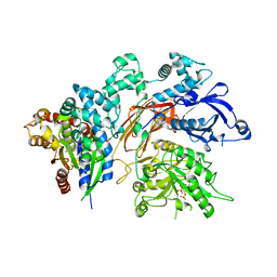
4L9I
DownloadVisualize
BU of 4l9i by Molmil
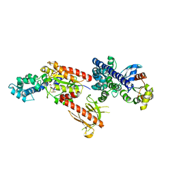
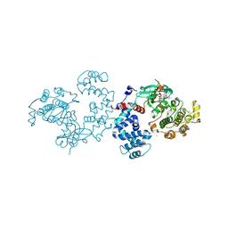
Bovine G Protein Coupled Receptor Kinase 1 in Complex with Paroxetine
Descriptor: 2-(N-MORPHOLINO)-ETHANESULFONIC ACID, CHLORIDE ION, Paroxetine, ...
Authors:Homan, K.T, Tesmer, J.J.G.
Deposit date:2013-06-18
Release date:2014-01-22
Last modified:2023-09-20
Method:X-RAY DIFFRACTION (2.32 Å)
Cite:Structural and functional analysis of g protein-coupled receptor kinase inhibition by paroxetine and a rationally designed analog.
Mol.Pharmacol., 85, 2014
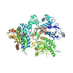
4MK0
DownloadVisualize
BU of 4mk0 by Molmil
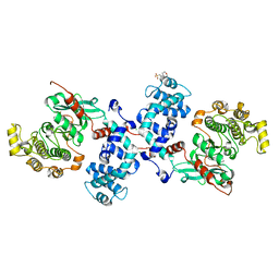
Crystal structure of G protein-coupled receptor kinase 2 in complex with a a rationally designed paroxetine derivative
Descriptor: 5-{[(3S,4R)-4-(4-fluorophenyl)piperidin-3-yl]methoxy}-1H-isoindol-1-one, Beta-adrenergic receptor kinase 1, GLYCEROL, ...
Authors:Homan, K.T, Tesmer, J.J.G.
Deposit date:2013-09-04
Release date:2014-01-22
Last modified:2024-02-28
Method:X-RAY DIFFRACTION (2.4 Å)
Cite:Structural and functional analysis of g protein-coupled receptor kinase inhibition by paroxetine and a rationally designed analog.
Mol.Pharmacol., 85, 2014
3C4W
DownloadVisualize
BU of 3c4w by Molmil
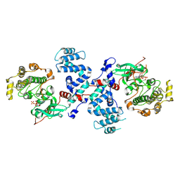
Crystal Structure of G protein coupled receptor kinase 1 bound to ATP and magnesium chloride at 2.7A
Descriptor: ADENOSINE-5'-TRIPHOSPHATE, CHLORIDE ION, MAGNESIUM ION, ...
Authors:Singh, P, Tesmer, J.J.G.
Deposit date:2008-01-30
Release date:2008-03-11
Last modified:2023-08-30
Method:X-RAY DIFFRACTION (2.7 Å)
Cite:Structures of rhodopsin kinase in different ligand states reveal key elements involved in G protein-coupled receptor kinase activation.
J.Biol.Chem., 283, 2008
3C4Z
DownloadVisualize
BU of 3c4z by Molmil
Crystal structure of G protein coupled receptor kinase 1 bound to ADP and magnesium chloride at 1.84A
Descriptor: ADENOSINE-5'-DIPHOSPHATE, CHLORIDE ION, MAGNESIUM ION, ...
Authors:Singh, P, Tesmer, J.J.G.
Deposit date:2008-01-30
Release date:2008-03-11
Last modified:2023-08-30
Method:X-RAY DIFFRACTION (1.84 Å)
Cite:Structures of rhodopsin kinase in different ligand states reveal key elements involved in G protein-coupled receptor kinase activation.
J.Biol.Chem., 283, 2008
6MVD
DownloadVisualize
BU of 6mvd by Molmil
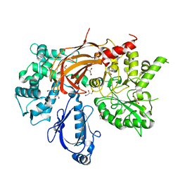
Crystal structure of Lecithin:cholesterol acyltransferase (LCAT) in complex with isopropyl dodec-11-enylfluorophosphonate (IDFP) and a small molecule activator
Descriptor: 2-acetamido-2-deoxy-beta-D-glucopyranose, 6-{4-[(4R)-4-hydroxy-6-oxo-4-(trifluoromethyl)-4,5,6,7-tetrahydro-2H-pyrazolo[3,4-b]pyridin-3-yl]piperidin-1-yl}-4-(trifluoromethyl)pyridine-3-carbonitrile, NICKEL (II) ION, ...
Authors:Manthei, K.A, Chang, L, Tesmer, J.J.G.
Deposit date:2018-10-25
Release date:2018-12-05
Last modified:2023-10-11
Method:X-RAY DIFFRACTION (3.1 Å)
Cite:Molecular basis for activation of lecithin:cholesterol acyltransferase by a compound that increases HDL cholesterol.
Elife, 7, 2018
3V5W
DownloadVisualize
BU of 3v5w by Molmil
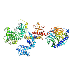
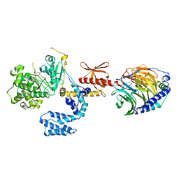
Human G Protein-Coupled Receptor Kinase 2 in Complex with Soluble Gbetagamma Subunits and Paroxetine
Descriptor: G-protein coupled receptor kinase 2, Guanine nucleotide-binding protein G(I)/G(S)/G(O) subunit gamma-2, Guanine nucleotide-binding protein G(I)/G(S)/G(T) subunit beta-1, ...
Authors:Thal, D.M, Tesmer, J.J.G.
Deposit date:2011-12-17
Release date:2012-09-26
Last modified:2023-09-13
Method:X-RAY DIFFRACTION (2.07 Å)
Cite:Paroxetine is a direct inhibitor of g protein-coupled receptor kinase 2 and increases myocardial contractility.
Acs Chem.Biol., 7, 2012
2I1K
DownloadVisualize
BU of 2i1k by Molmil
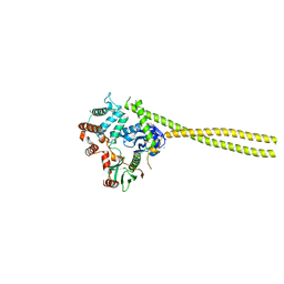
Moesin from Spodoptera frugiperda reveals the coiled-coil domain at 3.0 angstrom resolution
Descriptor: CHLORIDE ION, GLYCEROL, Moesin, ...
Authors:Nance, M.R, Tesmer, J.J.G.
Deposit date:2006-08-14
Release date:2006-12-19
Last modified:2024-02-21
Method:X-RAY DIFFRACTION (3 Å)
Cite:Self-masking in an Intact ERM-merlin Protein: An Active Role for the Central alpha-Helical Domain.
J.Mol.Biol., 365, 2007
2I1J
DownloadVisualize
BU of 2i1j by Molmil
Moesin from Spodoptera frugiperda at 2.1 angstroms resolution
Descriptor: CHLORIDE ION, GLYCEROL, Moesin, ...
Authors:Li, Q, Nance, M.R, Tesmer, J.J.G.
Deposit date:2006-08-14
Release date:2006-12-19
Last modified:2024-02-21
Method:X-RAY DIFFRACTION (2.1 Å)
Cite:Self-masking in an Intact ERM-merlin Protein: An Active Role for the Central alpha-Helical Domain.
J.Mol.Biol., 365, 2007
3C50
DownloadVisualize
BU of 3c50 by Molmil
Crystal Structure of G protein coupled receptor kinase 1 bound to ADP and magnesium chloride at 2.6A
Descriptor: 2-(N-MORPHOLINO)-ETHANESULFONIC ACID, ADENOSINE-5'-DIPHOSPHATE, CHLORIDE ION, ...
Authors:Singh, P, Tesmer, J.J.G.
Deposit date:2008-01-30
Release date:2008-03-11
Last modified:2023-08-30
Method:X-RAY DIFFRACTION (2.6 Å)
Cite:Structures of rhodopsin kinase in different ligand states reveal key elements involved in G protein-coupled receptor kinase activation.
J.Biol.Chem., 283, 2008
3C4Y
DownloadVisualize
BU of 3c4y by Molmil
Crystal Structure of Apo form of G protein coupled receptor kinase 1 at 7.51A
Descriptor: Rhodopsin kinase
Authors:Singh, P, Tesmer, J.J.G.
Deposit date:2008-01-30
Release date:2008-03-11
Last modified:2023-08-30
Method:X-RAY DIFFRACTION (7.509 Å)
Cite:Structures of rhodopsin kinase in different ligand states reveal key elements involved in G protein-coupled receptor kinase activation.
J.Biol.Chem., 283, 2008
3C51
DownloadVisualize
BU of 3c51 by Molmil
Crystal structure of G protein coupled receptor kinase 1 bound to ADP and magnesium chloride at 3.55A
Descriptor: ADENOSINE-5'-DIPHOSPHATE, BROMIDE ION, MAGNESIUM ION, ...
Authors:Singh, P, Tesmer, J.J.G.
Deposit date:2008-01-30
Release date:2008-03-11
Last modified:2023-08-30
Method:X-RAY DIFFRACTION (3.55 Å)
Cite:Structures of rhodopsin kinase in different ligand states reveal key elements involved in G protein-coupled receptor kinase activation.
J.Biol.Chem., 283, 2008
3C4X
DownloadVisualize
BU of 3c4x by Molmil
Crystal Structure of G protein coupled receptor kinase 1 bound to ATP and magnesium chloride at 2.9A
Descriptor: ADENOSINE-5'-TRIPHOSPHATE, CHLORIDE ION, MAGNESIUM ION, ...
Authors:Singh, P, Tesmer, J.J.G.
Deposit date:2008-01-30
Release date:2008-03-11
Last modified:2023-08-30
Method:X-RAY DIFFRACTION (2.9 Å)
Cite:Structures of rhodopsin kinase in different ligand states reveal key elements involved in G protein-coupled receptor kinase activation.
J.Biol.Chem., 283, 2008
1ZCB
DownloadVisualize
BU of 1zcb by Molmil
Crystal structure of G alpha 13 in complex with GDP
Descriptor: G alpha i/13, GUANOSINE-5'-DIPHOSPHATE
Authors:Nance, M.R, Tesmer, J.J.G.
Deposit date:2005-04-11
Release date:2005-11-15
Last modified:2023-08-23
Method:X-RAY DIFFRACTION (2 Å)
Cite:A new approach to producing functional G alpha subunits yields the activated and deactivated structures of G alpha(12/13) proteins.
Biochemistry, 45, 2006
1ZCA
DownloadVisualize
BU of 1zca by Molmil
Crystal structure of G alpha 12 in complex with GDP, Mg2+ and AlF4-
Descriptor: G alpha i/12, GUANOSINE-5'-DIPHOSPHATE, MAGNESIUM ION, ...
Authors:Nance, M.R, Tesmer, J.J.G.
Deposit date:2005-04-11
Release date:2005-11-15
Last modified:2023-08-23
Method:X-RAY DIFFRACTION (2.9 Å)
Cite:A new approach to producing functional G alpha subunits yields the activated and deactivated structures of G alpha(12/13) proteins.
Biochemistry, 45, 2006
3QR0
DownloadVisualize
BU of 3qr0 by Molmil
Crystal Structure of S. officinalis PLC21
Descriptor: CALCIUM ION, GLYCEROL, phospholipase C-beta (PLC-beta)
Authors:Lyon, A.M, Northup, J.K, Tesmer, J.J.G.
Deposit date:2011-02-16
Release date:2011-08-10
Last modified:2023-09-13
Method:X-RAY DIFFRACTION (2 Å)
Cite:An autoinhibitory helix in the C-terminal region of phospholipase C-beta mediates Galphaq activation.
Nat.Struct.Mol.Biol., 18, 2011
3QR1
DownloadVisualize
BU of 3qr1 by Molmil
Crystal Structure of L. pealei PLC21
Descriptor: CALCIUM ION, PHOSPHOLIPASE C-BETA (PLC-BETA)
Authors:Lyon, A.M, Suddala, K.C, Northup, J.K, Tesmer, J.J.G.
Deposit date:2011-02-16
Release date:2011-08-10
Last modified:2024-02-21
Method:X-RAY DIFFRACTION (3.2 Å)
Cite:An autoinhibitory helix in the C-terminal region of phospholipase C-beta mediates Galphaq activation.
Nat.Struct.Mol.Biol., 18, 2011
4QJ3
DownloadVisualize
BU of 4qj3 by Molmil
Structure of a fragment of human phospholipase C-beta3 delta472-559, in complex with Galphaq
Descriptor: 1-phosphatidylinositol 4,5-bisphosphate phosphodiesterase beta-3, CALCIUM ION, GUANOSINE-5'-DIPHOSPHATE, ...
Authors:Lyon, A.M, Tesmer, J.J.G.
Deposit date:2014-06-03
Release date:2014-10-22
Last modified:2023-09-20
Method:X-RAY DIFFRACTION (3 Å)
Cite:Molecular mechanisms of phospholipase C beta 3 autoinhibition.
Structure, 22, 2014
4PNK
DownloadVisualize
BU of 4pnk by Molmil
G protein-coupled receptor kinase 2 in complex with GSK180736A
Descriptor: (4S)-4-(4-fluorophenyl)-N-(2H-indazol-5-yl)-6-methyl-2-oxo-1,2,3,4-tetrahydropyrimidine-5-carboxamide, Beta-adrenergic receptor kinase 1, Guanine nucleotide-binding protein G(I)/G(S)/G(O) subunit gamma-2, ...
Authors:Homan, K.T, Larimore, K.M, Elkins, J, Knapp, S, Tesmer, J.J.G.
Deposit date:2014-05-23
Release date:2014-10-08
Last modified:2023-12-27
Method:X-RAY DIFFRACTION (2.56 Å)
Cite:Identification and structure-function analysis of subfamily selective g protein-coupled receptor kinase inhibitors.
Acs Chem.Biol., 10, 2015
4PNI
DownloadVisualize
BU of 4pni by Molmil
Bovine G protein-coupled receptor kinase 1 in complex with GSK2163632A
Descriptor: 3-[(2-{[1-(N,N-dimethylglycyl)-6-methoxy-4,4-dimethyl-1,2,3,4-tetrahydroquinolin-7-yl]amino}-7H-pyrrolo[2,3-d]pyrimidin-4-yl)amino]thiophene-2-carboxamide, CHLORIDE ION, Rhodopsin kinase
Authors:Homan, K.T, Tesmer, J.J.G.
Deposit date:2014-05-23
Release date:2014-10-08
Last modified:2023-12-27
Method:X-RAY DIFFRACTION (1.85 Å)
Cite:Identification and structure-function analysis of subfamily selective g protein-coupled receptor kinase inhibitors.
Acs Chem.Biol., 10, 2015
4QJ4
DownloadVisualize
BU of 4qj4 by Molmil
Structure of a fragment of human phospholipase C-beta3 delta472-569, bound to IP3 and in complex with Galphaq
Descriptor: 1-phosphatidylinositol 4,5-bisphosphate phosphodiesterase beta-3, CALCIUM ION, D-MYO-INOSITOL-1,4,5-TRIPHOSPHATE, ...
Authors:Lyon, A.M, Tesmer, J.J.G.
Deposit date:2014-06-03
Release date:2014-10-22
Last modified:2023-09-20
Method:X-RAY DIFFRACTION (3.3 Å)
Cite:Molecular mechanisms of phospholipase C beta 3 autoinhibition.
Structure, 22, 2014
4QJ5
DownloadVisualize
BU of 4qj5 by Molmil
Structure of a fragment of human phospholipase C-beta3 delta472-581, bound to IP3 and in complex with Galphaq
Descriptor: 1-phosphatidylinositol 4,5-bisphosphate phosphodiesterase beta-3, CALCIUM ION, D-MYO-INOSITOL-1,4,5-TRIPHOSPHATE, ...
Authors:Lyon, A.M, Tesmer, J.J.G.
Deposit date:2014-06-03
Release date:2014-10-22
Last modified:2023-09-20
Method:X-RAY DIFFRACTION (3.41 Å)
Cite:Molecular mechanisms of phospholipase C beta 3 autoinhibition.
Structure, 22, 2014
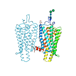
4BEZ
DownloadVisualize
BU of 4bez by Molmil
Night blindness causing G90D rhodopsin in the active conformation
Descriptor: ACETATE ION, PALMITIC ACID, RHODOPSIN, ...
Authors:Singhal, A, Ostermaier, M.K, Vishnivetskiy, S.A, Panneels, V, Homan, K.T, Tesmer, J.J.G, Veprintsev, D, Deupi, X, Gurevich, V.V, Schertler, G.F.X, Standfuss, J.
Deposit date:2013-03-12
Release date:2013-04-24
Last modified:2023-12-20
Method:X-RAY DIFFRACTION (3.3 Å)
Cite:Insights Into Congenital Stationary Night Blindness Based on the Structure of G90D Rhodopsin.
Embo Rep., 14, 2013

221051

數據於2024-06-12公開中

PDB statisticsPDBj update infoContact PDBjnumon