Loading
PDBj
MenuPDBj@FacebookPDBj@TwitterPDBj@YouTubewwPDB FoundationwwPDB
RCSB PDBPDBeBMRBAdv. SearchSearch help
Search by PDB author
3NL9
DownloadVisualize
BU of 3nl9 by Molmil
Crystal structure of a putative NTP pyrophosphohydrolase (Exig_1061) from EXIGUOBACTERIUM SP. 255-15 at 1.78 A resolution
Descriptor: 1,2-ETHANEDIOL, putative NTP pyrophosphohydrolase
Authors:Joint Center for Structural Genomics (JCSG)
Deposit date:2010-06-21
Release date:2010-07-21
Last modified:2023-02-01
Method:X-RAY DIFFRACTION (1.78 Å)
Cite:Structure of a putative NTP pyrophosphohydrolase: YP_001813558.1 from Exiguobacterium sibiricum 255-15.
Acta Crystallogr.,Sect.F, 66, 2010
3L5O
DownloadVisualize
BU of 3l5o by Molmil
Crystal structure of protein with unknown function from DUF364 family (ZP_00559375.1) from Desulfitobacterium hafniense DCB-2 at 2.01 A resolution
Descriptor: 1,2-ETHANEDIOL, CHLORIDE ION, IMIDAZOLE, ...
Authors:Joint Center for Structural Genomics, Joint Center for Structural Genomics (JCSG)
Deposit date:2009-12-22
Release date:2010-02-02
Last modified:2023-02-01
Method:X-RAY DIFFRACTION (2.01 Å)
Cite:Structure of the first representative of Pfam family PF04016 (DUF364) reveals enolase and Rossmann-like folds that combine to form a unique active site with a possible role in heavy-metal chelation.
Acta Crystallogr.,Sect.F, 66, 2010
1VR8
DownloadVisualize
BU of 1vr8 by Molmil
Crystal structure of GTP binding regulator (TM1622) from Thermotoga Maritima at 1.75 A resolution
Descriptor: AZIDE ION, GLYCEROL, GTP binding regulator
Authors:Joint Center for Structural Genomics (JCSG)
Deposit date:2005-02-16
Release date:2005-03-15
Last modified:2023-01-25
Method:X-RAY DIFFRACTION (1.75 Å)
Cite:Crystal structure of an ORFan protein (TM1622) from Thermotoga maritima at 1.75 A resolution reveals a fold similar to the Ran-binding protein Mog1p.
Proteins, 65, 2006
2GVI
DownloadVisualize
BU of 2gvi by Molmil
Crystal structure of a putative formylmethanofuran dehydrogenase subunit e (ta1109) from thermoplasma acidophilum at 1.87 A resolution
Descriptor: 1,2-ETHANEDIOL, ACETIC ACID, UNKNOWN LIGAND, ...
Authors:Joint Center for Structural Genomics (JCSG)
Deposit date:2006-05-02
Release date:2006-06-13
Last modified:2023-01-25
Method:X-RAY DIFFRACTION (1.87 Å)
Cite:Structures of three members of Pfam PF02663 (FmdE) implicated in microbial methanogenesis reveal a conserved alpha+beta core domain and an auxiliary C-terminal treble-clef zinc finger.
Acta Crystallogr.,Sect.F, 66, 2010
2HAG
DownloadVisualize
BU of 2hag by Molmil
Crystal structure of a putative dyp-type peroxidase protein (so_0740) from shewanella oneidensis at 2.75 A resolution
Descriptor: Melanin biosynthesis protein TyrA, putative
Authors:Joint Center for Structural Genomics (JCSG)
Deposit date:2006-06-12
Release date:2006-08-08
Last modified:2023-01-25
Method:X-RAY DIFFRACTION (2.75 Å)
Cite:Crystal structures of two novel dye-decolorizing peroxidases reveal a beta-barrel fold with a conserved heme-binding motif.
Proteins, 69, 2007
2GHR
DownloadVisualize
BU of 2ghr by Molmil
Crystal structure of homoserine o-succinyltransferase (NP_981826.1) from Bacillus cereus ATCC 10987 at 2.40 A resolution
Descriptor: Homoserine O-succinyltransferase, SULFATE ION
Authors:Joint Center for Structural Genomics (JCSG)
Deposit date:2006-03-27
Release date:2006-04-11
Last modified:2023-01-25
Method:X-RAY DIFFRACTION (2.4 Å)
Cite:Crystal structure of homoserine O-succinyltransferase from Bacillus cereus at 2.4 A resolution
Proteins, 68, 2007
2GLZ
DownloadVisualize
BU of 2glz by Molmil
Crystal structure of a formylmethanofuran dehydrogenase subunit e-like protein (dhaf_2992) from desulfitobacterium hafniense dcb-2 at 1.45 A resolution
Descriptor: 1,2-ETHANEDIOL, NICKEL (II) ION, ZINC ION, ...
Authors:Joint Center for Structural Genomics (JCSG)
Deposit date:2006-04-05
Release date:2006-04-18
Last modified:2023-01-25
Method:X-RAY DIFFRACTION (1.45 Å)
Cite:Structures of three members of Pfam PF02663 (FmdE) implicated in microbial methanogenesis reveal a conserved alpha+beta core domain and an auxiliary C-terminal treble-clef zinc finger.
Acta Crystallogr.,Sect.F, 66, 2010
2GVK
DownloadVisualize
BU of 2gvk by Molmil
Crystal structure of a dye-decolorizing peroxidase (DyP) from Bacteroides thetaiotaomicron VPI-5482 at 1.6 A resolution
Descriptor: 1,2-ETHANEDIOL, ACETIC ACID, CHLORIDE ION, ...
Authors:Joint Center for Structural Genomics (JCSG)
Deposit date:2006-05-02
Release date:2006-05-16
Last modified:2023-01-25
Method:X-RAY DIFFRACTION (1.6 Å)
Cite:Crystal structures of two novel dye-decolorizing peroxidases reveal a beta-barrel fold with a conserved heme-binding motif.
Proteins, 69, 2007
2F46
DownloadVisualize
BU of 2f46 by Molmil
Crystal structure of a putative phosphatase (nma1982) from neisseria meningitidis z2491 at 1.41 A resolution
Descriptor: CHLORIDE ION, UNKNOWN LIGAND, hypothetical protein
Authors:Joint Center for Structural Genomics (JCSG)
Deposit date:2005-11-22
Release date:2006-02-07
Last modified:2023-01-25
Method:X-RAY DIFFRACTION (1.41 Å)
Cite:Crystal structure of NMA1982 from Neisseria meningitidis at 1.5 A resolution provides a structural scaffold for nonclassical, eukaryotic-like phosphatases.
Proteins, 69, 2007
6YBQ
DownloadVisualize
BU of 6ybq by Molmil
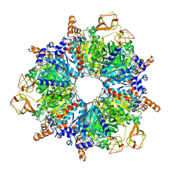
Engineered glycolyl-CoA carboxylase (quintuple mutant) with bound CoA
Descriptor: 5-(HEXAHYDRO-2-OXO-1H-THIENO[3,4-D]IMIDAZOL-6-YL)PENTANAL, COENZYME A, Propionyl-CoA carboxylase alpha subunit, ...
Authors:Schuller, J.M, Schuller, S.K, Zarzycki, J, Scheffen, M, Marchal, D.M, Erb, T.J.
Deposit date:2020-03-17
Release date:2020-10-28
Last modified:2021-05-12
Method:ELECTRON MICROSCOPY (1.96 Å)
Cite:A new-to-nature carboxylation module to improve natural and synthetic CO2 fixation
Nat Catal, 2021
6YBP
DownloadVisualize
BU of 6ybp by Molmil
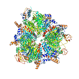
Propionyl-CoA carboxylase of Methylorubrum extorquens with bound CoA
Descriptor: 5-(HEXAHYDRO-2-OXO-1H-THIENO[3,4-D]IMIDAZOL-6-YL)PENTANAL, COENZYME A, Propionyl-CoA carboxylase alpha subunit, ...
Authors:Schuller, J.M, Schuller, S.K, Zarzycki, J, Scheffen, M, Marchal, D.M, Erb, T.J.
Deposit date:2020-03-17
Release date:2020-10-28
Last modified:2021-05-12
Method:ELECTRON MICROSCOPY (3.48 Å)
Cite:A new-to-nature carboxylation module to improve natural and synthetic CO2 fixation
Nat Catal, 2021
2JNY
DownloadVisualize
BU of 2jny by Molmil
Solution NMR structure of protein Uncharacterized BCR, Northeast Structural Genomics Consortium target CgR1
Descriptor: Uncharacterized BCR
Authors:Wu, Y, Liu, G, Zhang, Q, Chen, C, Nwosu, C, Cunningham, K, Ma, L, Xiao, R, Liu, J, Baran, M, Swapna, G, Acton, T, Rost, B, Montelione, G, Szyperski, T, Northeast Structural Genomics Consortium (NESG)
Deposit date:2007-02-14
Release date:2007-04-24
Last modified:2024-05-08
Method:SOLUTION NMR
Cite:Solution NMR structure of protein Uncharacterized BCR, Northeast Structural Genomics Consortium target CgR1
To be Published
5JJG
DownloadVisualize
BU of 5jjg by Molmil
Structure of magnesium-loaded ALG-2
Descriptor: ISOPROPYL ALCOHOL, MAGNESIUM ION, Pcalcium-binding protein ALG-2, ...
Authors:Tanner, J.J.
Deposit date:2016-04-23
Release date:2016-09-07
Last modified:2023-09-27
Method:X-RAY DIFFRACTION (1.72 Å)
Cite:EF5 Is the High-Affinity Mg(2+) Site in ALG-2.
Biochemistry, 55, 2016
2IIZ
DownloadVisualize
BU of 2iiz by Molmil
Crystal structure of putative melanin biosynthesis protein TyrA with bound heme (NP_716371.1) from Shewanella Oneidensis at 2.30 A resolution
Descriptor: 1,2-ETHANEDIOL, ISOPROPYL ALCOHOL, Melanin biosynthesis protein TyrA, ...
Authors:Joint Center for Structural Genomics (JCSG)
Deposit date:2006-09-28
Release date:2006-11-14
Last modified:2023-11-15
Method:X-RAY DIFFRACTION (2.3 Å)
Cite:Identification and structural characterization of heme binding in a novel dye-decolorizing peroxidase, TyrA.
Proteins, 69, 2007
2IAY
DownloadVisualize
BU of 2iay by Molmil
Crystal structure of a duf1831 family protein (lp2179) from lactobacillus plantarum at 1.20 A resolution
Descriptor: CHLORIDE ION, GLYCEROL, Hypothetical protein
Authors:Joint Center for Structural Genomics (JCSG)
Deposit date:2006-09-08
Release date:2006-10-10
Last modified:2023-01-25
Method:X-RAY DIFFRACTION (1.2 Å)
Cite:Structure of LP2179, the first representative of Pfam family PF08866, suggests a new fold with a role in amino-acid metabolism.
Acta Crystallogr.,Sect.F, 66, 2010
2ICH
DownloadVisualize
BU of 2ich by Molmil
CRYSTAL STRUCTURE OF A PUTATIVE ATTH (NE1406) FROM NITROSOMONAS EUROPAEA AT 2.00 A RESOLUTION
Descriptor: 2-[N-CYCLOHEXYLAMINO]ETHANE SULFONIC ACID, GLYCEROL, Putative AttH, ...
Authors:Joint Center for Structural Genomics (JCSG)
Deposit date:2006-09-12
Release date:2006-10-03
Last modified:2023-01-25
Method:X-RAY DIFFRACTION (2 Å)
Cite:Structure of the first representative of Pfam family PF09410 (DUF2006) reveals a structural signature of the calycin superfamily that suggests a role in lipid metabolism.
Acta Crystallogr.,Sect.F, 66, 2010
2H1T
DownloadVisualize
BU of 2h1t by Molmil
Crystal structure of a duf1089 family protein (pa1994) from pseudomonas aeruginosa at 1.80 A resolution
Descriptor: (4S)-2-METHYL-2,4-PENTANEDIOL, 1,2-ETHANEDIOL, Hypothetical protein
Authors:Joint Center for Structural Genomics (JCSG)
Deposit date:2006-05-16
Release date:2006-05-30
Last modified:2023-01-25
Method:X-RAY DIFFRACTION (1.8 Å)
Cite:The structure of the first representative of Pfam family PF06475 reveals a new fold with possible involvement in glycolipid metabolism.
Acta Crystallogr.,Sect.F, 66, 2010
4AR0
DownloadVisualize
BU of 4ar0 by Molmil
N0 domain of Neisseria meningitidis Pilus assembly protein PilQ
Descriptor: TYPE IV PILUS BIOGENESIS AND COMPETENCE PROTEIN PILQ
Authors:Phelan, M.M, Berry, J.L, Derrick, J.P, Lian, L.Y.
Deposit date:2012-04-20
Release date:2012-10-17
Last modified:2024-05-15
Method:SOLUTION NMR
Cite:Structure and Assembly of a Trans-Periplasmic Channel for Type Iv Pili in Neisseria Meningitidis.
Plos Pathog., 8, 2012
4AQZ
DownloadVisualize
BU of 4aqz by Molmil
B2 domain of Neisseria meningitidis Pilus assembly protein PilQ
Descriptor: TYPE IV PILUS BIOGENESIS AND COMPETENCE PROTEIN PILQ
Authors:Phelan, M.M, Berry, J.L, Derrick, J.P, Lian, L.Y.
Deposit date:2012-04-20
Release date:2012-10-17
Last modified:2024-06-19
Method:SOLUTION NMR
Cite:Structure and Assembly of a Trans-Periplasmic Channel for Type Iv Pili in Neisseria Meningitidis.
Plos Pathog., 8, 2012
1QW7
DownloadVisualize
BU of 1qw7 by Molmil
Structure of an Engineered Organophosphorous Hydrolase with Increased Activity Toward Hydrolysis of Phosphothiolate Bonds
Descriptor: COBALT (II) ION, DIETHYL 4-METHYLBENZYLPHOSPHONATE, Parathion hydrolase, ...
Authors:Mesecar, A.D, Grimsley, J.K, Holton, T, Wild, J.R.
Deposit date:2003-09-01
Release date:2004-11-30
Last modified:2023-11-15
Method:X-RAY DIFFRACTION (1.9 Å)
Cite:Structural and mutational studies of organophosphorus hydrolase reveal a cryptic and functional allosteric-binding site.
Arch.Biochem.Biophys., 442, 2005
2FEA
DownloadVisualize
BU of 2fea by Molmil
Crystal structure of MtnX phosphatase from Bacillus Subtilis at 2.00 A resolution
Descriptor: 1,2-ETHANEDIOL, 2-hydroxy-3-keto-5-methylthiopentenyl-1-phosphate phosphatase, MAGNESIUM ION, ...
Authors:Joint Center for Structural Genomics (JCSG)
Deposit date:2005-12-15
Release date:2005-12-27
Last modified:2023-01-25
Method:X-RAY DIFFRACTION (2 Å)
Cite:Crystal structure of MtnX phosphatase from Bacillus subtilis at 2.0 A resolution provides a structural basis for bipartite phosphomonoester hydrolysis of 2-hydroxy-3-keto-5-methylthiopentenyl-1-phosphate.
Proteins, 69, 2007
2HBW
DownloadVisualize
BU of 2hbw by Molmil
Crystal structure of a putative endopeptidase (ava_3396) from anabaena variabilis atcc 29413 at 1.05 A resolution
Descriptor: ACETATE ION, NLP/P60 protein, UNKNOWN LIGAND
Authors:Joint Center for Structural Genomics (JCSG)
Deposit date:2006-06-14
Release date:2006-08-08
Last modified:2023-01-25
Method:X-RAY DIFFRACTION (1.05 Å)
Cite:Structural Basis of Murein Peptide Specificity of a gamma-D-Glutamyl-L-Diamino Acid Endopeptidase.
Structure, 17, 2009
2HUJ
DownloadVisualize
BU of 2huj by Molmil
Crystal structure of a protein of uknown function (NP_471338.1) from Listeria innocua at 1.74 A resolution
Descriptor: Lin2004 protein
Authors:Joint Center for Structural Genomics (JCSG)
Deposit date:2006-07-26
Release date:2006-08-08
Last modified:2023-01-25
Method:X-RAY DIFFRACTION (1.74 Å)
Cite:Crystal structures of MW1337R and lin2004: representatives of a novel protein family that adopt a four-helical bundle fold.
Proteins, 71, 2008
6AHE
DownloadVisualize
BU of 6ahe by Molmil
Crystal structure of enoyl-ACP reductase from Acinetobacter baumannii in complex with NAD and AFN-1252
Descriptor: Enoyl-[acyl-carrier-protein] reductase [NADH], N-methyl-N-[(3-methyl-1-benzofuran-2-yl)methyl]-3-(7-oxo-5,6,7,8-tetrahydro-1,8-naphthyridin-3-yl)propanamide, NICOTINAMIDE-ADENINE-DINUCLEOTIDE
Authors:Rani, S.T, Nataraj, V, Laxminarasimhan, A, Thomas, A, Krishnamurthy, N.
Deposit date:2018-08-17
Release date:2019-08-21
Last modified:2023-11-22
Method:X-RAY DIFFRACTION (2.29 Å)
Cite:Ternary complex formation of AFN-1252 with Acinetobacter baumannii FabI and NADH: Crystallographic and biochemical studies.
Chem.Biol.Drug Des., 96, 2020
5EWI
DownloadVisualize
BU of 5ewi by Molmil
Crystal Structure of the Human Fab VRC38.01, an HIV-1 V1V2-Directed Neutralizing Antibody Isolated from Donor N90
Descriptor: VRC38.01 Heavy Chain, VRC38.01 Light Chain
Authors:Gorman, J, Kwong, P.D.
Deposit date:2015-11-20
Release date:2016-11-23
Last modified:2023-09-27
Method:X-RAY DIFFRACTION (1.5996 Å)
Cite:Virus-like Particles Identify an HIV V1V2 Apex-Binding Neutralizing Antibody that Lacks a Protruding Loop.
Immunity, 46, 2017

222415

數據於2024-07-10公開中

PDB statisticsPDBj update infoContact PDBjnumon