Loading
PDBj
MenuPDBj@FacebookPDBj@TwitterPDBj@YouTubewwPDB FoundationwwPDB
RCSB PDBPDBeBMRBAdv. SearchSearch help
Search by PDB author
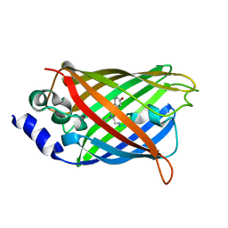
3DQU
DownloadVisualize
BU of 3dqu by Molmil
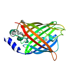
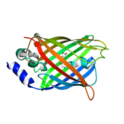
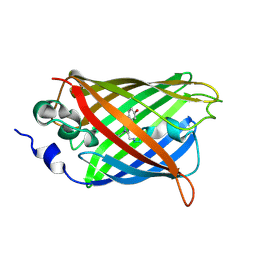
Structure of the Yellow Fluorescent Protein Citrine Frozen at 2000 Atmospheres Number 1: Structure 20 in a Series of 26 High Pressure Structures
Descriptor: Green fluorescent protein
Authors:Barstow, B, Kim, C.U.
Deposit date:2008-07-09
Release date:2008-09-23
Last modified:2024-10-30
Method:X-RAY DIFFRACTION (1.42 Å)
Cite:Alteration of citrine structure by hydrostatic pressure explains the accompanying spectral shift.
Proc.Natl.Acad.Sci.Usa, 105, 2008
3DQJ
DownloadVisualize
BU of 3dqj by Molmil
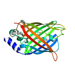
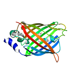
Structure of the Yellow Fluorescent Protein Citrine Frozen at 1000 Atmospheres Number 3: Structure 7 in a Series of 26 High Pressure Structures
Descriptor: Green fluorescent protein
Authors:Barstow, B, Kim, C.U.
Deposit date:2008-07-09
Release date:2008-09-23
Last modified:2023-11-15
Method:X-RAY DIFFRACTION (1.51 Å)
Cite:Alteration of citrine structure by hydrostatic pressure explains the accompanying spectral shift.
Proc.Natl.Acad.Sci.Usa, 105, 2008
3DQN
DownloadVisualize
BU of 3dqn by Molmil
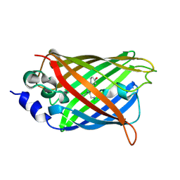
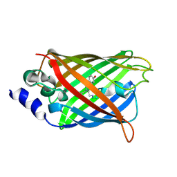
Structure of the Yellow Fluorescent Protein Citrine Frozen at 500 Atmospheres: Structure 3 in a Series of 26 High Pressure Structures
Descriptor: Green fluorescent protein
Authors:Barstow, B, Kim, C.U.
Deposit date:2008-07-09
Release date:2008-09-23
Last modified:2024-10-16
Method:X-RAY DIFFRACTION (1.44 Å)
Cite:Alteration of citrine structure by hydrostatic pressure explains the accompanying spectral shift.
Proc.Natl.Acad.Sci.Usa, 105, 2008
3DPW
DownloadVisualize
BU of 3dpw by Molmil
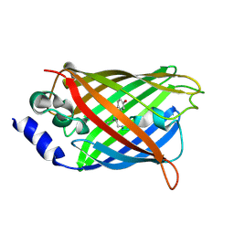
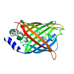
Structure of the Yellow Fluorescent Protein Citrine Frozen at 1 Atmosphere Number 1: Structure 1 in a Series of 26 High Pressure Structures
Descriptor: Green fluorescent protein
Authors:Barstow, B, Kim, C.U.
Deposit date:2008-07-09
Release date:2008-09-23
Last modified:2023-11-15
Method:X-RAY DIFFRACTION (1.59 Å)
Cite:Alteration of citrine structure by hydrostatic pressure explains the accompanying spectral shift.
Proc.Natl.Acad.Sci.Usa, 105, 2008
3DQ4
DownloadVisualize
BU of 3dq4 by Molmil
Structure of the Yellow Fluorescent Protein Citrine Frozen at 2000 Atmospheres Number 2: Structure 20 in a Series of 26 High Pressure Structures
Descriptor: Green fluorescent protein
Authors:Barstow, B, Kim, C.U.
Deposit date:2008-07-09
Release date:2008-09-23
Last modified:2023-11-15
Method:X-RAY DIFFRACTION (1.47 Å)
Cite:Alteration of citrine structure by hydrostatic pressure explains the accompanying spectral shift.
Proc.Natl.Acad.Sci.Usa, 105, 2008
3DQF
DownloadVisualize
BU of 3dqf by Molmil
Structure of the Yellow Fluorescent Protein Citrine Frozen at 1000 Atmospheres Number 6: Structure 10 in a Series of 26 High Pressure Structures
Descriptor: Green fluorescent protein
Authors:Barstow, B, Kim, C.U.
Deposit date:2008-07-09
Release date:2008-09-23
Last modified:2023-11-15
Method:X-RAY DIFFRACTION (1.46 Å)
Cite:Alteration of citrine structure by hydrostatic pressure explains the accompanying spectral shift.
Proc.Natl.Acad.Sci.Usa, 105, 2008
3DQO
DownloadVisualize
BU of 3dqo by Molmil
Structure of the Yellow Fluorescent Protein Citrine Frozen at 1 Atmosphere Number 2: Structure 2 in a Series of 26 High Pressure Structures
Descriptor: Green fluorescent protein
Authors:Barstow, B, Kim, C.U.
Deposit date:2008-07-09
Release date:2008-09-23
Last modified:2023-11-15
Method:X-RAY DIFFRACTION (1.5 Å)
Cite:Alteration of citrine structure by hydrostatic pressure explains the accompanying spectral shift.
Proc.Natl.Acad.Sci.Usa, 105, 2008
3DPX
DownloadVisualize
BU of 3dpx by Molmil
Structure of the Yellow Fluorescent Protein Citrine Frozen at 5000 Atmospheres: Structure 26 in a Series of 26 High Pressure Structures
Descriptor: Green fluorescent protein
Authors:Barstow, B, Kim, C.U.
Deposit date:2008-07-09
Release date:2008-09-23
Last modified:2024-10-30
Method:X-RAY DIFFRACTION (1.5 Å)
Cite:Alteration of citrine structure by hydrostatic pressure explains the accompanying spectral shift.
Proc.Natl.Acad.Sci.Usa, 105, 2008
3DQ5
DownloadVisualize
BU of 3dq5 by Molmil
Structure of the Yellow Fluorescent Protein Citrine Frozen at 1960 Atmospheres: Structure 19 in a Series of 26 High Pressure Structures
Descriptor: Green fluorescent protein
Authors:Barstow, B, Kim, C.U.
Deposit date:2008-07-09
Release date:2008-09-23
Last modified:2023-11-15
Method:X-RAY DIFFRACTION (1.5 Å)
Cite:Alteration of citrine structure by hydrostatic pressure explains the accompanying spectral shift.
Proc.Natl.Acad.Sci.Usa, 105, 2008
3DQE
DownloadVisualize
BU of 3dqe by Molmil
Structure of the Yellow Fluorescent Protein Citrine Frozen at 1250 Atmospheres Number 1: Structure 11 in a Series of 26 High Pressure Structures
Descriptor: Green fluorescent protein
Authors:Barstow, B, Kim, C.U.
Deposit date:2008-07-09
Release date:2008-09-23
Last modified:2023-11-15
Method:X-RAY DIFFRACTION (1.43 Å)
Cite:Alteration of citrine structure by hydrostatic pressure explains the accompanying spectral shift.
Proc.Natl.Acad.Sci.Usa, 105, 2008
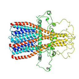
7RPM
DownloadVisualize
BU of 7rpm by Molmil
Structures of the Intracellular Domain and Transmembrane Domain of the Human alpha7 Nicotinic Acetylcholine Receptors
Descriptor: Neuronal acetylcholine receptor subunit alpha-7
Authors:Bondarenko, V, Chen, Q, Tang, P.
Deposit date:2021-08-03
Release date:2022-01-19
Last modified:2024-05-15
Method:SOLUTION NMR
Cite:Structures of highly flexible intracellular domain of human alpha 7 nicotinic acetylcholine receptor.
Nat Commun, 13, 2022
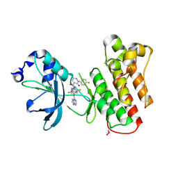
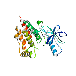
9C1R
DownloadVisualize
BU of 9c1r by Molmil
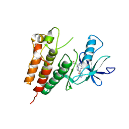
Crystal structure of mutant cMET D1228N kinase domain in complex with inhibitor compound 13
Descriptor: GLYCEROL, Hepatocyte growth factor receptor, N-(2,5-difluoro-4-{[(1s,3S)-3-(1-methyl-1H-pyrazol-3-yl)cyclobutyl][(8R)-pyrazolo[1,5-a]pyrazin-4-yl]amino}phenyl)-2-(5-fluoropyridin-2-yl)-3-oxo-2,3-dihydropyridazine-4-carboxamide
Authors:Simpson, H, Wu, W.-I, Mou, T.-C.
Deposit date:2024-05-29
Release date:2024-08-21
Last modified:2024-09-04
Method:X-RAY DIFFRACTION (1.59 Å)
Cite:Discovery of Pyrazolopyrazines as Selective, Potent, and Mutant-Active MET Inhibitors with Intracranial Efficacy.
J.Med.Chem., 67, 2024
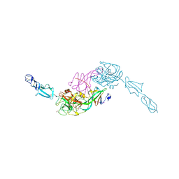
7PQO
DownloadVisualize
BU of 7pqo by Molmil
Catalytic fragment of MASP-1 in complex with P1 site mutant ecotin
Descriptor: Ecotin, GLYCEROL, Mannan-binding lectin serine protease 1, ...
Authors:Harmat, V, Fodor, K, Heja, D.
Deposit date:2021-09-17
Release date:2022-05-18
Last modified:2024-01-31
Method:X-RAY DIFFRACTION (3.39 Å)
Cite:Synergy of protease-binding sites within the ecotin homodimer is crucial for inhibition of MASP enzymes and for blocking lectin pathway activation.
J.Biol.Chem., 298, 2022
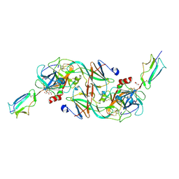
7PQN
DownloadVisualize
BU of 7pqn by Molmil
Catalytic fragment of MASP-2 in complex with ecotin
Descriptor: Ecotin, GLYCEROL, Mannan-binding lectin serine protease 2 A chain, ...
Authors:Harmat, V, Fodor, K, Heja, D.
Deposit date:2021-09-17
Release date:2022-05-18
Last modified:2024-01-31
Method:X-RAY DIFFRACTION (2.400015 Å)
Cite:Synergy of protease-binding sites within the ecotin homodimer is crucial for inhibition of MASP enzymes and for blocking lectin pathway activation.
J.Biol.Chem., 298, 2022
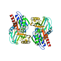
3CUK
DownloadVisualize
BU of 3cuk by Molmil
Crystal structure of human D-amino acid oxidase: bound to an inhibitor
Descriptor: 4H-furo[3,2-b]pyrrole-5-carboxylic acid, D-amino-acid oxidase, FLAVIN-ADENINE DINUCLEOTIDE
Authors:Prasad, S, Munshi, S.
Deposit date:2008-04-16
Release date:2008-07-22
Last modified:2023-08-30
Method:X-RAY DIFFRACTION (2.49 Å)
Cite:The discovery of fused pyrrole carboxylic acids as novel, potent D-amino acid oxidase (DAO) inhibitors.
Bioorg.Med.Chem.Lett., 18, 2008
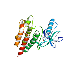
3PSD
DownloadVisualize
BU of 3psd by Molmil
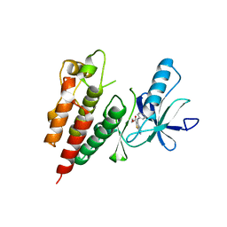
Non-oxime pyrazole based inhibitors of B-Raf kinase
Descriptor: 6-[1-(piperidin-4-yl)-3-(pyridin-4-yl)-1H-pyrazol-4-yl]indeno[1,2-c]pyrazole, B-RAF PROTO-ONCOGENE SERINE/THREONINE-PROTEIN KINASE
Authors:Morales, T, Vigers, G.P.A, Brandhuber, B.J.
Deposit date:2010-12-01
Release date:2011-05-18
Last modified:2023-09-06
Method:X-RAY DIFFRACTION (3.6 Å)
Cite:Non-oxime pyrazole based inhibitors of B-Raf kinase.
Bioorg.Med.Chem.Lett., 21, 2011
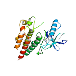
3D4Q
DownloadVisualize
BU of 3d4q by Molmil
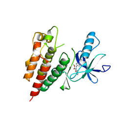
Pyrazole-based inhibitors of B-Raf kinase
Descriptor: (1E)-5-(1-piperidin-4-yl-3-pyridin-4-yl-1H-pyrazol-4-yl)-2,3-dihydro-1H-inden-1-one oxime, B-Raf proto-oncogene serine/threonine-protein kinase
Authors:Morales, T, Vigers, G.P.A, Brandhuber, B.J.
Deposit date:2008-05-14
Release date:2008-08-19
Last modified:2024-02-21
Method:X-RAY DIFFRACTION (2.8 Å)
Cite:Potent and selective pyrazole-based inhibitors of B-Raf kinase.
Bioorg.Med.Chem.Lett., 18, 2008
3PRF
DownloadVisualize
BU of 3prf by Molmil
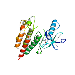
Crystal Structure of Human B-Raf Kinase Domain in Complex with a Non-Oxime Furopyridine Inhibitor
Descriptor: 2-chloro-5-{[2-(pyrimidin-2-yl)furo[2,3-c]pyridin-3-yl]amino}phenol, Serine/threonine-protein kinase B-raf
Authors:Voegtli, W.C, Vigers, G.P.A, Morales, T, Brandhuber, B.J.
Deposit date:2010-11-29
Release date:2011-02-02
Last modified:2024-02-21
Method:X-RAY DIFFRACTION (2.9 Å)
Cite:Non-oxime inhibitors of B-Raf(V600E) kinase.
Bioorg.Med.Chem.Lett., 21, 2011
3PRI
DownloadVisualize
BU of 3pri by Molmil
Crystal Structure of Human B-Raf Kinase in Complex with a Non-Oxime Furopyridine Inhibitor
Descriptor: 3-(4-{[2-(pyrimidin-2-yl)furo[2,3-c]pyridin-3-yl]amino}-1H-indazol-3-yl)propan-1-ol, Serine/threonine-protein kinase B-raf
Authors:Voegtli, W.C, Vigers, G.P.A, Morales, T, Brandhuber, B.J.
Deposit date:2010-11-29
Release date:2011-02-02
Last modified:2024-02-21
Method:X-RAY DIFFRACTION (3.5 Å)
Cite:Non-oxime inhibitors of B-Raf(V600E) kinase.
Bioorg.Med.Chem.Lett., 21, 2011
3PSB
DownloadVisualize
BU of 3psb by Molmil
Furo[2,3-c]pyridine-based Indanone Oximes as Potent and Selective B-Raf Inhibitors
Descriptor: B-RAF PROTO-ONCOGENE SERINE/THREONINE-PROTEIN KINASE, ethyl 3-{[1-(hydroxyamino)-2H-inden-5-yl]amino}thieno[2,3-c]pyridine-2-carboxylate
Authors:Morales, T, Vigers, G.P.A, Brandhuber, B.J.
Deposit date:2010-12-01
Release date:2011-01-19
Last modified:2023-09-06
Method:X-RAY DIFFRACTION (3.4 Å)
Cite:The Discovery of furo[2,3-c]pyridine-based indanone oximes as potent and selective B-Raf inhibitors.
Bioorg.Med.Chem.Lett., 21, 2011
3PPJ
DownloadVisualize
BU of 3ppj by Molmil
Human B-Raf Kinase in Complex with a Furopyridine Inhibitor
Descriptor: Serine/threonine-protein kinase B-raf, methyl 3-{[(5S)-1-(hydroxyamino)-5H-inden-5-yl]amino}furo[2,3-c]pyridine-2-carboxylate
Authors:Voegtli, W.C, Vigers, G.P.A, Morales, T, Brandhuber, B.J.
Deposit date:2010-11-24
Release date:2011-02-02
Last modified:2024-02-21
Method:X-RAY DIFFRACTION (3.7 Å)
Cite:Non-oxime inhibitors of B-Raf(V600E) kinase.
Bioorg.Med.Chem.Lett., 21, 2011
3PPK
DownloadVisualize
BU of 3ppk by Molmil
Human B-Raf Kinase in Complex with a Non-Oxime Furopyridine Inhibitor
Descriptor: 3-[(5-hydroxynaphthalen-2-yl)amino]-N-(pyrimidin-4-yl)furo[2,3-c]pyridine-2-carboxamide, Serine/threonine-protein kinase B-raf
Authors:Voegtli, W.C, Vigers, G.P.A, Morales, T, Brandhuber, B.J.
Deposit date:2010-11-24
Release date:2011-02-02
Last modified:2024-02-21
Method:X-RAY DIFFRACTION (3 Å)
Cite:Non-oxime inhibitors of B-Raf(V600E) kinase.
Bioorg.Med.Chem.Lett., 21, 2011
3AWT
DownloadVisualize
BU of 3awt by Molmil
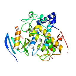
Crystal structure of Streptomyces tyrosinase in a complex with caddie soaked in a Cu(II)-containing solution for 20 hr: occupancy of Cu(II) is high
Descriptor: COPPER (II) ION, MelC, NITRATE ION, ...
Authors:Matoba, Y, Sugiyama, M.
Deposit date:2011-03-26
Release date:2011-06-29
Last modified:2023-11-01
Method:X-RAY DIFFRACTION (1.35 Å)
Cite:A molecular mechanism for copper transportation to tyrosinase that is assisted by a metallochaperone, caddie protein
J.Biol.Chem., 286, 2011
3AWY
DownloadVisualize
BU of 3awy by Molmil
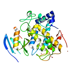
Crystal structure of Streptomyces tyrosinase in a complex with caddie M84L mutant soaked in a Cu(II)-containing solution for 80 hr
Descriptor: COPPER (II) ION, MelC, NITRATE ION, ...
Authors:Matoba, Y, Sugiyama, M.
Deposit date:2011-03-26
Release date:2011-06-29
Last modified:2023-11-01
Method:X-RAY DIFFRACTION (1.58 Å)
Cite:A molecular mechanism for copper transportation to tyrosinase that is assisted by a metallochaperone, caddie protein
J.Biol.Chem., 286, 2011
3AWW
DownloadVisualize
BU of 3aww by Molmil
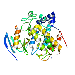
Crystal structure of Streptomyces tyrosinase in a complex with caddie soaked in a Cu(II)-containing solution for 80 hr: occupancy of CuA is high
Descriptor: COPPER (II) ION, MelC, NITRATE ION, ...
Authors:Matoba, Y, Sugiyama, M.
Deposit date:2011-03-26
Release date:2011-06-29
Last modified:2023-11-01
Method:X-RAY DIFFRACTION (1.35 Å)
Cite:A molecular mechanism for copper transportation to tyrosinase that is assisted by a metallochaperone, caddie protein
J.Biol.Chem., 286, 2011

226707

數據於2024-10-30公開中

PDB statisticsPDBj update infoContact PDBjnumon