Loading
PDBj
MenuPDBj@FacebookPDBj@TwitterPDBj@YouTubewwPDB FoundationwwPDB
RCSB PDBPDBeBMRBAdv. SearchSearch help
Search by PDB author
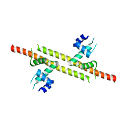
7VU8
DownloadVisualize
BU of 7vu8 by Molmil
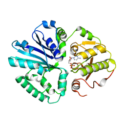
L7-Tir domain with bound ligand
Descriptor: 2',3'- cyclic AMP, Flax rust resistance protein
Authors:Tan, Y, Xu, C, Yu, D, Song, W, Wu, B, Schulze-Lefert, P, Chai, J.
Deposit date:2021-11-01
Release date:2022-06-01
Last modified:2022-07-06
Method:ELECTRON MICROSCOPY (3 Å)
Cite:TIR domains of plant immune receptors are 2',3'-cAMP/cGMP synthetases mediating cell death.
Cell, 185, 2022
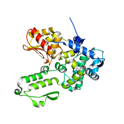
7VT1
DownloadVisualize
BU of 7vt1 by Molmil
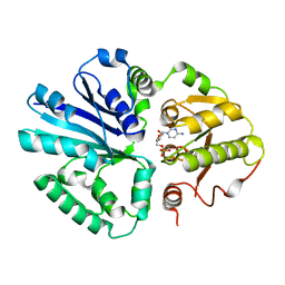
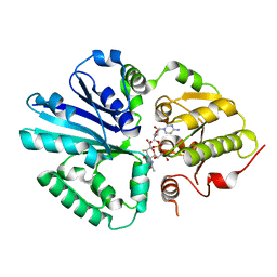
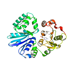
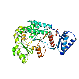
Acyltransferase from the 9th Module of Salinomycin Polyketide Synthase
Descriptor: Type I modular polyketide synthase
Authors:Feng, Y, Zhang, F, Zheng, J.
Deposit date:2021-10-27
Release date:2022-06-08
Last modified:2023-11-29
Method:X-RAY DIFFRACTION (2.5 Å)
Cite:Structural visualization of transient interactions between the cis-acting acyltransferase and acyl carrier protein of the salinomycin modular polyketide synthase.
Acta Crystallogr D Struct Biol, 78, 2022
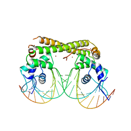
7VRS
DownloadVisualize
BU of 7vrs by Molmil
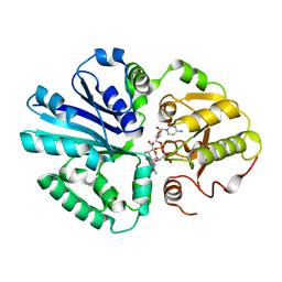
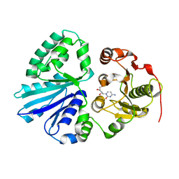
The complex of Acyltransferase and Acyl Carrier Protein Domains from module 9 of Salinomycin Polyketide Synthase
Descriptor: 1,1'-butane-1,4-diylbis(1H-pyrrole-2,5-dione), 4'-PHOSPHOPANTETHEINE, Type I modular polyketide synthase
Authors:Feng, Y, Zheng, J.
Deposit date:2021-10-24
Release date:2022-06-08
Last modified:2023-11-29
Method:X-RAY DIFFRACTION (2.6 Å)
Cite:Structural visualization of transient interactions between the cis-acting acyltransferase and acyl carrier protein of the salinomycin modular polyketide synthase.
Acta Crystallogr D Struct Biol, 78, 2022
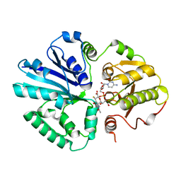
7YOW
DownloadVisualize
BU of 7yow by Molmil
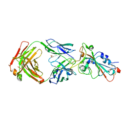
Crystal structure of SARS-CoV-2 omicron variant spike receptor-binding domain (RBD) in complex with NCV2SG48 Fab
Descriptor: Fab Heavy chain, Fab Light chain, SULFATE ION, ...
Authors:Yamamoto, A, Higashiura, A.
Deposit date:2022-08-02
Release date:2023-04-19
Last modified:2023-11-29
Method:X-RAY DIFFRACTION (3.3 Å)
Cite:Structural basis of spike RBM-specific human antibodies counteracting broad SARS-CoV-2 variants.
Commun Biol, 6, 2023
7WN2
DownloadVisualize
BU of 7wn2 by Molmil
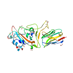
Crystal structure of SARS-CoV-2 spike receptor-binding domain (RBD) in complex with NCV2SG53 Fab
Descriptor: 2-acetamido-2-deoxy-beta-D-glucopyranose, Fab Heavy chain, Fab Light chain, ...
Authors:Yamamoto, A, Higashiura, A.
Deposit date:2022-01-17
Release date:2023-04-19
Last modified:2023-11-29
Method:X-RAY DIFFRACTION (2.35 Å)
Cite:Structural basis of spike RBM-specific human antibodies counteracting broad SARS-CoV-2 variants.
Commun Biol, 6, 2023
7WNB
DownloadVisualize
BU of 7wnb by Molmil
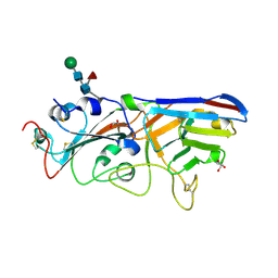
Crystal structure of SARS-CoV-2 spike receptor-binding domain (RBD) in complex with NCV2SG48 Fab
Descriptor: 2-acetamido-2-deoxy-beta-D-glucopyranose-(1-4)-2-acetamido-2-deoxy-beta-D-glucopyranose, Fab Heavy chain, Fab Light chain, ...
Authors:Yamamoto, A, Higashiura, A.
Deposit date:2022-01-18
Release date:2023-04-19
Method:X-RAY DIFFRACTION (2.18 Å)
Cite:Structural basis of spike RBM-specific human antibodies counteracting broad SARS-CoV-2 variants.
Commun Biol, 6, 2023
3V5Q
DownloadVisualize
BU of 3v5q by Molmil
Discovery of a selective TRK Inhibitor with efficacy in rodent cancer tumor models
Descriptor: 1-(3-{[(3Z)-2-oxo-3-(1H-pyrrol-2-ylmethylidene)-2,3-dihydro-1H-indol-6-yl]amino}phenyl)-3-[3-(trifluoromethyl)phenyl]urea, CHLORIDE ION, NT-3 growth factor receptor
Authors:Kreusch, A.
Deposit date:2011-12-16
Release date:2012-02-08
Last modified:2024-02-28
Method:X-RAY DIFFRACTION (2.2001 Å)
Cite:Discovery of GNF-5837, a Selective TRK Inhibitor with Efficacy in Rodent Cancer Tumor Models.
ACS Med Chem Lett, 3, 2012
7XKE
DownloadVisualize
BU of 7xke by Molmil
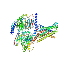
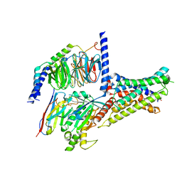
Cryo-EM structure of DHEA-ADGRG2-FL-Gs complex
Descriptor: 3-BETA-HYDROXY-5-ANDROSTEN-17-ONE, Adhesion G-protein coupled receptor G2, Guanine nucleotide-binding protein G(I)/G(S)/G(O) subunit gamma-2, ...
Authors:Guo, S.C, Xiao, P, Lin, H, Sun, J.P, Yu, X.
Deposit date:2022-04-19
Release date:2022-08-10
Last modified:2023-03-15
Method:ELECTRON MICROSCOPY (2.9 Å)
Cite:Structures of the ADGRG2-G s complex in apo and ligand-bound forms.
Nat.Chem.Biol., 18, 2022
7XKF
DownloadVisualize
BU of 7xkf by Molmil
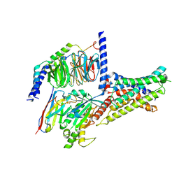
Cryo-EM structure of DHEA-ADGRG2-BT-Gs complex at lower state
Descriptor: 3-BETA-HYDROXY-5-ANDROSTEN-17-ONE, Adhesion G-protein coupled receptor G2, Guanine nucleotide-binding protein G(I)/G(S)/G(O) subunit gamma-2, ...
Authors:Guo, S.C, Xiao, P, Lin, H, Sun, J.P, Yu, X.
Deposit date:2022-04-19
Release date:2022-08-10
Last modified:2023-03-15
Method:ELECTRON MICROSCOPY (2.4 Å)
Cite:Structures of the ADGRG2-G s complex in apo and ligand-bound forms.
Nat.Chem.Biol., 18, 2022
7XKD
DownloadVisualize
BU of 7xkd by Molmil
Cryo-EM structure of DHEA-ADGRG2-BT-Gs complex
Descriptor: 3-BETA-HYDROXY-5-ANDROSTEN-17-ONE, Adhesion G-protein coupled receptor G2, Guanine nucleotide-binding protein G(I)/G(S)/G(O) subunit gamma-2, ...
Authors:Guo, S.C, Xiao, P, Lin, H, Sun, J.P, Yu, X.
Deposit date:2022-04-19
Release date:2022-08-10
Last modified:2023-03-15
Method:ELECTRON MICROSCOPY (2.4 Å)
Cite:Structures of the ADGRG2-G s complex in apo and ligand-bound forms.
Nat.Chem.Biol., 18, 2022
1AKP
DownloadVisualize
BU of 1akp by Molmil
SEQUENTIAL 1H,13C AND 15N NMR ASSIGNMENTS AND SOLUTION CONFORMATION OF APOKEDARCIDIN
Descriptor: APOKEDARCIDIN
Authors:Constantine, K.L, Colson, K.L, Wittekind, M, Friedrichs, M.S, Zein, N, Tuttle, J, Langley, D.R, Leet, J.E, Schroeder, D.R, Lam, K.S, Farmer II, B.T, Metzler, W.J, Bruccoleri, R.E, Mueller, L.
Deposit date:1994-06-20
Release date:1994-08-31
Last modified:2017-11-29
Method:SOLUTION NMR
Cite:Sequential 1H, 13C, and 15N NMR assignments and solution conformation of apokedarcidin.
Biochemistry, 33, 1994
6JNI
DownloadVisualize
BU of 6jni by Molmil
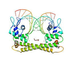
Crystal structure of the transcriptional regulator CadR from P. putida in complex with Zinc(II) and DNA
Descriptor: CadR, DNA (25-MER), ZINC ION
Authors:Liu, X.C, Gan, J.H, Chen, H.
Deposit date:2019-03-16
Release date:2019-09-25
Last modified:2023-11-22
Method:X-RAY DIFFRACTION (2.9 Å)
Cite:Selective cadmium regulation mediated by a cooperative binding mechanism in CadR.
Proc.Natl.Acad.Sci.USA, 116, 2019
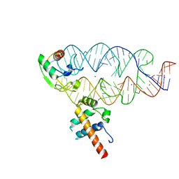
7X5K
DownloadVisualize
BU of 7x5k by Molmil
Tir-dsDNA complex, the initial binding state
Descriptor: DNA (43-MER), Flax rust resistance protein
Authors:Tan, Y, Xu, C, Yu, D, Song, W, Wu, B, Schulze-Lefert, P, Chai, J.
Deposit date:2022-03-04
Release date:2022-06-08
Last modified:2022-07-06
Method:ELECTRON MICROSCOPY (3.8 Å)
Cite:TIR domains of plant immune receptors are 2',3'-cAMP/cGMP synthetases mediating cell death.
Cell, 185, 2022
6JWE
DownloadVisualize
BU of 6jwe by Molmil
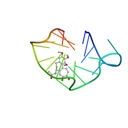
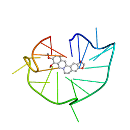
structure of RET G-quadruplex in complex with colchicine
Descriptor: DNA (5'-D(*GP*GP*GP*GP*CP*GP*GP*GP*GP*CP*GP*GP*GP*GP*CP*GP*GP*GP*GP*T)-3'), N-[(7S)-1,2,3,10-tetramethoxy-9-oxo-6,7-dihydro-5H-benzo[d]heptalen-7-yl]ethanamide
Authors:Wang, F, Wang, C, Liu, Y, Lan, W.X, Li, Y.M, Wang, R.X, Cao, C.
Deposit date:2019-04-20
Release date:2020-02-26
Last modified:2024-05-15
Method:SOLUTION NMR
Cite:Colchicine selective interaction with oncogene RET G-quadruplex revealed by NMR.
Chem.Commun.(Camb.), 56, 2020
6JWD
DownloadVisualize
BU of 6jwd by Molmil
structure of RET G-quadruplex in complex with berberine
Descriptor: BERBERINE, DNA (5'-D(*GP*GP*GP*GP*CP*GP*GP*GP*GP*CP*GP*GP*GP*GP*CP*GP*GP*GP*GP*T)-3')
Authors:Wang, F, Wang, C, Liu, Y, Lan, W.X, Li, Y.M, Wang, R.X, Cao, C.
Deposit date:2019-04-19
Release date:2020-04-22
Last modified:2024-05-15
Method:SOLUTION NMR
Cite:Colchicine selective interaction with oncogene RET G-quadruplex revealed by NMR.
Chem.Commun.(Camb.), 56, 2020
1FSA
DownloadVisualize
BU of 1fsa by Molmil
THE T-STATE STRUCTURE OF LYS 42 TO ALA MUTANT OF THE PIG KIDNEY FRUCTOSE 1,6-BISPHOSPHATASE EXPRESSED IN E. COLI
Descriptor: 6-O-phosphono-beta-D-fructofuranose, ADENOSINE MONOPHOSPHATE, FRUCTOSE 1,6-BISPHOSPHATASE, ...
Authors:Lu, G, Stec, B, Giroux, E, Kantrowitz, E.R.
Deposit date:1996-08-24
Release date:1997-09-04
Last modified:2024-02-07
Method:X-RAY DIFFRACTION (2.3 Å)
Cite:Evidence for an active T-state pig kidney fructose 1,6-bisphosphatase: interface residue Lys-42 is important for allosteric inhibition and AMP cooperativity.
Protein Sci., 5, 1996
1RDZ
DownloadVisualize
BU of 1rdz by Molmil
T-STATE STRUCTURE OF THE ARG 243 TO ALA MUTANT OF PIG KIDNEY FRUCTOSE 1,6-BISPHOSPHATASE EXPRESSED IN E. COLI
Descriptor: 6-O-phosphono-beta-D-fructofuranose, ADENOSINE MONOPHOSPHATE, FRUCTOSE 1,6-BISPHOSPHATASE
Authors:Stec, B, Abraham, R, Giroux, E, Kantrowitz, E.R.
Deposit date:1996-05-17
Release date:1997-01-11
Last modified:2024-02-14
Method:X-RAY DIFFRACTION (2.05 Å)
Cite:Crystal structures of the active site mutant (Arg-243-->Ala) in the T and R allosteric states of pig kidney fructose-1,6-bisphosphatase expressed in Escherichia coli.
Protein Sci., 5, 1996
6JGW
DownloadVisualize
BU of 6jgw by Molmil
Crystal structure of the transcriptional regulator CadR from P. putida in complex with DNA
Descriptor: CadR, DNA (27-MER)
Authors:Liu, X.C, Gan, J.H, Chen, H.
Deposit date:2019-02-15
Release date:2019-09-25
Last modified:2023-11-22
Method:X-RAY DIFFRACTION (2.8 Å)
Cite:Selective cadmium regulation mediated by a cooperative binding mechanism in CadR.
Proc.Natl.Acad.Sci.USA, 116, 2019
1RDX
DownloadVisualize
BU of 1rdx by Molmil
R-STATE STRUCTURE OF THE ARG 243 TO ALA MUTANT OF PIG KIDNEY FRUCTOSE 1,6-BISPHOSPHATASE EXPRESSED IN E. COLI
Descriptor: 6-O-phosphono-beta-D-fructofuranose, FRUCTOSE 1,6-BISPHOSPHATASE
Authors:Stec, B, Abraham, R, Giroux, E, Kantrowitz, E.R.
Deposit date:1996-05-17
Release date:1997-01-11
Last modified:2024-02-14
Method:X-RAY DIFFRACTION (2.75 Å)
Cite:Crystal structures of the active site mutant (Arg-243-->Ala) in the T and R allosteric states of pig kidney fructose-1,6-bisphosphatase expressed in Escherichia coli.
Protein Sci., 5, 1996
1RDY
DownloadVisualize
BU of 1rdy by Molmil
T-STATE STRUCTURE OF THE ARG 243 TO ALA MUTANT OF PIG KIDNEY FRUCTOSE 1,6-BISPHOSPHATASE EXPRESSED IN E. COLI
Descriptor: 6-O-phosphono-beta-D-fructofuranose, ADENOSINE MONOPHOSPHATE, FRUCTOSE 1,6-BISPHOSPHATASE
Authors:Stec, B, Abraham, R, Giroux, E, Kantrowitz, E.R.
Deposit date:1996-05-17
Release date:1997-01-11
Last modified:2021-11-03
Method:X-RAY DIFFRACTION (2.2 Å)
Cite:Crystal structures of the active site mutant (Arg-243-->Ala) in the T and R allosteric states of pig kidney fructose-1,6-bisphosphatase expressed in Escherichia coli.
Protein Sci., 5, 1996
3C2H
DownloadVisualize
BU of 3c2h by Molmil
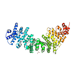
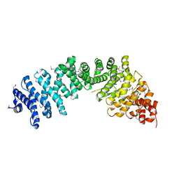
Crystal Structure of SYS-1 at 2.6A resolution
Descriptor: CITRATE ANION, GLYCEROL, Sys-1 protein
Authors:Liu, J, Phillips, B.T, Amaya, M.F, Kimble, J, Xu, W.
Deposit date:2008-01-25
Release date:2008-05-20
Last modified:2024-02-21
Method:X-RAY DIFFRACTION (2.6 Å)
Cite:The C. elegans SYS-1 protein is a bona fide beta-catenin.
Dev.Cell, 14, 2008
7CCF
DownloadVisualize
BU of 7ccf by Molmil
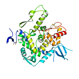
Mechanism insights on steroselective oxidation of phosphorylated ethylphenols with cytochrome P450 CreJ
Descriptor: (3-ethylphenyl) dihydrogen phosphate, Cytochrome P450, PROTOPORPHYRIN IX CONTAINING FE
Authors:Dong, S, Du, L, Li, S.Y, Feng, Y.G.
Deposit date:2020-06-17
Release date:2021-03-17
Last modified:2023-11-29
Method:X-RAY DIFFRACTION (2.8 Å)
Cite:Structural Basis for Selective Oxidation of Phosphorylated Ethylphenols by Cytochrome P450 Monooxygenase CreJ.
Appl.Environ.Microbiol., 87, 2021
3C2G
DownloadVisualize
BU of 3c2g by Molmil
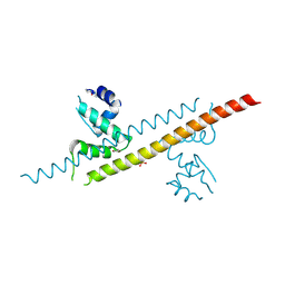
Crystal complex of SYS-1/POP-1 at 2.5A resolution
Descriptor: Pop-1 8-residue peptide, Sys-1 protein
Authors:Liu, J, Phillips, B.T, Amaya, M.F, Kimble, J, Xu, W.
Deposit date:2008-01-24
Release date:2008-05-20
Last modified:2017-10-25
Method:X-RAY DIFFRACTION (2.5 Å)
Cite:The C. elegans SYS-1 protein is a bona fide beta-catenin.
Dev.Cell, 14, 2008
7CJS
DownloadVisualize
BU of 7cjs by Molmil
structure of aquaporin
Descriptor: Aquaporin NIP2-1, CHOLESTEROL HEMISUCCINATE, SODIUM ION, ...
Authors:Saitoh, Y, Ma, J.F, Suga, M.
Deposit date:2020-07-13
Release date:2021-11-03
Last modified:2023-11-29
Method:X-RAY DIFFRACTION (1.8 Å)
Cite:Structural basis for high selectivity of a rice silicon channel Lsi1.
Nat Commun, 12, 2021
7D64
DownloadVisualize
BU of 7d64 by Molmil
The crystal structure of SARS-CoV-2 3CLpro with Zinc
Descriptor: 3C-like proteinase, ZINC ION
Authors:Mao, Z.W, Xia, W, Tao, X.
Deposit date:2020-09-29
Release date:2021-09-29
Last modified:2023-11-29
Method:X-RAY DIFFRACTION (2.450116 Å)
Cite:Inhibition of SARS-CoV-2 replication by zinc gluconate in combination with hinokitiol.
J.Inorg.Biochem., 231, 2022

221371

數據於2024-06-19公開中

PDB statisticsPDBj update infoContact PDBjnumon