Loading
PDBj
MenuPDBj@FacebookPDBj@X(formerly Twitter)PDBj@BlueSkyPDBj@YouTubewwPDB FoundationwwPDB
RCSB PDBPDBeBMRBAdv. SearchSearch help
Search by PDB author
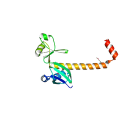
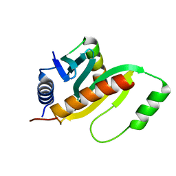
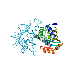
1JYO
DownloadVisualize
BU of 1jyo by Molmil
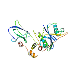
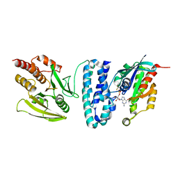
Structure of the Salmonella Virulence Effector SptP in Complex with its Secretion Chaperone SicP
Descriptor: SicP, protein tyrosine phosphatase SptP
Authors:Stebbins, C.E, Galan, J.E.
Deposit date:2001-09-12
Release date:2001-11-07
Last modified:2024-10-16
Method:X-RAY DIFFRACTION (1.9 Å)
Cite:Maintenance of an unfolded polypeptide by a cognate chaperone in bacterial type III secretion.
Nature, 414, 2001
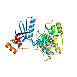
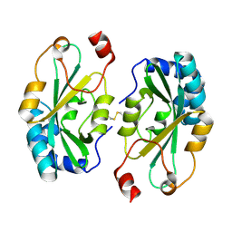
1VCB
DownloadVisualize
BU of 1vcb by Molmil
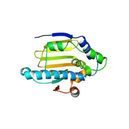
THE VHL-ELONGINC-ELONGINB STRUCTURE
Descriptor: PROTEIN (ELONGIN B), PROTEIN (ELONGIN C), PROTEIN (VHL)
Authors:Stebbins, C.E, Kaelin, W.G, Pavletich, N.P.
Deposit date:1999-03-13
Release date:1999-04-21
Last modified:2023-12-27
Method:X-RAY DIFFRACTION (2.7 Å)
Cite:Structure of the VHL-ElonginC-ElonginB complex: implications for VHL tumor suppressor function.
Science, 284, 1999
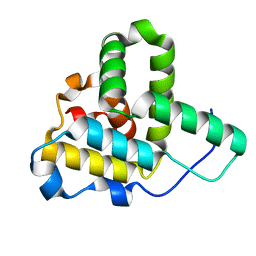
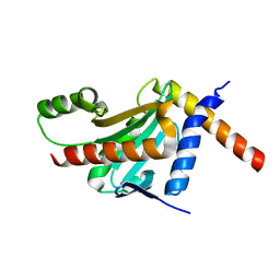
1YET
DownloadVisualize
BU of 1yet by Molmil
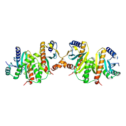
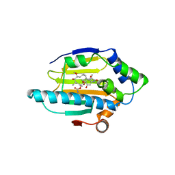
GELDANAMYCIN BOUND TO THE HSP90 GELDANAMYCIN-BINDING DOMAIN
Descriptor: GELDANAMYCIN, HEAT SHOCK PROTEIN 90
Authors:Stebbins, C.E, Russo, A.A, Pavletich, N.P.
Deposit date:1997-03-25
Release date:1998-04-22
Last modified:2024-02-14
Method:X-RAY DIFFRACTION (1.9 Å)
Cite:Crystal structure of an Hsp90-geldanamycin complex: targeting of a protein chaperone by an antitumor agent.
Cell(Cambridge,Mass.), 89, 1997
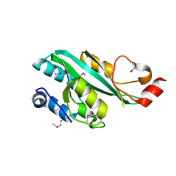
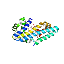
1YER
DownloadVisualize
BU of 1yer by Molmil
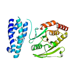
HUMAN HSP90 GELDANAMYCIN-BINDING DOMAIN, "CLOSED" CONFORMATION
Descriptor: HEAT SHOCK PROTEIN 90
Authors:Stebbins, C.E, Russo, A.A, Pavletich, N.P.
Deposit date:1997-03-25
Release date:1998-04-22
Last modified:2024-02-14
Method:X-RAY DIFFRACTION (1.65 Å)
Cite:Crystal structure of an Hsp90-geldanamycin complex: targeting of a protein chaperone by an antitumor agent.
Cell(Cambridge,Mass.), 89, 1997
1YES
DownloadVisualize
BU of 1yes by Molmil
HUMAN HSP90 GELDANAMYCIN-BINDING DOMAIN, "OPEN" CONFORMATION
Descriptor: HEAT SHOCK PROTEIN 90
Authors:Stebbins, C.E, Russo, A.A, Pavletich, N.P.
Deposit date:1997-03-25
Release date:1998-04-22
Last modified:2024-02-14
Method:X-RAY DIFFRACTION (2.2 Å)
Cite:Crystal structure of an Hsp90-geldanamycin complex: targeting of a protein chaperone by an antitumor agent.
Cell(Cambridge,Mass.), 89, 1997
1G4U
DownloadVisualize
BU of 1g4u by Molmil
CRYSTAL STRUCTURE OF THE SALMONELLA TYROSINE PHOSPHATASE AND GTPASE ACTIVATING PROTEIN SPTP BOUND TO RAC1
Descriptor: ALUMINUM FLUORIDE, GUANOSINE-5'-DIPHOSPHATE, MAGNESIUM ION, ...
Authors:Stebbins, C.E, Galan, J.E.
Deposit date:2000-10-28
Release date:2001-01-24
Last modified:2024-02-07
Method:X-RAY DIFFRACTION (2.3 Å)
Cite:Modulation of host signaling by a bacterial mimic: structure of the Salmonella effector SptP bound to Rac1.
Mol.Cell, 6, 2000
1G4W
DownloadVisualize
BU of 1g4w by Molmil
CRYSTAL STRUCTURE OF THE SALMONELLA TYROSINE PHOSPHATASE AND GTPASE ACTIVATING PROTEIN SPTP
Descriptor: PROTEIN TYROSINE PHOSPHATASE SPTP
Authors:Stebbins, C.E, Galan, J.E.
Deposit date:2000-10-28
Release date:2001-01-24
Last modified:2024-02-07
Method:X-RAY DIFFRACTION (2.2 Å)
Cite:Modulation of host signaling by a bacterial mimic: structure of the Salmonella effector SptP bound to Rac1.
Mol.Cell, 6, 2000
6ELC
DownloadVisualize
BU of 6elc by Molmil
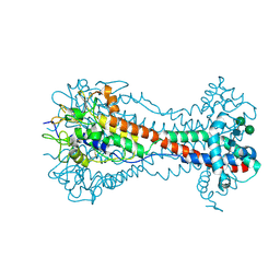
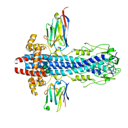
Crystal Structure of O-linked Glycosylated VSG3
Descriptor: Variant surface glycoprotein, alpha-D-glucopyranose, alpha-D-mannopyranose-(1-6)-beta-D-mannopyranose-(1-4)-2-acetamido-2-deoxy-beta-D-glucopyranose-(1-4)-2-acetamido-2-deoxy-beta-D-glucopyranose
Authors:Stebbins, C.E.
Deposit date:2017-09-28
Release date:2018-07-11
Last modified:2024-10-16
Method:X-RAY DIFFRACTION (1.41 Å)
Cite:African trypanosomes evade immune clearance by O-glycosylation of the VSG surface coat.
Nat Microbiol, 3, 2018
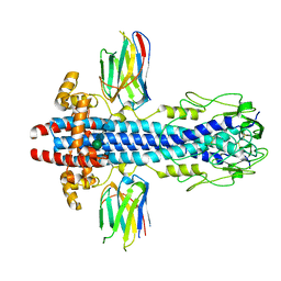
7AQX
DownloadVisualize
BU of 7aqx by Molmil
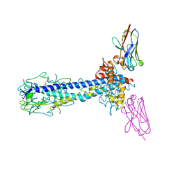
Co-Crystal Structure of Variant Surface Glycoprotein VSG2 in complex with Nanobody VSG2(NB9)
Descriptor: CITRIC ACID, Nanobody VSG2(NB9), SODIUM ION, ...
Authors:Stebbins, C.E, Hempelmann, A.
Deposit date:2020-10-23
Release date:2021-11-03
Last modified:2024-10-23
Method:X-RAY DIFFRACTION (1.499 Å)
Cite:Nanobody-mediated macromolecular crowding induces membrane fission and remodeling in the African trypanosome.
Cell Rep, 37, 2021
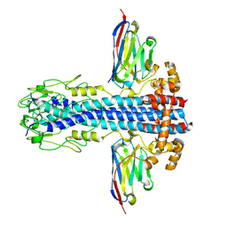
7AQY
DownloadVisualize
BU of 7aqy by Molmil
Co-Crystal Structure of Variant Surface Glycoprotein VSG2 in complex with Nanobody VSG2(NB11)
Descriptor: Nanobody VSG2(NB11), Variant surface glycoprotein MITAT 1.2, beta-D-mannopyranose-(1-4)-2-acetamido-2-deoxy-beta-D-glucopyranose-(1-4)-2-acetamido-2-deoxy-beta-D-glucopyranose
Authors:Stebbins, C.E, Hempelmann, A, VanStraaten, M.
Deposit date:2020-10-23
Release date:2021-11-03
Last modified:2024-11-20
Method:X-RAY DIFFRACTION (1.9 Å)
Cite:Nanobody-mediated macromolecular crowding induces membrane fission and remodeling in the African trypanosome.
Cell Rep, 37, 2021
7AR0
DownloadVisualize
BU of 7ar0 by Molmil
Co-Crystal Structure of Variant Surface Glycoprotein VSG2 in complex with Nanobody VSG2(NB19)
Descriptor: Nanobody VSG2(NB19), Variant surface glycoprotein MITAT 1.2, beta-D-mannopyranose-(1-4)-2-acetamido-2-deoxy-beta-D-glucopyranose-(1-4)-2-acetamido-2-deoxy-beta-D-glucopyranose
Authors:Stebbins, C.E, Hempelmann, A.
Deposit date:2020-10-23
Release date:2021-11-03
Last modified:2024-11-13
Method:X-RAY DIFFRACTION (2.139 Å)
Cite:Nanobody-mediated macromolecular crowding induces membrane fission and remodeling in the African trypanosome.
Cell Rep, 37, 2021
7AQZ
DownloadVisualize
BU of 7aqz by Molmil
Co-Crystal Structure of Variant Surface Glycoprotein VSG2 in complex with Nanobody VSG2(NB14)
Descriptor: CITRIC ACID, Nanobody VSG2(NB14), SODIUM ION, ...
Authors:Stebbins, C.E, Hempelmann, A, VanStraaten, M.
Deposit date:2020-10-23
Release date:2021-11-03
Last modified:2024-11-13
Method:X-RAY DIFFRACTION (1.3 Å)
Cite:Nanobody-mediated macromolecular crowding induces membrane fission and remodeling in the African trypanosome.
Cell Rep, 37, 2021
3EFY
DownloadVisualize
BU of 3efy by Molmil
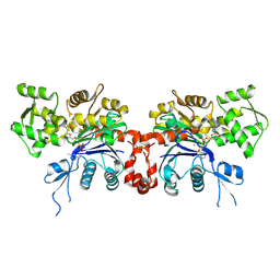
Structure of the Cyclomodulin Cif from Pathogenic Escherichia coli
Descriptor: Cif (Cell cycle inhibiting factor)
Authors:Stebbins, C.E, Hsu, Y.
Deposit date:2008-09-10
Release date:2008-10-28
Last modified:2024-02-21
Method:X-RAY DIFFRACTION (1.7 Å)
Cite:Structure of the cyclomodulin Cif from pathogenic Escherichia coli.
J.Mol.Biol., 384, 2008
1GRJ
DownloadVisualize
BU of 1grj by Molmil
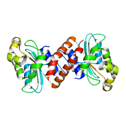
GREA TRANSCRIPT CLEAVAGE FACTOR FROM ESCHERICHIA COLI
Descriptor: GREA PROTEIN
Authors:Darst, S.A, Stebbins, C.E.
Deposit date:1995-05-02
Release date:1995-07-10
Last modified:2024-02-07
Method:X-RAY DIFFRACTION (2.2 Å)
Cite:Crystal structure of the GreA transcript cleavage factor from Escherichia coli.
Nature, 373, 1995
4G29
DownloadVisualize
BU of 4g29 by Molmil
Structure of the Catalytic Domain of the Salmonella Virulence Factor SseI
Descriptor: Secreted effector protein sseI
Authors:Stebbins, C.E, Bhaskaran, S.S.
Deposit date:2012-07-11
Release date:2012-11-28
Last modified:2024-11-20
Method:X-RAY DIFFRACTION (1.7 Å)
Cite:Structure of the catalytic domain of the Salmonella virulence factor SseI.
Acta Crystallogr.,Sect.D, 68, 2012
4GF3
DownloadVisualize
BU of 4gf3 by Molmil
Structure of a SycH-YopH Chaperone-Effector Complex
Descriptor: Putative yopH targeting protein, Tyrosine-protein phosphatase yopH
Authors:Stebbins, C.E, Vujanac, M.
Deposit date:2012-08-02
Release date:2013-03-27
Last modified:2024-02-28
Method:X-RAY DIFFRACTION (1.9 Å)
Cite:Context-dependent protein folding of a virulence peptide in the bacterial and host environments: structure of an SycH-YopH chaperone-effector complex.
Acta Crystallogr.,Sect.D, 69, 2013
3IEC
DownloadVisualize
BU of 3iec by Molmil
Helicobacter pylori CagA Inhibits PAR1/MARK Family Kinases by Mimicking Host Substrates
Descriptor: Cytotoxicity-associated immunodominant antigen, Serine/threonine-protein kinase MARK2
Authors:Stebbins, C.E, Nesic, D, Miller, M.
Deposit date:2009-07-22
Release date:2009-12-08
Last modified:2024-02-21
Method:X-RAY DIFFRACTION (2.2 Å)
Cite:Helicobacter pylori CagA inhibits PAR1-MARK family kinases by mimicking host substrates.
Nat.Struct.Mol.Biol., 17, 2010
3LW9
DownloadVisualize
BU of 3lw9 by Molmil
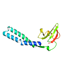
Structure of a Cytoplasmic Domain of Salmonella InvA
Descriptor: Invasion protein invA
Authors:Stebbins, C.E, Lilic, M, Quezada, C.M.
Deposit date:2010-02-23
Release date:2010-05-26
Last modified:2024-11-20
Method:X-RAY DIFFRACTION (1.85 Å)
Cite:A conserved domain in type III secretion links the cytoplasmic domain of InvA to elements of the basal body.
Acta Crystallogr.,Sect.D, 66, 2010
1Q5Z
DownloadVisualize
BU of 1q5z by Molmil
Crystal Structure of the C-terminal Actin Binding Domain of Salmonella Invasion Protein A (SipA)
Descriptor: SipA
Authors:Stebbins, C.E, Lilic, M, Galkin, V.E, Orlova, A, VanLoock, M.S, Egelman, E.H.
Deposit date:2003-08-11
Release date:2003-10-07
Last modified:2024-02-14
Method:X-RAY DIFFRACTION (1.8 Å)
Cite:Salmonella SipA polymerizes actin by stapling filaments with nonglobular protein arms.
Science, 301, 2003
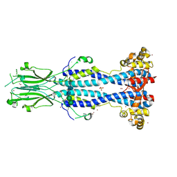
6Z8H
DownloadVisualize
BU of 6z8h by Molmil
Crystal structure of Variant Surface Glycoprotein VSG13
Descriptor: 2-acetamido-2-deoxy-beta-D-glucopyranose, SULFATE ION, Variant surface glycoprotein MITat 1.13, ...
Authors:Stebbins, C.E, Hempelmann, A, Van Straaten, M, Zeelen, J.
Deposit date:2020-06-02
Release date:2021-03-17
Last modified:2024-10-23
Method:X-RAY DIFFRACTION (1.38 Å)
Cite:Structure of trypanosome coat protein VSGsur and function in suramin resistance.
Nat Microbiol, 6, 2021
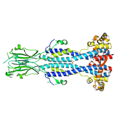
6Z8G
DownloadVisualize
BU of 6z8g by Molmil
Crystal structure of VSG13 soaked in 0.5 M used to phase VSG13 to solve the structure.
Descriptor: BROMIDE ION, SULFATE ION, Variant surface glycoprotein MITat 1.13, ...
Authors:Stebbins, C.E, Hempelmann, A, Van Straaten, M, Zeelen, J.
Deposit date:2020-06-02
Release date:2021-03-17
Last modified:2024-11-06
Method:X-RAY DIFFRACTION (1.56 Å)
Cite:Structure of trypanosome coat protein VSGsur and function in suramin resistance.
Nat Microbiol, 6, 2021
4IRV
DownloadVisualize
BU of 4irv by Molmil
Structure of the Helicobacter pylori CagA Oncogene Bound to the Human Tumor Suppressor Apoptosis-stimulating Protein of p53-2
Descriptor: Apoptosis-stimulating of p53 protein 2, Cytotoxicity-associated immunodominant antigen
Authors:Stebbins, C.E, Nesic, D.
Deposit date:2013-01-15
Release date:2014-01-15
Last modified:2024-10-16
Method:X-RAY DIFFRACTION (2.04 Å)
Cite:Structure of the Helicobacter pylori CagA oncoprotein bound to the human tumor suppressor ASPP2.
Proc.Natl.Acad.Sci.USA, 111, 2014
4G6T
DownloadVisualize
BU of 4g6t by Molmil
Structure of the HopA1-SchA Chaperone-Effector Complex
Descriptor: Type III chaperone protein ShcA, Type III effector HopA1
Authors:Stebbins, C.E, Janjusevic, R, Quezada, C.M.
Deposit date:2012-07-19
Release date:2013-06-05
Last modified:2024-02-28
Method:X-RAY DIFFRACTION (1.56 Å)
Cite:Structure of the HopA1(21-102)-ShcA chaperone-effector complex of Pseudomonas syringae reveals conservation of a virulence factor binding motif from animal to plant pathogens.
J.Bacteriol., 195, 2013
4G2B
DownloadVisualize
BU of 4g2b by Molmil
Structure of the Catalytic Domain of the Salmonella Virulence Factor SseI
Descriptor: Secreted effector protein sseI
Authors:Stebbins, C.E, Bhaskaran, S.S.
Deposit date:2012-07-11
Release date:2012-11-28
Last modified:2024-10-30
Method:X-RAY DIFFRACTION (2.05 Å)
Cite:Structure of the catalytic domain of the Salmonella virulence factor SseI.
Acta Crystallogr.,Sect.D, 68, 2012
2GWK
DownloadVisualize
BU of 2gwk by Molmil
SpvB ADP-ribosylated actin: orthorhombic crystal form
Descriptor: ADENOSINE-5'-TRIPHOSPHATE, Actin, alpha skeletal muscle, ...
Authors:Stebbins, C.E, Margarit, S.M.
Deposit date:2006-05-04
Release date:2006-08-29
Last modified:2011-07-13
Method:X-RAY DIFFRACTION (2 Å)
Cite:A steric antagonism of actin polymerization by a salmonella virulence protein.
Structure, 14, 2006


123>

227933

数据于2024-11-27公开中

PDB statisticsPDBj update infoContact PDBjnumon