Loading
PDBj
MenuPDBj@FacebookPDBj@X(formerly Twitter)PDBj@BlueSkyPDBj@YouTubewwPDB FoundationwwPDB
RCSB PDBPDBeBMRBAdv. SearchSearch help
Search by PDB author
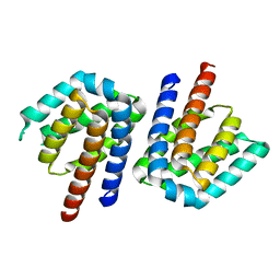
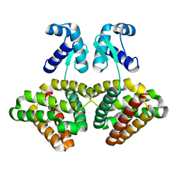
3SXM
DownloadVisualize
BU of 3sxm by Molmil
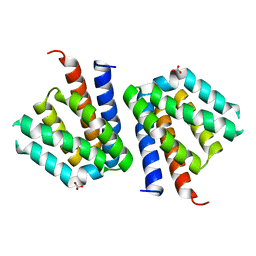
Metal-free FCD domain of TM0439 a putative transcriptional regulator
Descriptor: BICARBONATE ION, Transcriptional regulator, GntR family
Authors:Czelakowski, G.P, Derewenda, Z.S, Integrated Center for Structure and Function Innovation (ISFI)
Deposit date:2011-07-15
Release date:2011-11-16
Last modified:2023-09-13
Method:X-RAY DIFFRACTION (1.55 Å)
Cite:Metal-free FCD domain of TM0439 a putative transcriptional regulator
To be Published
3SXY
DownloadVisualize
BU of 3sxy by Molmil
Metal-free full-length structure of Tm0439, a metal-binding FCD family transcriptional regulator
Descriptor: Transcriptional regulator, GntR family
Authors:Czelakowski, G.P, Derewenda, Z.S, Integrated Center for Structure and Function Innovation (ISFI)
Deposit date:2011-07-15
Release date:2011-11-16
Last modified:2023-09-13
Method:X-RAY DIFFRACTION (1.647 Å)
Cite:Metal-free full-length structure of Tm0439, a metal-binding FCD family transcriptional regulator
To be Published
3SXZ
DownloadVisualize
BU of 3sxz by Molmil
Metal-free FCD domain of TM0439 a putative transcriptional regulator
Descriptor: Transcriptional regulator, GntR family
Authors:Czelakowski, G.P, Derewenda, Z.S, Integrated Center for Structure and Function Innovation (ISFI)
Deposit date:2011-07-15
Release date:2011-11-16
Last modified:2023-09-13
Method:X-RAY DIFFRACTION (2.322 Å)
Cite:Metal-free FCD domain of TM0439 a putative transcriptional regulator
To be Published
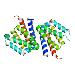
3SXK
DownloadVisualize
BU of 3sxk by Molmil
Zn2+-bound FCD domain of TM0439, a putative transcriptional regulator
Descriptor: BICARBONATE ION, SULFATE ION, Transcriptional regulator, ...
Authors:Czelakowski, G.P, Derewenda, Z.S, Integrated Center for Structure and Function Innovation (ISFI)
Deposit date:2011-07-15
Release date:2011-11-16
Last modified:2023-09-13
Method:X-RAY DIFFRACTION (1.63 Å)
Cite:Zn2+-bound FCD domain of TM0439, a putative transcriptional regulator
To be Published
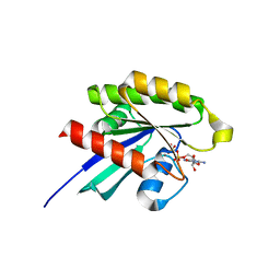
6P0J
DownloadVisualize
BU of 6p0j by Molmil
Crystal structure of GDP-bound human RalA
Descriptor: CALCIUM ION, GUANOSINE-5'-DIPHOSPHATE, Ras-related protein Ral-A
Authors:Bum-Erdene, K, Gonzalez-Gutierrez, G, Liu, D, Meroueh, S.O.
Deposit date:2019-05-17
Release date:2020-03-04
Last modified:2023-10-11
Method:X-RAY DIFFRACTION (1.31 Å)
Cite:Small-molecule covalent bond formation at tyrosine creates a binding site and inhibits activation of Ral GTPases.
Proc.Natl.Acad.Sci.USA, 117, 2020
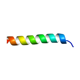
6PHJ
DownloadVisualize
BU of 6phj by Molmil
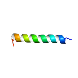
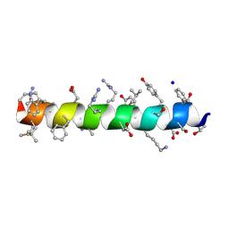
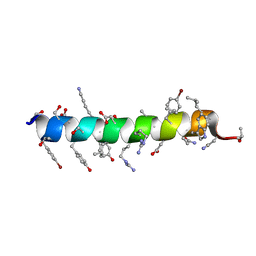
Crystal structure of native glucagon in space group P213 at 1.99 A resolution
Descriptor: Glucagon
Authors:Mroz, P.A, Gonzalez-Gutierrez, G, DiMarchi, R.D.
Deposit date:2019-06-25
Release date:2020-07-01
Last modified:2023-10-11
Method:X-RAY DIFFRACTION (1.99 Å)
Cite:High resolution X-ray structure of glucagon and selected stereo-inversed analogs in novel crystallographic packing arrangement.
To Be Published
6PHO
DownloadVisualize
BU of 6pho by Molmil
Crystal structure of glucagon analog with selenomethionine substitutions at position 1 and 27 in space group I41 at 1.42 A resolution
Descriptor: Glucagon
Authors:Mroz, P.A, Gonzalez-Gutierrez, G, DiMarchi, R.D.
Deposit date:2019-06-25
Release date:2020-07-01
Last modified:2024-10-09
Method:X-RAY DIFFRACTION (1.42 Å)
Cite:High resolution X-ray structure of glucagon and selected stereo-inversed analogs in novel crystallographic packing arrangement.
To Be Published
6PHK
DownloadVisualize
BU of 6phk by Molmil
Crystal structure of glucagon analog with mono-stereoinversion at position 21 (D-Asp21) in space group I41 at 1.18 A resolution
Descriptor: Glucagon
Authors:Mroz, P.A, Gonzalez-Gutierrez, G, DiMarchi, R.D.
Deposit date:2019-06-25
Release date:2020-07-01
Last modified:2024-10-16
Method:X-RAY DIFFRACTION (1.18 Å)
Cite:High resolution X-ray structure of glucagon and selected stereo-inversed analogs in novel crystallographic packing arrangement.
To Be Published
6PHP
DownloadVisualize
BU of 6php by Molmil
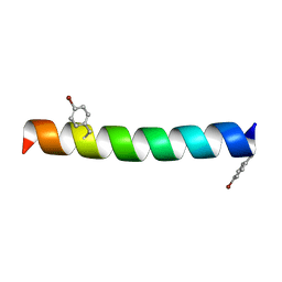
Crystal structure of glucagon analog with 4-bromo-phenylalanine substitutions at position 6 and 22 in space group I41 at 1.65 A resolution
Descriptor: Glucagon
Authors:Mroz, P.A, Gonzalez-Gutierrez, G, DiMarchi, R.D.
Deposit date:2019-06-25
Release date:2020-07-01
Last modified:2023-11-15
Method:X-RAY DIFFRACTION (1.65 Å)
Cite:High resolution X-ray structure of glucagon and selected stereo-inversed analogs in novel crystallographic packing arrangement.
To Be Published
6PQ7
DownloadVisualize
BU of 6pq7 by Molmil
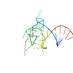
Structure of the iMango-III fluorescent aptamer at room temperature.
Descriptor: MAGNESIUM ION, POTASSIUM ION, RNA (37-MER), ...
Authors:Trachman III, R.J, Ferre-D'Amare, A.R.
Deposit date:2019-07-08
Release date:2019-07-31
Last modified:2023-10-11
Method:X-RAY DIFFRACTION (3 Å)
Cite:Co-crystal structure of the iMango-III fluorescent RNA aptamer using an X-ray free-electron laser.
Acta Crystallogr.,Sect.F, 75, 2019
6PHN
DownloadVisualize
BU of 6phn by Molmil
Crystal structure of glucagon analog composed of D-amino acids with mono-stereoinversion at position 23 (L-Val23) in space group I41 at 1.33 A resolution
Descriptor: D-glucagon L-Val23, SODIUM ION
Authors:Mroz, P.A, Gonzalez-Gutierrez, G, DiMarchi, R.D.
Deposit date:2019-06-25
Release date:2020-07-01
Last modified:2024-10-30
Method:X-RAY DIFFRACTION (1.33 Å)
Cite:High resolution X-ray structure of glucagon and selected stereo-inversed analogs in novel crystallographic packing arrangement.
To Be Published
6PHI
DownloadVisualize
BU of 6phi by Molmil
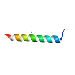
Crystal structure of native glucagon in space group I41 at 1.1 A resolution
Descriptor: CHLORIDE ION, Glucagon
Authors:Mroz, P.A, Gonzalez-Gutierrez, G, DiMarchi, R.D.
Deposit date:2019-06-25
Release date:2020-07-01
Last modified:2023-10-11
Method:X-RAY DIFFRACTION (1.1 Å)
Cite:High resolution X-ray structure of glucagon and selected stereo-inversed analogs in novel crystallographic packing arrangement.
To Be Published
6PHM
DownloadVisualize
BU of 6phm by Molmil
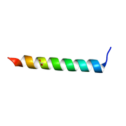
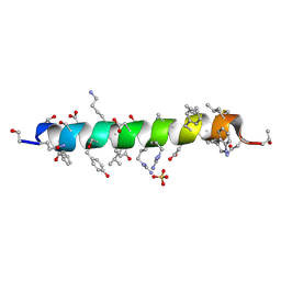
Crystal structure of glucagon analog fully composed of D-amino acids in space group I41 at 1.1 A resolution
Descriptor: D-glucagon, SULFATE ION
Authors:Mroz, P.A, Gonzalez-Gutierrez, G, DiMarchi, R.D.
Deposit date:2019-06-25
Release date:2020-07-01
Last modified:2024-10-23
Method:X-RAY DIFFRACTION (1.1 Å)
Cite:High resolution X-ray structure of glucagon and selected stereo-inversed analogs in novel crystallographic packing arrangement.
To Be Published
6PHL
DownloadVisualize
BU of 6phl by Molmil
Crystal structure of glucagon analog with mono-stereoinversion at position 23 (D-Val23) in space group I41 at 1.44 A resolution
Descriptor: Glucagon
Authors:Mroz, P.A, Gonzalez-Gutierrez, G, DiMarchi, R.D.
Deposit date:2019-06-25
Release date:2020-07-01
Last modified:2024-10-16
Method:X-RAY DIFFRACTION (1.443 Å)
Cite:High resolution X-ray structure of glucagon and selected stereo-inversed analogs in novel crystallographic packing arrangement.
To Be Published
6PHQ
DownloadVisualize
BU of 6phq by Molmil
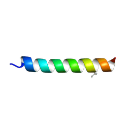
Crystal structure of glucagon analog fully composed of D-amino acids with 4-bromo-D-phenylalanine substitutions at position 6 and 22 in space group I41 at 1.1 A resolution
Descriptor: D-glucagon D-BrPhe 6,22
Authors:Mroz, P.A, Gonzalez-Gutierrez, G, DiMarchi, R.D.
Deposit date:2019-06-25
Release date:2020-07-01
Last modified:2023-11-15
Method:X-RAY DIFFRACTION (1.32 Å)
Cite:High resolution X-ray structure of glucagon and selected stereo-inversed analogs in novel crystallographic packing arrangement.
To Be Published
6P7M
DownloadVisualize
BU of 6p7m by Molmil
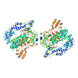
Cryo-EM structure of LbCas12a-crRNA: AcrVA4 (1:2 complex)
Descriptor: Cas12a, MAGNESIUM ION, anti-CRISPR VA4, ...
Authors:Knott, G.J, Liu, J.J, Doudna, J.A.
Deposit date:2019-06-06
Release date:2019-08-21
Last modified:2024-03-20
Method:ELECTRON MICROSCOPY (3 Å)
Cite:Structural basis for AcrVA4 inhibition of specific CRISPR-Cas12a.
Elife, 8, 2019
6P7N
DownloadVisualize
BU of 6p7n by Molmil
Cryo-EM structure of LbCas12a-crRNA: AcrVA4 (2:2 complex)
Descriptor: Cas12a, MAGNESIUM ION, anti-CRISPR VA4, ...
Authors:Knott, G.J, Liu, J.J, Doudna, J.A.
Deposit date:2019-06-06
Release date:2019-08-21
Last modified:2023-08-16
Method:ELECTRON MICROSCOPY (4.9 Å)
Cite:Structural basis for AcrVA4 inhibition of specific CRISPR-Cas12a.
Elife, 8, 2019
6PW8
DownloadVisualize
BU of 6pw8 by Molmil
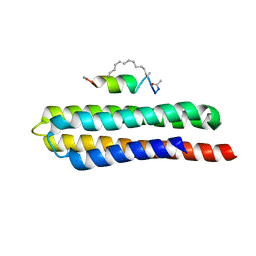
Hydrocarbon-Stapled Paxillin Peptide Bound to the Focal Adhesion Targeting (FAT) Domain of the Focal Adhesion Kinase (FAK)
Descriptor: CHLORIDE ION, Focal adhesion kinase 1, SP3, ...
Authors:Thifault, D.G, Fromme, P, Martin-Garcia, J.M.
Deposit date:2019-07-22
Release date:2020-07-22
Last modified:2023-11-15
Method:X-RAY DIFFRACTION (1.95 Å)
Cite:Stapled Peptide Ligand Bound to the Focal Adhesion Targeting (FAT) Domain of the Focal Adhesion Kinase (FAK)
To Be Published
7OS2
DownloadVisualize
BU of 7os2 by Molmil
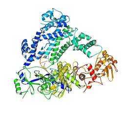
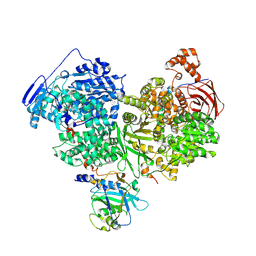
Cryo-EM structure of Brr2 in complex with Jab1/MPN and C9ORF78
Descriptor: Pre-mRNA-processing-splicing factor 8, Telomere length and silencing protein 1 homolog, U5 small nuclear ribonucleoprotein 200 kDa helicase
Authors:Bergfort, A, Hilal, T, Weber, G, Wahl, M.C.
Deposit date:2021-06-07
Release date:2022-02-23
Last modified:2024-09-25
Method:ELECTRON MICROSCOPY (2.76 Å)
Cite:The intrinsically disordered TSSC4 protein acts as a helicase inhibitor, placeholder and multi-interaction coordinator during snRNP assembly and recycling.
Nucleic Acids Res., 50, 2022
7OS1
DownloadVisualize
BU of 7os1 by Molmil
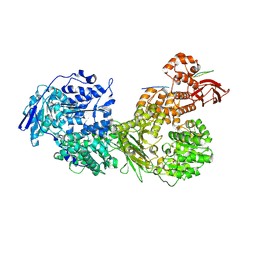
Cryo-EM structure of Brr2 in complex with Fbp21
Descriptor: U5 small nuclear ribonucleoprotein 200 kDa helicase, WW domain-binding protein 4
Authors:Bergfort, A, Hilal, T, Weber, G, Wahl, M.C.
Deposit date:2021-06-07
Release date:2022-02-23
Last modified:2024-07-17
Method:ELECTRON MICROSCOPY (3.3 Å)
Cite:The intrinsically disordered TSSC4 protein acts as a helicase inhibitor, placeholder and multi-interaction coordinator during snRNP assembly and recycling.
Nucleic Acids Res., 50, 2022
7PV9
DownloadVisualize
BU of 7pv9 by Molmil
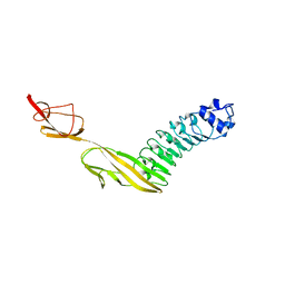
Listeria monocytogene InlB (internalin B) residues 36-392 (internalin domain and B-repeat)
Descriptor: Internalin B
Authors:Geerds, C, Niemann, H.H.
Deposit date:2021-10-01
Release date:2022-01-26
Last modified:2024-01-31
Method:X-RAY DIFFRACTION (3.3 Å)
Cite:A recurring packing contact in crystals of InlB pinpoints functional binding sites in the internalin domain and the B repeat.
Acta Crystallogr D Struct Biol, 78, 2022
4FE1
DownloadVisualize
BU of 4fe1 by Molmil
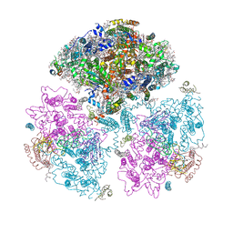
Improving the Accuracy of Macromolecular Structure Refinement at 7 A Resolution
Descriptor: 1,2-DIPALMITOYL-PHOSPHATIDYL-GLYCEROLE, 1,2-DISTEAROYL-MONOGALACTOSYL-DIGLYCERIDE, BETA-CAROTENE, ...
Authors:Fromme, R, Adams, P.D, Fromme, P, Levitt, M, Schroeder, G.F, Brunger, A.T.
Deposit date:2012-05-29
Release date:2012-08-15
Last modified:2024-11-20
Method:X-RAY DIFFRACTION (4.9228 Å)
Cite:Improving the accuracy of macromolecular structure refinement at 7 A resolution.
Structure, 20, 2012
7PPM
DownloadVisualize
BU of 7ppm by Molmil
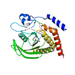
SHP2 catalytic domain in complex with IRS1 (889-901) phosphopeptide (pSer-892, pTyr-896)
Descriptor: GLYCEROL, Insulin receptor substrate 1, Tyrosine-protein phosphatase non-receptor type 11,Tyrosine-protein phosphatase non-receptor type 11
Authors:Sok, P, Zeke, A, Remenyi, A.
Deposit date:2021-09-14
Release date:2022-09-07
Last modified:2024-11-20
Method:X-RAY DIFFRACTION (1.48 Å)
Cite:Structural insights into the pSer/pThr dependent regulation of the SHP2 tyrosine phosphatase in insulin and CD28 signaling.
Nat Commun, 13, 2022
7PPN
DownloadVisualize
BU of 7ppn by Molmil
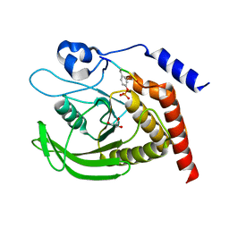
SHP2 catalytic domain in complex with CD28 (183-198) phosphopeptide (pTyr-191, p-Thr-195)
Descriptor: GLYCEROL, T-cell-specific surface glycoprotein CD28, Tyrosine-protein phosphatase non-receptor type 11,Tyrosine-protein phosphatase non-receptor type 11
Authors:Sok, P, Zeke, A, Remenyi, A.
Deposit date:2021-09-14
Release date:2022-09-07
Last modified:2024-11-13
Method:X-RAY DIFFRACTION (1.9 Å)
Cite:Structural insights into the pSer/pThr dependent regulation of the SHP2 tyrosine phosphatase in insulin and CD28 signaling.
Nat Commun, 13, 2022
7PPL
DownloadVisualize
BU of 7ppl by Molmil
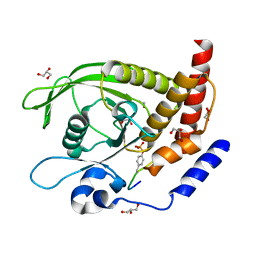
SHP2 catalytic domain in complex with IRS1 (625-639) phosphopeptide (pTyr-632, pSer-636)
Descriptor: ETHANOL, GLYCEROL, Insulin receptor substrate 1, ...
Authors:Sok, P, Zeke, A, Remenyi, A.
Deposit date:2021-09-14
Release date:2022-09-07
Last modified:2024-10-23
Method:X-RAY DIFFRACTION (1.53 Å)
Cite:Structural insights into the pSer/pThr dependent regulation of the SHP2 tyrosine phosphatase in insulin and CD28 signaling.
Nat Commun, 13, 2022

227561

数据于2024-11-20公开中

PDB statisticsPDBj update infoContact PDBjnumon