Loading
PDBj
MenuPDBj@FacebookPDBj@TwitterPDBj@YouTubewwPDB FoundationwwPDB
RCSB PDBPDBeBMRBAdv. SearchSearch help
Search by PDB author
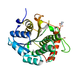
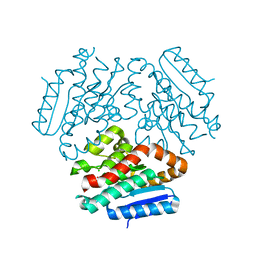
3R1J
DownloadVisualize
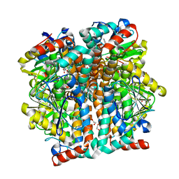
BU of 3r1j by Molmil
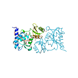
Crystal structure of Alpha-ketoglutarate-dependent taurine dioxygenase from Mycobacterium avium, native form
Descriptor: 1,2-ETHANEDIOL, Alpha-ketoglutarate-dependent taurine dioxygenase, CHLORIDE ION, ...
Authors:Seattle Structural Genomics Center for Infectious Disease (SSGCID)
Deposit date:2011-03-10
Release date:2011-04-06
Last modified:2023-09-13
Method:X-RAY DIFFRACTION (2.05 Å)
Cite:Increasing the structural coverage of tuberculosis drug targets.
Tuberculosis (Edinb), 95, 2015
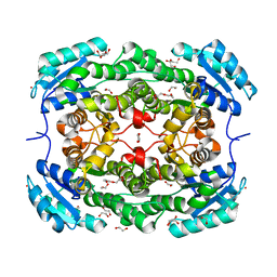
3R6H
DownloadVisualize
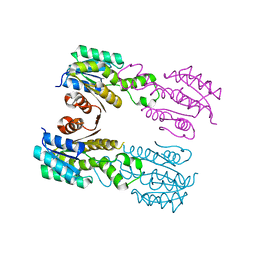
BU of 3r6h by Molmil
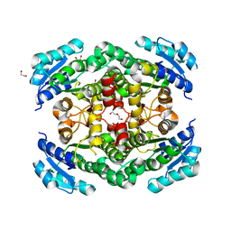
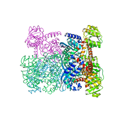
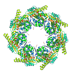
Crystal structure of an enoyl-CoA hydratase (ECHA3) from Mycobacterium marinum
Descriptor: CHLORIDE ION, Enoyl-CoA hydratase, EchA3, ...
Authors:Seattle Structural Genomics Center for Infectious Disease (SSGCID)
Deposit date:2011-03-21
Release date:2011-06-08
Last modified:2023-09-13
Method:X-RAY DIFFRACTION (1.75 Å)
Cite:Increasing the structural coverage of tuberculosis drug targets.
Tuberculosis (Edinb), 95, 2015
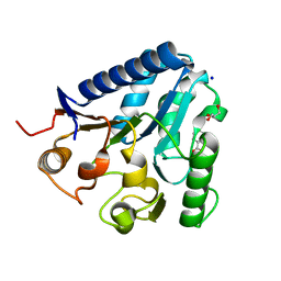
3RD5
DownloadVisualize
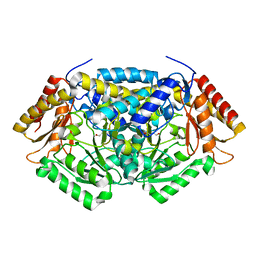
BU of 3rd5 by Molmil
Crystal structure of a putative uncharacterized protein from Mycobacterium Paratuberculosis
Descriptor: 1,2-ETHANEDIOL, 4-(2-HYDROXYETHYL)-1-PIPERAZINE ETHANESULFONIC ACID, MYPAA.01249.C
Authors:Seattle Structural Genomics Center for Infectious Disease (SSGCID)
Deposit date:2011-03-31
Release date:2011-04-20
Last modified:2024-04-03
Method:X-RAY DIFFRACTION (1.5 Å)
Cite:Increasing the structural coverage of tuberculosis drug targets.
Tuberculosis (Edinb), 95, 2015
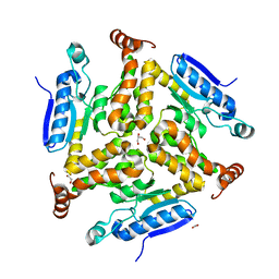
3RIH
DownloadVisualize
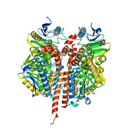
BU of 3rih by Molmil
Crystal structure of a putative short chain dehydrogenase or reductase from Mycobacterium abscessus
Descriptor: 1,2-ETHANEDIOL, 1-METHOXY-2-[2-(2-METHOXY-ETHOXY]-ETHANE, POTASSIUM ION, ...
Authors:Seattle Structural Genomics Center for Infectious Disease (SSGCID)
Deposit date:2011-04-13
Release date:2011-04-20
Last modified:2023-09-13
Method:X-RAY DIFFRACTION (2.15 Å)
Cite:Increasing the structural coverage of tuberculosis drug targets.
Tuberculosis (Edinb), 95, 2015
3TX2
DownloadVisualize
BU of 3tx2 by Molmil
Structure of a Probable 6-phosphogluconolactonase from Mycobacterium abscessus
Descriptor: BENZOIC ACID, GLYCEROL, Probable 6-phosphogluconolactonase, ...
Authors:Seattle Structural Genomics Center for Infectious Disease (SSGCID)
Deposit date:2011-09-22
Release date:2011-10-05
Last modified:2023-09-13
Method:X-RAY DIFFRACTION (1.5 Å)
Cite:Increasing the structural coverage of tuberculosis drug targets.
Tuberculosis (Edinb), 95, 2015
3TRR
DownloadVisualize
BU of 3trr by Molmil
Crystal structure of a probable enoyl-CoA hydratase/isomerase from Mycobacterium abscessus
Descriptor: 1,2-ETHANEDIOL, DI(HYDROXYETHYL)ETHER, GLYCEROL, ...
Authors:Seattle Structural Genomics Center for Infectious Disease (SSGCID)
Deposit date:2011-09-09
Release date:2011-09-21
Last modified:2023-09-13
Method:X-RAY DIFFRACTION (2.09 Å)
Cite:Increasing the structural coverage of tuberculosis drug targets.
Tuberculosis (Edinb), 95, 2015
3QK8
DownloadVisualize
BU of 3qk8 by Molmil
Crystal structure of enoyl-coA hydratase EchA15 from Mycobacterium marinum in complex with an unknown ligand
Descriptor: 1,2-ETHANEDIOL, CHLORIDE ION, Enoyl-CoA hydratase EchA15, ...
Authors:Seattle Structural Genomics Center for Infectious Disease (SSGCID)
Deposit date:2011-01-31
Release date:2011-02-09
Last modified:2023-09-13
Method:X-RAY DIFFRACTION (1.6 Å)
Cite:Increasing the structural coverage of tuberculosis drug targets.
Tuberculosis (Edinb), 95, 2015
3QIV
DownloadVisualize
BU of 3qiv by Molmil
Crystal structure of a putative short-chain dehydrogenase or 3-oxoacyl-[acyl-carrier-protein] reductase from Mycobacterium paratuberculosis ATCC BAA-968 / K-10
Descriptor: short-chain dehydrogenase or 3-oxoacyl-[acyl-carrier-protein] reductase
Authors:Seattle Structural Genomics Center for Infectious Disease (SSGCID)
Deposit date:2011-01-27
Release date:2011-02-16
Last modified:2023-09-13
Method:X-RAY DIFFRACTION (2.25 Å)
Cite:Increasing the structural coverage of tuberculosis drug targets.
Tuberculosis (Edinb), 95, 2015
3Q8N
DownloadVisualize
BU of 3q8n by Molmil
Crystal structure of 4-aminobutyrate transaminase from Mycobacterium smegmatis
Descriptor: 4-aminobutyrate transaminase, 4-oxobutanoic acid
Authors:Seattle Structural Genomics Center for Infectious Disease (SSGCID)
Deposit date:2011-01-06
Release date:2011-02-09
Last modified:2023-09-13
Method:X-RAY DIFFRACTION (2.05 Å)
Cite:Increasing the structural coverage of tuberculosis drug targets.
Tuberculosis (Edinb), 95, 2015
3QKA
DownloadVisualize
BU of 3qka by Molmil
Crystal structure of enoyl-CoA hydratase EchA5 from Mycobacterium marinum
Descriptor: Enoyl-CoA hydratase, EchA5
Authors:Seattle Structural Genomics Center for Infectious Disease (SSGCID)
Deposit date:2011-01-31
Release date:2011-02-09
Last modified:2023-09-13
Method:X-RAY DIFFRACTION (2.15 Å)
Cite:Increasing the structural coverage of tuberculosis drug targets.
Tuberculosis (Edinb), 95, 2015
3U7J
DownloadVisualize
BU of 3u7j by Molmil
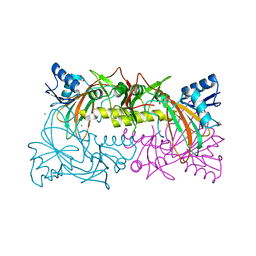
Crystal structure of Ribose-5-phosphate isomerase A from Burkholderia thailandensis
Descriptor: 1,2-ETHANEDIOL, POTASSIUM ION, Ribose-5-phosphate isomerase A, ...
Authors:Seattle Structural Genomics Center for Infectious Disease (SSGCID)
Deposit date:2011-10-13
Release date:2011-11-16
Last modified:2023-09-13
Method:X-RAY DIFFRACTION (2.1 Å)
Cite:Combining functional and structural genomics to sample the essential Burkholderia structome.
Plos One, 8, 2013
3TZQ
DownloadVisualize
BU of 3tzq by Molmil
Crystal structure of a short-chain type dehydrogenase/reductase from Mycobacterium marinum
Descriptor: 1,2-ETHANEDIOL, DI(HYDROXYETHYL)ETHER, GLYCEROL, ...
Authors:Seattle Structural Genomics Center for Infectious Disease (SSGCID)
Deposit date:2011-09-27
Release date:2011-10-05
Last modified:2023-09-13
Method:X-RAY DIFFRACTION (2.5 Å)
Cite:Increasing the structural coverage of tuberculosis drug targets.
Tuberculosis (Edinb), 95, 2015
3U9N
DownloadVisualize
BU of 3u9n by Molmil
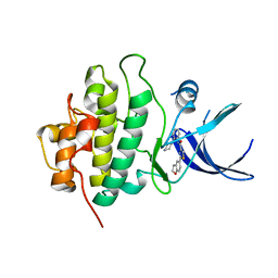
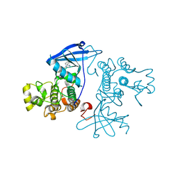
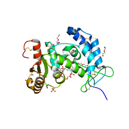
X-ray crystal structure of compound 1 bound to human CHK1 kinase domain
Descriptor: 2-(2,3-dihydro-1-benzofuran-5-yl)-N-[2-(piperazin-1-yl)phenyl]-1,3-thiazole-4-carboxamide, Serine/threonine-protein kinase Chk1
Authors:Fischmann, T.O.
Deposit date:2011-10-19
Release date:2012-01-25
Last modified:2024-02-28
Method:X-RAY DIFFRACTION (1.85 Å)
Cite:Discovery of a Novel Series of CHK1 Kinase Inhibitors with Distinctive Hinge Binding Mode
ACS MED.CHEM.LETT., 2012
3UIR
DownloadVisualize
BU of 3uir by Molmil
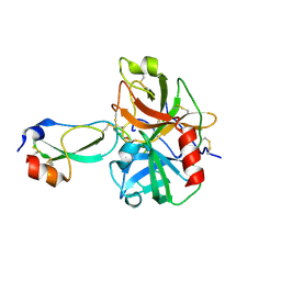
Crystal structure of the plasmin-textilinin-1 complex
Descriptor: Plasmin light chain B, SULFATE ION, Textilinin-1
Authors:Guddat, L.W, Millers, E.K, de jersey, J, Lavin, M.F, Masci, P.M.
Deposit date:2011-11-05
Release date:2012-12-26
Last modified:2013-07-17
Method:X-RAY DIFFRACTION (2.777 Å)
Cite:The structure of human microplasmin in complex with textilinin-1, an aprotinin-like inhibitor from the Australian brown snake.
Plos One, 8, 2013
7NWK
DownloadVisualize
BU of 7nwk by Molmil
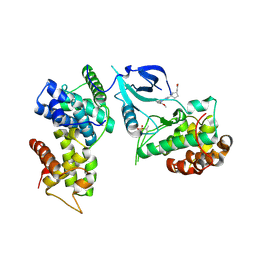
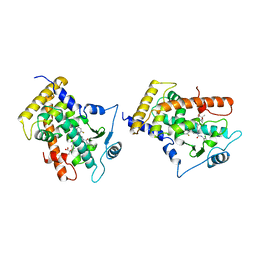
Crystal structure of CDK9-Cyclin T1 bound by compound 6
Descriptor: Cyclin-T1, Cyclin-dependent kinase 9, N-((1R,3R)-3-(7-(4-fluoro-2-methoxyphenyl)-3H-imidazo[4,5-b]pyridin-2-yl)cyclopentyl)acetamide
Authors:Collie, G.W, Ferguson, A.D.
Deposit date:2021-03-16
Release date:2021-10-27
Last modified:2024-05-01
Method:X-RAY DIFFRACTION (2.81 Å)
Cite:Discovery of a Series of 7-Azaindoles as Potent and Highly Selective CDK9 Inhibitors for Transient Target Engagement.
J.Med.Chem., 64, 2021
6F5M
DownloadVisualize
BU of 6f5m by Molmil
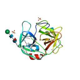
Crystal structure of highly glycosylated human leukocyte elastase in complex with a thiazolidinedione inhibitor
Descriptor: 5-[[4-[[(2~{S})-4-methyl-1-oxidanylidene-1-[(2-propylphenyl)amino]pentan-2-yl]carbamoyl]phenyl]methyl]-2-oxidanylidene-1,3-thiazol-1-ium-4-olate, ACETATE ION, Neutrophil elastase, ...
Authors:Hochscherf, J, Pietsch, M, Tieu, W, Kuan, K, Hautmann, S, Abell, A, Guetschow, M, Niefind, K.
Deposit date:2017-12-01
Release date:2018-08-08
Last modified:2024-01-17
Method:X-RAY DIFFRACTION (2.7 Å)
Cite:Crystal structure of highly glycosylated human leukocyte elastase in complex with an S2' site binding inhibitor.
Acta Crystallogr F Struct Biol Commun, 74, 2018
7NRU
DownloadVisualize
BU of 7nru by Molmil
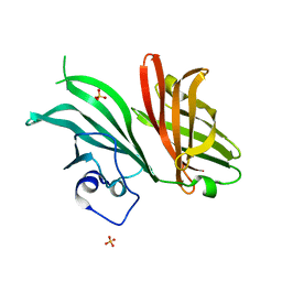
Structure of a natural chimera of meningococcal factor H binding protein belonging to NL096 strain
Descriptor: Factor H binding protein variant 1-2,3.x, SULFATE ION
Authors:Veggi, D, Malito, E, Bottomley, M.J.
Deposit date:2021-03-04
Release date:2022-06-08
Last modified:2024-01-31
Method:X-RAY DIFFRACTION (1.21998358 Å)
Cite:Structural characterization of a cross-protective natural chimera of factor H binding protein from meningococcal serogroup B strain NL096.
Comput Struct Biotechnol J, 20, 2022
7OXB
DownloadVisualize
BU of 7oxb by Molmil
Crystal structure of EGFR double mutant (T790M/L858R) in complex with compound 6.
Descriptor: 2-[2-(3-methoxyphenyl)pyrimidin-4-yl]-1'-prop-2-enoyl-spiro[5,6-dihydro-1~{H}-pyrrolo[3,2-c]pyridine-7,4'-piperidine]-4-one, Epidermal growth factor receptor
Authors:Collie, G.W.
Deposit date:2021-06-22
Release date:2021-10-20
Method:X-RAY DIFFRACTION (2.56 Å)
Cite:Discovery and optimization of covalent EGFR T790M/L858R mutant inhibitors".
Bioorg.Med.Chem.Lett., 2021
6VUA
DownloadVisualize
BU of 6vua by Molmil
X-ray structure of human CD38 catalytic domain with 2'-Cl-araNAD+
Descriptor: (4S)-2-METHYL-2,4-PENTANEDIOL, ADP-ribosyl cyclase/cyclic ADP-ribose hydrolase 1, DI(HYDROXYETHYL)ETHER, ...
Authors:Dai, Z, Zhang, X.N, Nasertorabi, F, Han, G.W, Stevens, R.C, Zhang, Y.
Deposit date:2020-02-14
Release date:2020-06-03
Last modified:2023-10-11
Method:X-RAY DIFFRACTION (1.5 Å)
Cite:Synthesis of site-specific antibody-drug conjugates by ADP-ribosyl cyclases.
Sci Adv, 6, 2020
2GWX
DownloadVisualize
BU of 2gwx by Molmil
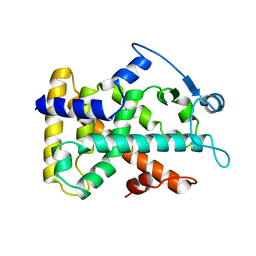
MOLECULAR RECOGNITION OF FATTY ACIDS BY PEROXISOME PROLIFERATOR-ACTIVATED RECEPTORS
Descriptor: PROTEIN (PPAR-DELTA)
Authors:Xu, H.E, Lambert, M.H, Montana, V.G, Park, D.J, Blanchard, S, Brown, P, Sternbach, D, Lehmann, J, Bruce, G.W, Willson, T.M, Kliewer, S.A, Milburn, M.V.
Deposit date:1999-03-11
Release date:2000-03-11
Last modified:2023-12-27
Method:X-RAY DIFFRACTION (2.3 Å)
Cite:Molecular recognition of fatty acids by peroxisome proliferator-activated receptors.
Mol.Cell, 3, 1999
1F60
DownloadVisualize
BU of 1f60 by Molmil
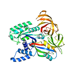
CRYSTAL STRUCTURE OF THE YEAST ELONGATION FACTOR COMPLEX EEF1A:EEF1BA
Descriptor: ELONGATION FACTOR EEF1A, ELONGATION FACTOR EEF1BA
Authors:Andersen, G.R, Pedersen, L, Valente, L, Kinzy, T.G, Nyborg, J.
Deposit date:2000-06-19
Release date:2000-07-04
Last modified:2024-02-07
Method:X-RAY DIFFRACTION (1.67 Å)
Cite:Structural basis for nucleotide exchange and competition with tRNA in the yeast elongation factor complex eEF1A:eEF1Balpha.
Mol.Cell, 6, 2000
1FIE
DownloadVisualize
BU of 1fie by Molmil
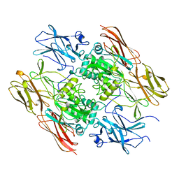
RECOMBINANT HUMAN COAGULATION FACTOR XIII
Descriptor: COAGULATION FACTOR XIII
Authors:Yee, V.C, Teller, D.C.
Deposit date:1996-08-24
Release date:1997-02-12
Last modified:2024-02-07
Method:X-RAY DIFFRACTION (2.5 Å)
Cite:Structural evidence that the activation peptide is not released upon thrombin cleavage of factor XIII.
Thromb.Res., 78, 1995
1GWX
DownloadVisualize
BU of 1gwx by Molmil
MOLECULAR RECOGNITION OF FATTY ACIDS BY PEROXISOME PROLIFERATOR-ACTIVATED RECEPTORS
Descriptor: 2-(4-{3-[1-[2-(2-CHLORO-6-FLUORO-PHENYL)-ETHYL]-3-(2,3-DICHLORO-PHENYL)-UREIDO]-PROPYL}-PHENOXY)-2-METHYL-PROPIONIC ACID, PROTEIN (PPAR-DELTA)
Authors:Xu, H.E, Lambert, M.H, Montana, V.G, Park, D.J, Blanchard, S, Brown, P, Sternbach, D, Lehmann, J, Bruce, G.W, Willson, T.M, Kliewer, S.A, Milburn, M.V.
Deposit date:1999-03-17
Release date:2000-03-17
Last modified:2023-12-27
Method:X-RAY DIFFRACTION (2.5 Å)
Cite:Molecular recognition of fatty acids by peroxisome proliferator-activated receptors.
Mol.Cell, 3, 1999
1HRD
DownloadVisualize
BU of 1hrd by Molmil
GLUTAMATE DEHYDROGENASE
Descriptor: GLUTAMATE DEHYDROGENASE
Authors:Britton, K.L, Baker, P.J, Stillman, T.J, Rice, D.W.
Deposit date:1996-04-03
Release date:1997-03-12
Last modified:2024-02-07
Method:X-RAY DIFFRACTION (1.96 Å)
Cite:The structure of Pyrococcus furiosus glutamate dehydrogenase reveals a key role for ion-pair networks in maintaining enzyme stability at extreme temperatures.
Structure, 3, 1995
2EU1
DownloadVisualize
BU of 2eu1 by Molmil
Crystal structure of the chaperonin GroEL-E461K
Descriptor: GROEL
Authors:Cabo-Bilbao, A, Spinelli, S, Sot, B, Agirre, J, Mechaly, A.E, Muga, A, Guerin, D.M.A.
Deposit date:2005-10-28
Release date:2006-08-29
Last modified:2023-08-23
Method:X-RAY DIFFRACTION (3.29 Å)
Cite:Crystal structure of the temperature-sensitive and allosteric-defective chaperonin GroEL(E461K).
J.Struct.Biol., 155, 2006

223790

数据于2024-08-14公开中

PDB statisticsPDBj update infoContact PDBjnumon