Loading
PDBj
MenuPDBj@FacebookPDBj@TwitterPDBj@YouTubewwPDB FoundationwwPDB
RCSB PDBPDBeBMRBAdv. SearchSearch help
Search by PDB author
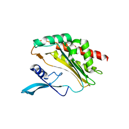
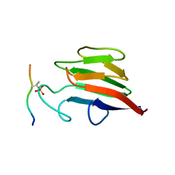
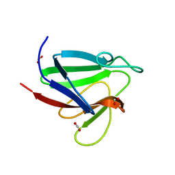
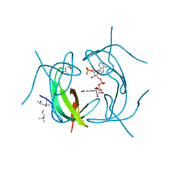
2RK1
DownloadVisualize
BU of 2rk1 by Molmil
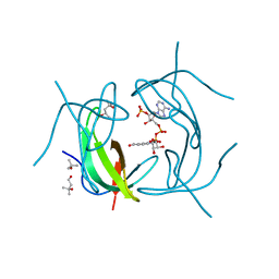
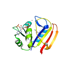
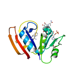
DHFR R67 Complexed with NADP and dihydrofolate
Descriptor: (4R)-2-METHYLPENTANE-2,4-DIOL, DIHYDROFOLIC ACID, Dihydrofolate reductase type 2, ...
Authors:Krahn, J.M, London, R.E.
Deposit date:2007-10-16
Release date:2008-06-03
Last modified:2023-08-30
Method:X-RAY DIFFRACTION (1.26 Å)
Cite:Crystal structure of a type II dihydrofolate reductase catalytic ternary complex.
Biochemistry, 46, 2007
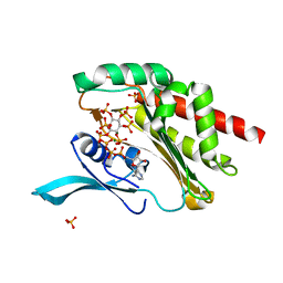
2RK2
DownloadVisualize
BU of 2rk2 by Molmil
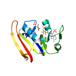
DHFR R-67 complexed with NADP
Descriptor: (4R)-2-METHYLPENTANE-2,4-DIOL, Dihydrofolate reductase type 2, NADP NICOTINAMIDE-ADENINE-DINUCLEOTIDE PHOSPHATE
Authors:Krahn, J.M, London, R.E.
Deposit date:2007-10-16
Release date:2008-06-03
Last modified:2023-08-30
Method:X-RAY DIFFRACTION (1.9 Å)
Cite:Crystal structure of a type II dihydrofolate reductase catalytic ternary complex.
Biochemistry, 46, 2007
4O4B
DownloadVisualize
BU of 4o4b by Molmil
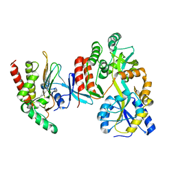
Crystal Structure of an Inositol hexakisphosphate kinase EhIP6KA as a fusion protein with maltose binding protein
Descriptor: Extracellular solute-binding protein family 1, Inositol hexakisphosphate kinase
Authors:Wang, H, Shears, S.B.
Deposit date:2013-12-18
Release date:2014-06-18
Last modified:2023-09-20
Method:X-RAY DIFFRACTION (1.8 Å)
Cite:IP6K structure and the molecular determinants of catalytic specificity in an inositol phosphate kinase family.
Nat Commun, 5, 2014
4O4E
DownloadVisualize
BU of 4o4e by Molmil
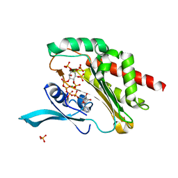
Crystal Structure of an Inositol hexakisphosphate kinase EhIP6KA in complexed with ATP and Ins(1,3,4,5,6)P5
Descriptor: ADENOSINE-5'-TRIPHOSPHATE, Inositol hexakisphosphate kinase, MAGNESIUM ION, ...
Authors:Wang, H, Shears, S.B.
Deposit date:2013-12-18
Release date:2014-06-18
Last modified:2023-09-20
Method:X-RAY DIFFRACTION (1.9 Å)
Cite:IP6K structure and the molecular determinants of catalytic specificity in an inositol phosphate kinase family.
Nat Commun, 5, 2014
4O4D
DownloadVisualize
BU of 4o4d by Molmil
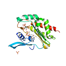
Crystal Structure of an Inositol hexakisphosphate kinase EhIP6KA in complexed with ATP and Ins(1,4,5)P3
Descriptor: ACETATE ION, ADENOSINE-5'-TRIPHOSPHATE, D-MYO-INOSITOL-1,4,5-TRIPHOSPHATE, ...
Authors:Wang, H, Shears, S.B.
Deposit date:2013-12-18
Release date:2014-06-18
Last modified:2023-09-20
Method:X-RAY DIFFRACTION (2.1 Å)
Cite:IP6K structure and the molecular determinants of catalytic specificity in an inositol phosphate kinase family.
Nat Commun, 5, 2014
4O4C
DownloadVisualize
BU of 4o4c by Molmil
Crystal Structure of an Inositol hexakisphosphate kinase apo-EhIP6KA
Descriptor: Inositol hexakisphosphate kinase
Authors:Wang, H, Shears, S.B.
Deposit date:2013-12-18
Release date:2014-06-18
Last modified:2023-09-20
Method:X-RAY DIFFRACTION (2.2 Å)
Cite:IP6K structure and the molecular determinants of catalytic specificity in an inositol phosphate kinase family.
Nat Commun, 5, 2014
4O4F
DownloadVisualize
BU of 4o4f by Molmil
Crystal Structure of an Inositol hexakisphosphate kinase EhIP6KA in complexed with ATP and InsP6
Descriptor: ADENOSINE-5'-TRIPHOSPHATE, INOSITOL HEXAKISPHOSPHATE, Inositol hexakisphosphate kinase, ...
Authors:Wang, H, Shears, S.B.
Deposit date:2013-12-18
Release date:2014-06-18
Last modified:2023-09-20
Method:X-RAY DIFFRACTION (1.7 Å)
Cite:IP6K structure and the molecular determinants of catalytic specificity in an inositol phosphate kinase family.
Nat Commun, 5, 2014
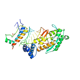
4QOZ
DownloadVisualize
BU of 4qoz by Molmil
Crystal structure of the histone mRNA stem-loop, stem-loop binding protein (phosphorylated), and 3'hExo ternary complex
Descriptor: 3'-5' exoribonuclease 1, Histone RNA hairpin-binding protein, histone mRNA stem-loop
Authors:Tan, D, Tong, L.
Deposit date:2014-06-21
Release date:2014-07-23
Last modified:2024-10-16
Method:X-RAY DIFFRACTION (2.304 Å)
Cite:Molecular mechanisms for the regulation of histone mRNA stem-loop-binding protein by phosphorylation.
Proc.Natl.Acad.Sci.USA, 111, 2014
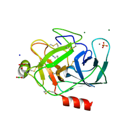
2A31
DownloadVisualize
BU of 2a31 by Molmil
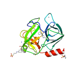
Trypsin in complex with borate
Descriptor: BORATE ION, CALCIUM ION, GUANIDINE-3-PROPANOL, ...
Authors:Transue, T.R, Gabel, S.A, London, R.E.
Deposit date:2005-06-23
Release date:2006-07-04
Last modified:2024-10-16
Method:X-RAY DIFFRACTION (1.25 Å)
Cite:NMR and crystallographic characterization of adventitious borate binding by trypsin.
Bioconjug.Chem., 17, 2006
2A32
DownloadVisualize
BU of 2a32 by Molmil
Trypsin in complex with benzene boronic acid
Descriptor: 3-CYCLOHEXYL-1-PROPYLSULFONIC ACID, BORATE ION, CALCIUM ION, ...
Authors:Transue, T.R, Gabel, S.A, London, R.E.
Deposit date:2005-06-23
Release date:2006-07-04
Last modified:2023-08-23
Method:X-RAY DIFFRACTION (1.5 Å)
Cite:NMR and crystallographic characterization of adventitious borate binding by trypsin.
Bioconjug.Chem., 17, 2006
5UII
DownloadVisualize
BU of 5uii by Molmil
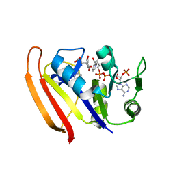
structure of DHFR with bound buformin and NADP
Descriptor: CALCIUM ION, Dihydrofolate reductase, N-butyl-N'-(diaminomethylidene)guanidine, ...
Authors:Pedersen, L.C, London, R.E.
Deposit date:2017-01-14
Release date:2018-01-31
Last modified:2023-10-25
Method:X-RAY DIFFRACTION (1.351 Å)
Cite:A Structural Basis for Biguanide Activity.
Biochemistry, 56, 2017
5UIP
DownloadVisualize
BU of 5uip by Molmil
structure of DHFR with bound DAP, p-ABG and NADP
Descriptor: Dihydrofolate reductase, N-(4-aminobenzene-1-carbonyl)-L-glutamic acid, NADP NICOTINAMIDE-ADENINE-DINUCLEOTIDE PHOSPHATE, ...
Authors:Pedersen, L.C, London, R.E.
Deposit date:2017-01-14
Release date:2017-11-22
Last modified:2023-10-04
Method:X-RAY DIFFRACTION (1.9 Å)
Cite:A Structural Basis for Biguanide Activity.
Biochemistry, 56, 2017
5UIO
DownloadVisualize
BU of 5uio by Molmil
structure of DHFR with bound DAP, p-ABG and NADP
Descriptor: BETA-MERCAPTOETHANOL, Dihydrofolate reductase, FORMIC ACID, ...
Authors:Pedersen, L.C, London, R.E.
Deposit date:2017-01-14
Release date:2017-11-22
Last modified:2023-10-04
Method:X-RAY DIFFRACTION (1.929 Å)
Cite:A Structural Basis for Biguanide Activity.
Biochemistry, 56, 2017
5U6Z
DownloadVisualize
BU of 5u6z by Molmil
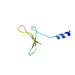
Crystal Structure of Xenopus laevis Apex2 C-terminal Znf-GRF Domain
Descriptor: DNA-(apurinic or apyrimidinic site) lyase, SULFATE ION, ZINC ION
Authors:Wallace, B.D, Williams, R.S.
Deposit date:2016-12-09
Release date:2017-01-11
Last modified:2024-03-06
Method:X-RAY DIFFRACTION (2.6 Å)
Cite:APE2 Zf-GRF facilitates 3'-5' resection of DNA damage following oxidative stress.
Proc. Natl. Acad. Sci. U.S.A., 114, 2017
5UIH
DownloadVisualize
BU of 5uih by Molmil
structure of DHFR with bound phenformin and NADP
Descriptor: 1,2-ETHANEDIOL, BETA-MERCAPTOETHANOL, Dihydrofolate reductase, ...
Authors:Pedersen, L.C, London, R.E.
Deposit date:2017-01-14
Release date:2017-11-22
Last modified:2023-10-04
Method:X-RAY DIFFRACTION (1.647 Å)
Cite:A Structural Basis for Biguanide Activity.
Biochemistry, 56, 2017
1RES
DownloadVisualize
BU of 1res by Molmil
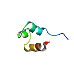
DETERMINATION OF THE STRUCTURE OF THE DNA BINDING DOMAIN OF GAMMA DELTA RESOLVASE IN SOLUTION
Descriptor: GAMMA DELTA-RESOLVASE
Authors:Mullen, G.P.
Deposit date:1994-06-29
Release date:1994-11-30
Last modified:2024-05-22
Method:SOLUTION NMR
Cite:Determination of the structure of the DNA binding domain of gamma delta resolvase in solution.
Protein Sci., 3, 1994
1RET
DownloadVisualize
BU of 1ret by Molmil
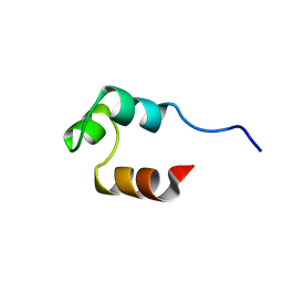
DETERMINATION OF THE STRUCTURE OF THE DNA BINDING DOMAIN OF GAMMA DELTA RESOLVASE IN SOLUTION
Descriptor: GAMMA DELTA-RESOLVASE
Authors:Mullen, G.P.
Deposit date:1994-06-29
Release date:1994-11-30
Last modified:2024-05-22
Method:SOLUTION NMR
Cite:Determination of the structure of the DNA binding domain of gamma delta resolvase in solution.
Protein Sci., 3, 1994
5W7Y
DownloadVisualize
BU of 5w7y by Molmil
Crystal Structure of FHA domain of human APLF in complex with XRCC1 monophosphorylated mutated peptide
Descriptor: Aprataxin and PNK-like factor, DNA repair protein XRCC1
Authors:Pedersen, L.C, Kim, K, London, R.E.
Deposit date:2017-06-21
Release date:2018-05-02
Last modified:2024-10-16
Method:X-RAY DIFFRACTION (2.1 Å)
Cite:Characterization of the APLF FHA-XRCC1 phosphopeptide interaction and its structural and functional implications.
Nucleic Acids Res., 45, 2017
5W7W
DownloadVisualize
BU of 5w7w by Molmil
Crystal Structure of FHA domain of human APLF
Descriptor: Aprataxin and PNK-like factor, FORMIC ACID, SODIUM ION
Authors:Pedersen, L.C, Kim, K, London, R.E.
Deposit date:2017-06-21
Release date:2018-05-02
Last modified:2023-10-04
Method:X-RAY DIFFRACTION (1.348 Å)
Cite:Characterization of the APLF FHA-XRCC1 phosphopeptide interaction and its structural and functional implications.
Nucleic Acids Res., 45, 2017
<123


226707

건을2024-10-30부터공개중

PDB statisticsPDBj update infoContact PDBjnumon