Loading
PDBj
メニューPDBj@FacebookPDBj@X(formerly Twitter)PDBj@BlueSkyPDBj@YouTubewwPDB FoundationwwPDB
RCSB PDBPDBeBMRBAdv. SearchSearch help
Search by PDB author
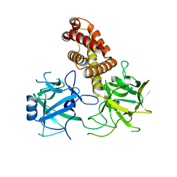
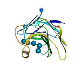
5DZE
DownloadVisualize
BU of 5dze by Molmil
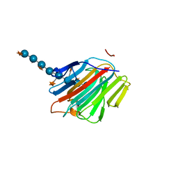
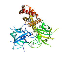
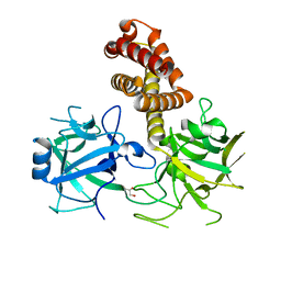
Crystal Structure of the catalytic nucleophile mutant of VvEG16 in complex with cellotetraose
分子名称: beta-D-glucopyranose, beta-D-glucopyranose-(1-4)-beta-D-glucopyranose-(1-4)-beta-D-glucopyranose-(1-4)-alpha-D-glucopyranose, endo-glucanase
著者McGregor, N.G.S, Tung, C.C, Van Petegem, F, Brumer, H.
登録日2015-09-25
公開日2016-09-21
最終更新日2024-11-13
実験手法X-RAY DIFFRACTION (0.97 Å)
主引用文献Crystallographic insight into the evolutionary origins of xyloglucan endotransglycosylases and endohydrolases.
Plant J., 89, 2017
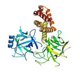
5DZG
DownloadVisualize
BU of 5dzg by Molmil
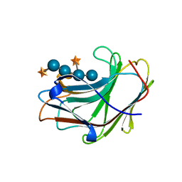
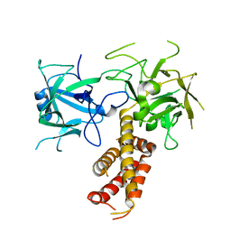
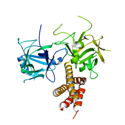
Crystal Structure of the catalytic nucleophile mutant of VvEG16 in complex with a xyloglucan tetradecasaccharide
分子名称: VvEG16, endo-glucanase, alpha-D-xylopyranose-(1-6)-beta-D-glucopyranose-(1-4)-[alpha-D-xylopyranose-(1-6)]beta-D-glucopyranose-(1-4)-[alpha-D-xylopyranose-(1-6)]beta-D-glucopyranose-(1-4)-alpha-D-glucopyranose, ...
著者McGregor, N.G.S, Tung, C.C, Van Petegem, F, Brumer, H.
登録日2015-09-25
公開日2016-09-21
最終更新日2023-09-27
実験手法X-RAY DIFFRACTION (1.79 Å)
主引用文献Crystallographic insight into the evolutionary origins of xyloglucan endotransglycosylases and endohydrolases.
Plant J., 89, 2017
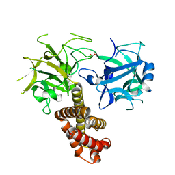
5SV8
DownloadVisualize
BU of 5sv8 by Molmil
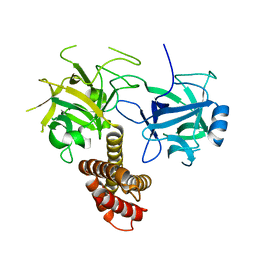
Crystal Structure of the catalytic nucleophile and surface cysteine mutant of VvEG16 in complex with a xyloglucan oligosaccharide
分子名称: alpha-D-xylopyranose-(1-6)-beta-D-glucopyranose-(1-4)-beta-D-glucopyranose-(1-4)-[alpha-D-xylopyranose-(1-6)]beta-D-glucopyranose-(1-4)-beta-D-glucopyranose, probable xyloglucan endotransglucosylase/hydrolase protein 19
著者McGregor, N.G.S, Tung, C.C, Van Petegem, F, Brumer, H.
登録日2016-08-05
公開日2016-09-21
最終更新日2023-10-04
実験手法X-RAY DIFFRACTION (1.588 Å)
主引用文献Crystallographic insight into the evolutionary origins of xyloglucan endotransglycosylases and endohydrolases.
Plant J., 89, 2017
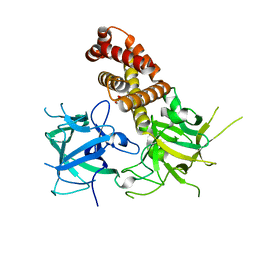
5DZF
DownloadVisualize
BU of 5dzf by Molmil
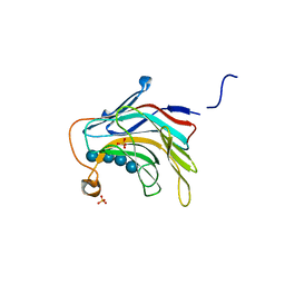
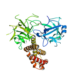
Crystal Structure of the catalytic nucleophile mutant of VvEG16 in complex with a mixed-linkage glucan octasaccharide
分子名称: SULFATE ION, beta-D-glucopyranose, beta-D-glucopyranose-(1-3)-beta-D-glucopyranose-(1-4)-beta-D-glucopyranose-(1-4)-alpha-D-glucopyranose, ...
著者McGregor, N.G.S, Tung, C.C, Van Petegem, F, Brumer, H.
登録日2015-09-25
公開日2016-09-21
最終更新日2023-09-27
実験手法X-RAY DIFFRACTION (1.65 Å)
主引用文献Crystallographic insight into the evolutionary origins of xyloglucan endotransglycosylases and endohydrolases.
Plant J., 89, 2017
6MUD
DownloadVisualize
BU of 6mud by Molmil
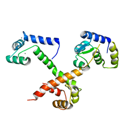
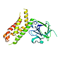
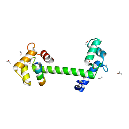
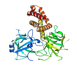
Voltage-gated sodium channel NaV1.5 C-terminal domain in complex with Ca2+/Calmodulin
分子名称: CALCIUM ION, Calmodulin-1, Sodium channel protein type 5 subunit alpha
著者Gardill, B.R, Tung, C.C, Van Petegem, F.
登録日2018-10-23
公開日2019-05-08
最終更新日2024-03-13
実験手法X-RAY DIFFRACTION (2.69 Å)
主引用文献Crystal structures of Ca2+-calmodulin bound to NaVC-terminal regions suggest role for EF-hand domain in binding and inactivation.
Proc.Natl.Acad.Sci.USA, 116, 2019
6B28
DownloadVisualize
BU of 6b28 by Molmil
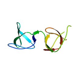
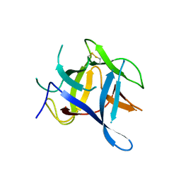
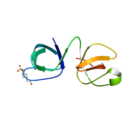
Crystal structure of human STAC2 Tandem SH3 Domains Q347I (296-411)
分子名称: SH3 and cysteine-rich domain-containing protein 2
著者Wong King Yuen, S.M, Tung, C.C, Van Petegem, F.
登録日2017-09-19
公開日2017-10-25
最終更新日2024-03-13
実験手法X-RAY DIFFRACTION (2.55 Å)
主引用文献Structural insights into binding of STAC proteins to voltage-gated calcium channels.
Proc. Natl. Acad. Sci. U.S.A., 114, 2017
4I96
DownloadVisualize
BU of 4i96 by Molmil
Crystal structure of the N-terminal two domains of the skeletal muscle ryanodine receptor (rabbit RyR1) residues 217-536
分子名称: Ryanodine receptor 1
著者Kimlicka, L, Tung, C.C, Van Petegem, F.
登録日2012-12-04
公開日2013-02-20
最終更新日2023-09-20
実験手法X-RAY DIFFRACTION (2.73 Å)
主引用文献Disease mutations in the ryanodine receptor N-terminal region couple to a mobile intersubunit interface.
Nat Commun, 4, 2013
4L4H
DownloadVisualize
BU of 4l4h by Molmil
Crystal structure of mouse Ryanodine Receptor isoform 2 (RyR2) 1-547
分子名称: CHLORIDE ION, DI(HYDROXYETHYL)ETHER, Ryanodine receptor 2, ...
著者Kimlicka, L, Tung, C.C, Van Petegem, F.
登録日2013-06-07
公開日2013-07-24
最終更新日2023-09-20
実験手法X-RAY DIFFRACTION (2 Å)
主引用文献Crystal structure of a ryanodine receptor domain
To be Published
2XOA
DownloadVisualize
BU of 2xoa by Molmil
Crystal Structure of the N-terminal three domains of the skeletal muscle Ryanodine Receptor (RyR1)
分子名称: RYANODINE RECEPTOR 1
著者Tung, C, Lobo, P.A, Kimlicka, L, Van Petegem, F.
登録日2010-08-10
公開日2010-11-10
最終更新日2024-05-08
実験手法X-RAY DIFFRACTION (2.5 Å)
主引用文献The Amino-Terminal Disease Hotspot of Ryanodine Receptors Forms a Cytoplasmic Vestibule.
Nature, 468, 2010
4DJC
DownloadVisualize
BU of 4djc by Molmil
1.35 A crystal structure of the NaV1.5 DIII-IV-Ca/CaM complex
分子名称: CALCIUM ION, Calmodulin, ISOPROPYL ALCOHOL, ...
著者Sarhan, M.F, Tung, C.-C, Van Petegem, F, Ahern, C.A.
登録日2012-02-01
公開日2012-02-22
最終更新日2024-02-28
実験手法X-RAY DIFFRACTION (1.35 Å)
主引用文献Crystallographic basis for calcium regulation of sodium channels.
Proc.Natl.Acad.Sci.USA, 109, 2012
3QR5
DownloadVisualize
BU of 3qr5 by Molmil
Structure of the first domain of a cardiac Ryanodine Receptor mutant with exon 3 deleted
分子名称: Cardiac Ca2+ release channel
著者Lobo, P.A, Van Petegem, F.
登録日2011-02-17
公開日2011-06-08
最終更新日2023-07-26
実験手法X-RAY DIFFRACTION (2.3 Å)
主引用文献The deletion of exon 3 in the cardiac ryanodine receptor is rescued by beta strand switching.
Structure, 19, 2011
6MUE
DownloadVisualize
BU of 6mue by Molmil
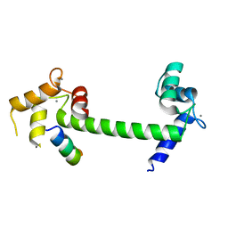
Voltage-gated sodium channel NaV1.4 IQ domain in complex with Ca2+/Calmodulin
分子名称: CALCIUM ION, Calmodulin-1, Sodium channel protein type 4 subunit alpha
著者Gardill, B.R, Van Petegem, F.
登録日2018-10-23
公開日2019-05-08
最終更新日2023-10-11
実験手法X-RAY DIFFRACTION (1.9 Å)
主引用文献Crystal structures of Ca2+-calmodulin bound to NaVC-terminal regions suggest role for EF-hand domain in binding and inactivation.
Proc.Natl.Acad.Sci.USA, 116, 2019
6B26
DownloadVisualize
BU of 6b26 by Molmil
Crystal structure of human STAC2 Tandem SH3 Domains (296-411)
分子名称: SH3 and cysteine-rich domain-containing protein 2
著者Wong King Yuen, S.M, Van Petegem, F.
登録日2017-09-19
公開日2017-10-25
最終更新日2023-10-04
実験手法X-RAY DIFFRACTION (1.2 Å)
主引用文献Structural insights into binding of STAC proteins to voltage-gated calcium channels.
Proc. Natl. Acad. Sci. U.S.A., 114, 2017
6B25
DownloadVisualize
BU of 6b25 by Molmil
Crystal structure of human STAC1 Tandem SH3 Domains (288-402)
分子名称: 4-(2-HYDROXYETHYL)-1-PIPERAZINE ETHANESULFONIC ACID, SH3 and cysteine-rich domain-containing protein
著者Wong King Yuen, S.M, Van Petegem, F.
登録日2017-09-19
公開日2017-10-25
最終更新日2024-03-13
実験手法X-RAY DIFFRACTION (2.39 Å)
主引用文献Structural insights into binding of STAC proteins to voltage-gated calcium channels.
Proc. Natl. Acad. Sci. U.S.A., 114, 2017
6B29
DownloadVisualize
BU of 6b29 by Molmil
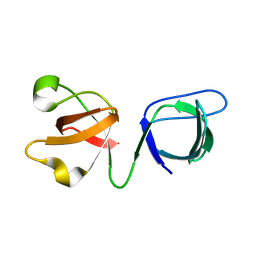
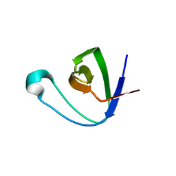
Crystal structure of the second SH3 domain of STAC3 (309-364)
分子名称: SH3 and cysteine-rich domain-containing protein 3
著者Wong King Yuen, S.M, Van Petegem, F.
登録日2017-09-19
公開日2017-10-25
最終更新日2024-03-13
実験手法X-RAY DIFFRACTION (1.3 Å)
主引用文献Structural insights into binding of STAC proteins to voltage-gated calcium channels.
Proc. Natl. Acad. Sci. U.S.A., 114, 2017
6B27
DownloadVisualize
BU of 6b27 by Molmil
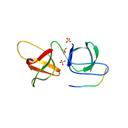
Crystal structure of human STAC2 Tandem SH3 Domains (296-411) in complex with a CaV1.1 II-III loop peptide
分子名称: CHLORIDE ION, SH3 and cysteine-rich domain-containing protein 2, SULFATE ION, ...
著者Wong King Yuen, S.M, Van Petegem, F.
登録日2017-09-19
公開日2017-10-25
最終更新日2024-03-13
実験手法X-RAY DIFFRACTION (1.73 Å)
主引用文献Structural insights into binding of STAC proteins to voltage-gated calcium channels.
Proc. Natl. Acad. Sci. U.S.A., 114, 2017
4I2S
DownloadVisualize
BU of 4i2s by Molmil
Crystal structure of rabbit ryanodine receptor 1 (residues 1-536) disease mutant I404M
分子名称: GLYCEROL, Ryanodine receptor 1
著者Kimlicka, L, Van Petegem, F.
登録日2012-11-22
公開日2013-02-20
最終更新日2023-09-20
実験手法X-RAY DIFFRACTION (2.5 Å)
主引用文献Disease mutations in the ryanodine receptor N-terminal region couple to a mobile intersubunit interface.
Nat Commun, 4, 2013
4I8M
DownloadVisualize
BU of 4i8m by Molmil
Crystal structure of rabbit Ryanodine receptor 1 (residues 1-536) disease mutant V219I
分子名称: GLYCEROL, Ryanodine receptor 1
著者Kimlicka, L, Van Petegem, F.
登録日2012-12-03
公開日2013-02-20
最終更新日2023-09-20
実験手法X-RAY DIFFRACTION (2.8 Å)
主引用文献Disease mutations in the ryanodine receptor N-terminal region couple to a mobile intersubunit interface.
Nat Commun, 4, 2013
4I6I
DownloadVisualize
BU of 4i6i by Molmil
Crystal structure of rabbit ryanodine receptor 1 (residues 1-559) disease mutant R45C
分子名称: Ryanodine receptor 1
著者Kimlicka, L, Van Petegem, F.
登録日2012-11-29
公開日2013-02-20
最終更新日2023-09-20
実験手法X-RAY DIFFRACTION (2.5 Å)
主引用文献Disease mutations in the ryanodine receptor N-terminal region couple to a mobile intersubunit interface.
Nat Commun, 4, 2013
4I37
DownloadVisualize
BU of 4i37 by Molmil
Crystal structure of rabbit ryanodine receptor 1 (residues 1-536) disease mutant R402G
分子名称: Ryanodine receptor 1
著者Kimlicka, L, Van Petegem, F.
登録日2012-11-25
公開日2013-02-20
最終更新日2023-09-20
実験手法X-RAY DIFFRACTION (2.95 Å)
主引用文献Disease mutations in the ryanodine receptor N-terminal region couple to a mobile intersubunit interface.
Nat Commun, 4, 2013
4I7I
DownloadVisualize
BU of 4i7i by Molmil
Crystal structure of rabbit ryanodine receptor 1 (residues 1-536) disease mutant L14R
分子名称: GLYCEROL, Ryanodine receptor 1
著者Kimlicka, L, Van Petegem, F.
登録日2012-11-30
公開日2013-02-20
最終更新日2023-09-20
実験手法X-RAY DIFFRACTION (2.9 Å)
主引用文献Disease mutations in the ryanodine receptor N-terminal region couple to a mobile intersubunit interface.
Nat Commun, 4, 2013
4I1E
DownloadVisualize
BU of 4i1e by Molmil
Crystal structure of Rabbit Ryanodine Receptor 1 (residues 1-536) disease mutant G249R
分子名称: Ryanodine receptor 1
著者Kimlicka, L, Van Petegem, F.
登録日2012-11-20
公開日2013-02-20
最終更新日2024-02-28
実験手法X-RAY DIFFRACTION (2.4 Å)
主引用文献Disease mutations in the ryanodine receptor N-terminal region couple to a mobile intersubunit interface.
Nat Commun, 4, 2013
4I0Y
DownloadVisualize
BU of 4i0y by Molmil
CRYSTAL STRUCTURE OF RABBIT RYANODINE RECEPTOR 1 (RESIDUES 1-536) DISEASE MUTANT C36R
分子名称: GLYCEROL, Ryanodine receptor 1
著者Van Petegem, F, Kimlicka, L.
登録日2012-11-19
公開日2013-02-20
最終更新日2024-02-28
実験手法X-RAY DIFFRACTION (2.8 Å)
主引用文献Disease mutations in the ryanodine receptor N-terminal region couple to a mobile intersubunit interface.
Nat Commun, 4, 2013
4I3N
DownloadVisualize
BU of 4i3n by Molmil
Crystal structure of rabbit ryanodine receptor 1 (residues 1-536) disease mutant D61N
分子名称: GLYCEROL, Ryanodine receptor 1
著者Kimlicka, L, Van Petegem, F.
登録日2012-11-26
公開日2013-02-20
最終更新日2023-09-20
実験手法X-RAY DIFFRACTION (2.95 Å)
主引用文献Disease mutations in the ryanodine receptor N-terminal region couple to a mobile intersubunit interface.
Nat Commun, 4, 2013
4L4I
DownloadVisualize
BU of 4l4i by Molmil
Crystal structure of mouse Ryanodine Receptor isoform 2 (RyR2) 1-547 disease mutant R420Q
分子名称: GLYCEROL, Ryanodine receptor 2
著者Kimlicka, L, Van Petegem, F.
登録日2013-06-07
公開日2013-07-24
最終更新日2023-09-20
実験手法X-RAY DIFFRACTION (2.15 Å)
主引用文献Crystal structure of ryanodine receptor disease mutant
To be Published

227344

件を2024-11-13に公開中

PDB statisticsPDBj update infoContact PDBjnumon