3LUC
 
 | |
3LUJ
 | |
6CFN
 | |
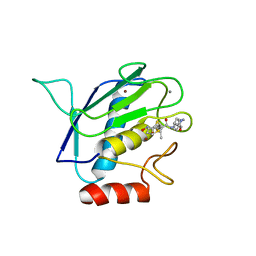
3QX8
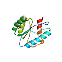
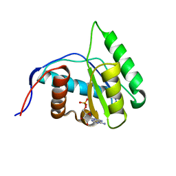
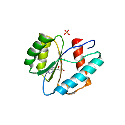
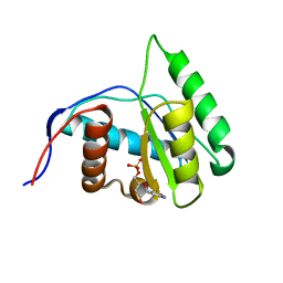
 | | Crystal structure of MID domain from hAGO2 in complex with m7GpppG | | 分子名称: | 7-METHYL-GUANOSINE-5'-TRIPHOSPHATE-5'-GUANOSINE, Protein argonaute-2 | | 著者 | Frank, F, Fabian, M.R, Stepinski, J, Jemielity, J, Darzynkiewicz, E, Sonenberg, N, Nagar, B. | | 登録日 | 2011-03-01 | | 公開日 | 2011-04-20 | | 最終更新日 | 2023-09-13 | | 実験手法 | X-RAY DIFFRACTION (2.3 Å) | | 主引用文献 | Structural analysis of 5'-mRNA-cap interactions with the human AGO2 MID domain.
Embo Rep., 12, 2011
|
|
3QX9
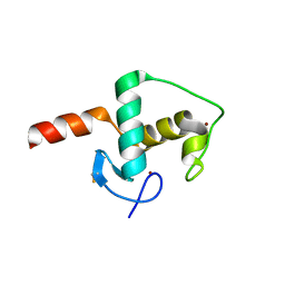
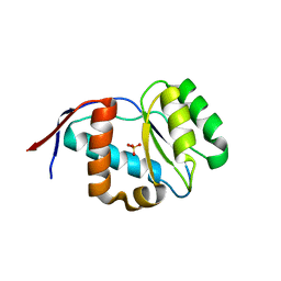
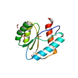
 | | Crystal structure of MID domain from hAGO2 in complex with ATP | | 分子名称: | ADENOSINE-5'-TRIPHOSPHATE, Protein argonaute-2 | | 著者 | Frank, F, Fabian, M.R, Stepinski, J, Jemielity, J, Darzynkiewicz, E, Sonenberg, N, Nagar, B. | | 登録日 | 2011-03-01 | | 公開日 | 2011-04-20 | | 最終更新日 | 2024-02-21 | | 実験手法 | X-RAY DIFFRACTION (2 Å) | | 主引用文献 | Structural analysis of 5'-mRNA-cap interactions with the human AGO2 MID domain.
Embo Rep., 12, 2011
|
|
4G0Y
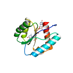
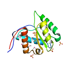
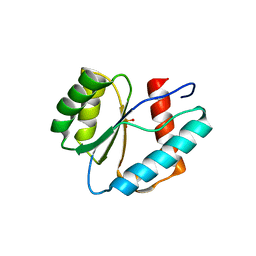
 | | Crystal structure of Arabidopsis AGO1 in complex with AMP | | 分子名称: | ADENOSINE MONOPHOSPHATE, Protein argonaute 1 | | 著者 | Frank, F, Hauver, J, Sonenberg, N, Nagar, B. | | 登録日 | 2012-07-10 | | 公開日 | 2012-07-25 | | 最終更新日 | 2024-02-28 | | 実験手法 | X-RAY DIFFRACTION (1.65 Å) | | 主引用文献 | Arabidopsis Argonaute MID domains use their nucleotide specificity loop to sort small RNAs.
Embo J., 31, 2012
|
|
4G0O
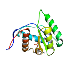
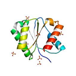
 | | Crystal structure of Arabidopsis thaliana AGO5 MID domain | | 分子名称: | Protein argonaute 5, SULFATE ION | | 著者 | Frank, F, Hauver, J, Sonenberg, N, Nagar, B. | | 登録日 | 2012-07-09 | | 公開日 | 2012-07-25 | | 最終更新日 | 2024-02-28 | | 実験手法 | X-RAY DIFFRACTION (2.186 Å) | | 主引用文献 | Arabidopsis Argonaute MID domains use their nucleotide specificity loop to sort small RNAs.
Embo J., 31, 2012
|
|
4G0X
 | | Crystal Structure of Arabidopsis thaliana AGO1 MID domain | | 分子名称: | Protein argonaute 1, SULFATE ION | | 著者 | Frank, F, Hauver, J, Sonenberg, N, Nagar, B. | | 登録日 | 2012-07-10 | | 公開日 | 2012-07-25 | | 最終更新日 | 2012-09-12 | | 実験手法 | X-RAY DIFFRACTION (1.35 Å) | | 主引用文献 | Arabidopsis Argonaute MID domains use their nucleotide specificity loop to sort small RNAs.
Embo J., 31, 2012
|
|
4G0P
 | | Crystal Structure of Arabidopsis thaliana AGO1 MID domain in complex with UMP | | 分子名称: | Protein argonaute 1, URIDINE-5'-MONOPHOSPHATE | | 著者 | Frank, F, Hauver, J, Sonenberg, N, Nagar, B. | | 登録日 | 2012-07-09 | | 公開日 | 2012-07-25 | | 最終更新日 | 2024-02-28 | | 実験手法 | X-RAY DIFFRACTION (1.8 Å) | | 主引用文献 | Arabidopsis Argonaute MID domains use their nucleotide specificity loop to sort small RNAs.
Embo J., 31, 2012
|
|
3LUD
 | |
3LUK
 | |
3LUH
 | |
3LUG
 | |
4G0Z
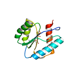
 | | Crystal structure of Arabidopsis thaliana AGO1 MID domain in complex with GMP | | 分子名称: | GUANOSINE-5'-MONOPHOSPHATE, Protein argonaute 1 | | 著者 | Frank, F, Hauver, J, Sonenberg, N, Nagar, B. | | 登録日 | 2012-07-10 | | 公開日 | 2012-07-25 | | 最終更新日 | 2024-02-28 | | 実験手法 | X-RAY DIFFRACTION (1.75 Å) | | 主引用文献 | Arabidopsis Argonaute MID domains use their nucleotide specificity loop to sort small RNAs.
Embo J., 31, 2012
|
|
4G0M
 | | Crystal structure of Arabidopsis thaliana AGO2 MID domain | | 分子名称: | Protein argonaute 2, SULFATE ION | | 著者 | Frank, F, Hauver, J, Sonenberg, N, Nagar, B. | | 登録日 | 2012-07-09 | | 公開日 | 2012-07-25 | | 最終更新日 | 2024-02-28 | | 実験手法 | X-RAY DIFFRACTION (2.306 Å) | | 主引用文献 | Arabidopsis Argonaute MID domains use their nucleotide specificity loop to sort small RNAs.
Embo J., 31, 2012
|
|
4G0Q
 | | Crystal structure of Arabidopsis thaliana AGO1 MID domain in complex with CMP | | 分子名称: | CYTIDINE-5'-MONOPHOSPHATE, Protein argonaute 1 | | 著者 | Frank, F, Hauver, J, Sonenberg, N, Nagar, B. | | 登録日 | 2012-07-09 | | 公開日 | 2012-07-25 | | 最終更新日 | 2024-02-28 | | 実験手法 | X-RAY DIFFRACTION (1.8 Å) | | 主引用文献 | Arabidopsis Argonaute MID domains use their nucleotide specificity loop to sort small RNAs.
Embo J., 31, 2012
|
|
4IUL
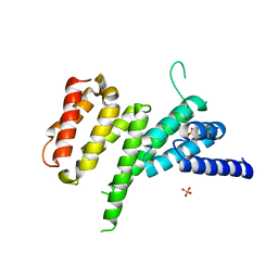 | | MIF4G domain of DAP5 | | 分子名称: | Eukaryotic translation initiation factor 4 gamma 2, SULFATE ION | | 著者 | Frank, F, Virgili, G, Feoktistova, K, Sawicki, M, Sonenberg, N, Fraser, C, Nagar, B. | | 登録日 | 2013-01-21 | | 公開日 | 2013-03-27 | | 最終更新日 | 2023-09-20 | | 実験手法 | X-RAY DIFFRACTION (2.3 Å) | | 主引用文献 | Structural Analysis of the DAP5 MIF4G Domain and Its Interaction with eIF4A.
Structure, 21, 2013
|
|
4J8S
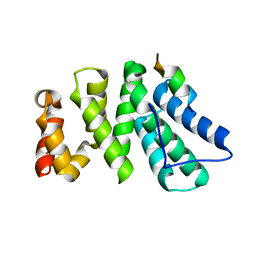 | | Crystal structure of human CNOT1 MIF4G domain in complex with a TTP peptide | | 分子名称: | CCR4-NOT transcription complex subunit 1, Tristetraprolin | | 著者 | Frank, F, Fabian, M.R, Rouya, C, Siddiqui, N, Lai, W.S, Karetnikov, A, Blackshear, P.J, Sonenberg, N, Nagar, B. | | 登録日 | 2013-02-14 | | 公開日 | 2013-05-08 | | 最終更新日 | 2024-02-28 | | 実験手法 | X-RAY DIFFRACTION (1.55 Å) | | 主引用文献 | Structural basis for the recruitment of the human CCR4-NOT deadenylase complex by tristetraprolin.
Nat.Struct.Mol.Biol., 20, 2013
|
|
2OIQ
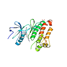 | | Crystal Structure of chicken c-Src kinase domain in complex with the cancer drug imatinib. | | 分子名称: | 4-(4-METHYL-PIPERAZIN-1-YLMETHYL)-N-[4-METHYL-3-(4-PYRIDIN-3-YL-PYRIMIDIN-2-YLAMINO)-PHENYL]-BENZAMIDE, GLYCEROL, Proto-oncogene tyrosine-protein kinase Src | | 著者 | Seeliger, M.A, Nagar, B, Frank, F, Cao, X, Henderson, M.N, Kuriyan, J. | | 登録日 | 2007-01-11 | | 公開日 | 2007-03-20 | | 最終更新日 | 2023-08-30 | | 実験手法 | X-RAY DIFFRACTION (2.07 Å) | | 主引用文献 | c-Src Binds to the Cancer Drug Imatinib with an Inactive Abl/c-Kit Conformation and a Distributed Thermodynamic Penalty.
Structure, 15, 2007
|
|
1RM8
 | | Crystal structure of the catalytic domain of MMP-16/MT3-MMP: Characterization of MT-MMP specific features | | 分子名称: | 4-(N-HYDROXYAMINO)-2R-ISOBUTYL-2S-(2-THIENYLTHIOMETHYL)SUCCINYL-L-PHENYLALANINE-N-METHYLAMIDE, CALCIUM ION, Matrix metalloproteinase-16, ... | | 著者 | Lang, R, Braun, M, Sounni, N.E, Noel, A, Frankenne, F, Foidart, J.-M, Bode, W, Maskos, K. | | 登録日 | 2003-11-27 | | 公開日 | 2004-03-09 | | 最終更新日 | 2023-08-23 | | 実験手法 | X-RAY DIFFRACTION (1.8 Å) | | 主引用文献 | Crystal structure of the catalytic domain of MMP-16/MT3-MMP: characterization of MT-MMP specific features.
J.Mol.Biol., 336, 2004
|
|
7A0X
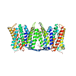 | | Structure of dimeric sodium proton antiporter NhaA, at pH 6.0, crystallized with chimeric Fab antibodies | | 分子名称: | 1,2-DIACYL-GLYCEROL-3-SN-PHOSPHATE, 1,2-Distearoyl-sn-glycerophosphoethanolamine, CALCIUM ION, ... | | 著者 | Fippel, A, Lentes, C.J, Mir, S.H, Wirth, C, Hunte, C. | | 登録日 | 2020-08-11 | | 公開日 | 2022-02-23 | | 最終更新日 | 2024-01-31 | | 実験手法 | X-RAY DIFFRACTION (2.37 Å) | | 主引用文献 | Molecular determinants for substrate uptake in electrogenic sodium/proton antiporters
To Be Published
|
|
7A0W
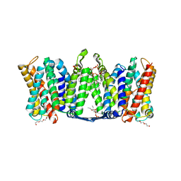 | | Structure of dimeric sodium proton antiporter NhaA, at pH 8.5, crystallized with chimeric Fab antibodies | | 分子名称: | 1,2-DIACYL-GLYCEROL-3-SN-PHOSPHATE, CARDIOLIPIN, CHLORIDE ION, ... | | 著者 | Fippel, A, Gross, N.M, Mir, S.H, Wirth, C, Hunte, C. | | 登録日 | 2020-08-11 | | 公開日 | 2022-02-23 | | 最終更新日 | 2024-10-16 | | 実験手法 | X-RAY DIFFRACTION (2.04 Å) | | 主引用文献 | Molecular determinants for substrate uptake in electrogenic sodium/proton antiporters
To Be Published
|
|
7A0Y
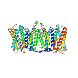 | | Structure of dimeric sodium proton antiporter NhaA K300R variant, at pH 8.2, crystallized with chimeric Fab antibodies | | 分子名称: | 1,2-DIACYL-GLYCEROL-3-SN-PHOSPHATE, 1,2-Distearoyl-sn-glycerophosphoethanolamine, CARDIOLIPIN, ... | | 著者 | Fippel, A, Mir, S.H, Lentes, C.J, Wirth, C, Hunte, C. | | 登録日 | 2020-08-11 | | 公開日 | 2022-02-23 | | 最終更新日 | 2024-01-31 | | 実験手法 | X-RAY DIFFRACTION (2.45 Å) | | 主引用文献 | Molecular determinants for substrate uptake in electrogenic sodium/proton antiporters
To Be Published
|
|
6EU6
 | | Sensor Amt Protein | | 分子名称: | 2-(N-MORPHOLINO)-ETHANESULFONIC ACID, AMMONIUM ION, Ammonium transporter, ... | | 著者 | Pflueger, T, Hernandez, C, Andrade, S.L.A. | | 登録日 | 2017-10-28 | | 公開日 | 2018-01-24 | | 最終更新日 | 2024-01-17 | | 実験手法 | X-RAY DIFFRACTION (1.98 Å) | | 主引用文献 | Signaling ammonium across membranes through an ammonium sensor histidine kinase.
Nat Commun, 9, 2018
|
|
8GDR
 | |


