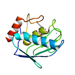
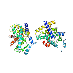
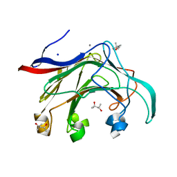
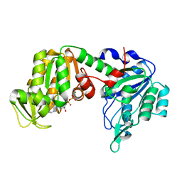
5HFQ
 
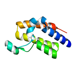 | | Crystal structure of the second bromodomain Q443H mutant of human BRD2 | | 分子名称: | Bromodomain-containing protein 2 | | 著者 | Tallant, C, Lori, C, Pasquo, A, Chiaraluce, R, Consalvi, V, Fonseca, M, von Delft, F, Arrowsmith, C.H, Edwards, A.M, Bountra, C, Knapp, S. | | 登録日 | 2016-01-07 | | 公開日 | 2016-01-20 | | 最終更新日 | 2024-01-10 | | 実験手法 | X-RAY DIFFRACTION (1.4 Å) | | 主引用文献 | Crystal structure of the second bromodomain Q443H mutant of human BRD2
To Be Published
|
|
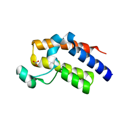
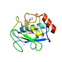
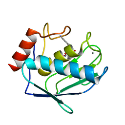
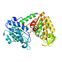
5HEM
 | | Crystal structure of the N-terminus D161Y bromodomain mutant of human BRD2 | | 分子名称: | 1,2-ETHANEDIOL, ACETATE ION, Bromodomain-containing protein 2, ... | | 著者 | Tallant, C, Lori, C, Pasquo, A, Chiaraluce, R, Consalvi, V, Newman, J.A, von Delft, F, Arrowsmith, C.H, Edwards, A.M, Bountra, C, Knapp, S. | | 登録日 | 2016-01-06 | | 公開日 | 2016-01-20 | | 最終更新日 | 2024-01-10 | | 実験手法 | X-RAY DIFFRACTION (1.65 Å) | | 主引用文献 | Crystal structure of the N-terminus D161Y bromodomain mutant of human BRD2
To Be Published
|
|
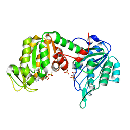
5HFR
 | | Crystal structure of the second bromodomain H395R mutant of human BRD3 | | 分子名称: | 1,2-ETHANEDIOL, Bromodomain-containing protein 3, NITRATE ION | | 著者 | Tallant, C, Lori, C, Pasquo, A, Chiaraluce, R, Consalvi, V, Fonseca, M, von Delft, F, Arrowsmith, C.H, Edwards, A.M, Bountra, C, Knapp, S. | | 登録日 | 2016-01-07 | | 公開日 | 2016-01-20 | | 最終更新日 | 2024-01-10 | | 実験手法 | X-RAY DIFFRACTION (1.7 Å) | | 主引用文献 | Crystal structure of the second bromodomain H395R mutant of human BRD3
To Be Published
|
|
5HEL
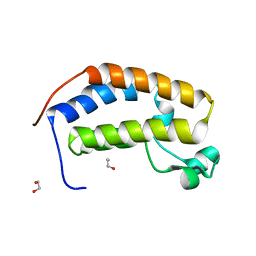 | | Crystal structure of the N-terminus Y153H bromodomain mutant of human BRD2 | | 分子名称: | 1,2-ETHANEDIOL, Bromodomain-containing protein 2 | | 著者 | Tallant, C, Lori, C, Pasquo, A, Chiaraluce, R, Consalvi, V, Newman, J.A, von Delft, F, Arrowsmith, C.H, Edwards, A.M, Bountra, C, Knapp, S. | | 登録日 | 2016-01-06 | | 公開日 | 2016-01-20 | | 最終更新日 | 2024-01-10 | | 実験手法 | X-RAY DIFFRACTION (1.45 Å) | | 主引用文献 | Crystal structure of the N-terminus Y153H bromodomain mutant of human BRD2
To Be Published
|
|
5HEN
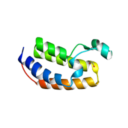 | | Crystal structure of the N-terminus R100L bromodomain mutant of human BRD2 | | 分子名称: | 1,2-ETHANEDIOL, Bromodomain-containing protein 2 | | 著者 | Tallant, C, Lori, C, Pasquo, A, Chiaraluce, R, Consalvi, V, Newman, J.A, von Delft, F, Arrowsmith, C.H, Edwards, A.M, Bountra, C, Knapp, S. | | 登録日 | 2016-01-06 | | 公開日 | 2016-01-20 | | 最終更新日 | 2024-01-10 | | 実験手法 | X-RAY DIFFRACTION (1.79 Å) | | 主引用文献 | Crystal structure of the N-terminus R100L bromodomain mutant of human BRD2
To Be Published
|
|
1ZVX
 | | Crystal structure of the complex between MMP-8 and a phosphonate inhibitor (R-enantiomer) | | 分子名称: | (1R)-1-{[(4'-METHOXY-1,1'-BIPHENYL-4-YL)SULFONYL]AMINO}-2-METHYLPROPYLPHOSPHONIC ACID, CALCIUM ION, Neutrophil collagenase, ... | | 著者 | Pochetti, G, Gavuzzo, E, Campestre, C, Agamennone, M, Tortorella, P, Consalvi, V, Gallina, C, Hiller, O, Tschesche, H, Tucker, P.A, Mazza, F. | | 登録日 | 2005-06-03 | | 公開日 | 2006-05-16 | | 最終更新日 | 2023-08-23 | | 実験手法 | X-RAY DIFFRACTION (1.87 Å) | | 主引用文献 | Structural insight into the stereoselective inhibition of MMP-8 by enantiomeric sulfonamide phosphonates.
J.Med.Chem., 49, 2006
|
|
1ZS0
 | | Crystal structure of the complex between MMP-8 and a phosphonate inhibitor (S-enantiomer) | | 分子名称: | (1S)-1-{[(4'-METHOXY-1,1'-BIPHENYL-4-YL)SULFONYL]AMINO}-2-METHYLPROPYLPHOSPHONIC ACID, 2-(N-MORPHOLINO)-ETHANESULFONIC ACID, CALCIUM ION, ... | | 著者 | Pochetti, G, Gavuzzo, E, Campestre, C, Agamennone, M, Tortorella, P, Consalvi, V, Gallina, C, Hiller, O, Tschesche, H, Tucker, P.A, Mazza, F. | | 登録日 | 2005-05-23 | | 公開日 | 2006-05-02 | | 最終更新日 | 2023-08-23 | | 実験手法 | X-RAY DIFFRACTION (1.56 Å) | | 主引用文献 | Structural insight into the stereoselective inhibition of MMP-8 by enantiomeric sulfonamide phosphonates.
J.Med.Chem., 49, 2006
|
|
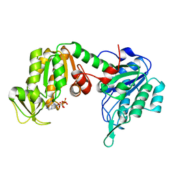
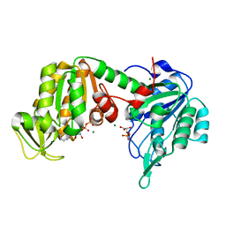
4L98
 | | Crystal structure of the complex of F360L PPARgamma mutant with the ligand LT175 | | 分子名称: | (2S)-2-(biphenyl-4-yloxy)-3-phenylpropanoic acid, Peroxisome proliferator-activated receptor gamma | | 著者 | Pochetti, G, Montanari, R, Consalvi, V, Chiaraluce, R, Pasquo, A, Capelli, D, Loiodice, F, Laghezza, A, Lori, C. | | 登録日 | 2013-06-18 | | 公開日 | 2014-06-25 | | 最終更新日 | 2023-09-20 | | 実験手法 | X-RAY DIFFRACTION (2.28 Å) | | 主引用文献 | Structural basis of the transactivation deficiency of the human PPAR gamma F360L mutant associated with familial partial lipodystrophy.
Acta Crystallogr.,Sect.D, 70, 2014
|
|
4L96
 | | Structure of the complex between the F360L PPARgamma mutant and the ligand LT175 (space group I222) | | 分子名称: | (2S)-2-(biphenyl-4-yloxy)-3-phenylpropanoic acid, Peroxisome proliferator-activated receptor gamma | | 著者 | Pochetti, G, Montanari, R, Capelli, D, Chiaraluce, R, Consalvi, V, Pasquo, A, Lori, C, Laghezza, A, Loiodice, F. | | 登録日 | 2013-06-18 | | 公開日 | 2014-06-25 | | 最終更新日 | 2023-09-20 | | 実験手法 | X-RAY DIFFRACTION (2.38 Å) | | 主引用文献 | Structural basis of the transactivation deficiency of the human PPAR gamma F360L mutant associated with familial partial lipodystrophy.
Acta Crystallogr.,Sect.D, 70, 2014
|
|
4O8F
 | | Crystal Structure of the complex between PPARgamma mutant R357A and rosiglitazone | | 分子名称: | 2,4-THIAZOLIDIINEDIONE, 5-[[4-[2-(METHYL-2-PYRIDINYLAMINO)ETHOXY]PHENYL]METHYL]-(9CL), Peroxisome proliferator-activated receptor gamma | | 著者 | Pochetti, G, Montanari, R, Capelli, D, Chiaraluce, R, Consalvi, V, Lori, C, Loiodice, F, Laghezza, A, Pasquo, A, Cervoni, L, Aschi, M. | | 登録日 | 2013-12-27 | | 公開日 | 2014-07-23 | | 最終更新日 | 2023-09-20 | | 実験手法 | X-RAY DIFFRACTION (2.6 Å) | | 主引用文献 | Structural basis of the transactivation deficiency of the human PPAR gamma F360L mutant associated with familial partial lipodystrophy.
Acta Crystallogr.,Sect.D, 70, 2014
|
|
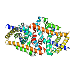
1GTM
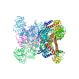 | | STRUCTURE OF GLUTAMATE DEHYDROGENASE | | 分子名称: | GLUTAMATE DEHYDROGENASE, SULFATE ION | | 著者 | Yip, K.S.P, Stillman, T.J, Britton, K.L, Pasquo, A, Rice, D.W. | | 登録日 | 1996-08-22 | | 公開日 | 1997-01-11 | | 最終更新日 | 2024-02-07 | | 実験手法 | X-RAY DIFFRACTION (2.2 Å) | | 主引用文献 | The structure of Pyrococcus furiosus glutamate dehydrogenase reveals a key role for ion-pair networks in maintaining enzyme stability at extreme temperatures.
Structure, 3, 1995
|
|
5O7D
 | | The X-ray structure of human R38M phosphoglycerate kinase 1 mutant | | 分子名称: | ADENOSINE-5'-DIPHOSPHATE, Phosphoglycerate kinase 1 | | 著者 | Ilari, A, Fiorillo, A, Cipollone, A, Petrosino, M. | | 登録日 | 2017-06-08 | | 公開日 | 2018-06-20 | | 最終更新日 | 2024-01-17 | | 実験手法 | X-RAY DIFFRACTION (1.84 Å) | | 主引用文献 | The phosphoglycerate kinase 1 variants found in carcinoma cells display different catalytic activity and conformational stability compared to the native enzyme.
PLoS ONE, 13, 2018
|
|
2IY4
 | | X-ray structure of Dps from Listeria monocytogenes | | 分子名称: | FE (III) ION, NON-HEME IRON-CONTAINING FERRITIN | | 著者 | Ilari, A, Bellapadrona, G, Stefanini, S, Chiancone, E. | | 登録日 | 2006-07-12 | | 公開日 | 2007-01-02 | | 最終更新日 | 2023-12-13 | | 実験手法 | X-RAY DIFFRACTION (2.31 Å) | | 主引用文献 | The Mutations Lys 114 --> Gln and Asp 126 --> Asn Disrupt an Intersubunit Salt Bridge and Convert Listeria Innocua Dps Into its Natural Mutant Listeria Monocytogenes Dps. Effects on Protein Stability at Low Ph.
Proteins, 66, 2007
|
|
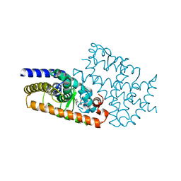
2VY0
 | | The X-ray structure of endo-beta-1,3-glucanase from Pyrococcus furiosus | | 分子名称: | (4R)-2-METHYLPENTANE-2,4-DIOL, (4S)-2-METHYL-2,4-PENTANEDIOL, CALCIUM ION, ... | | 著者 | Ilari, A, Fiorillo, A. | | 登録日 | 2008-07-15 | | 公開日 | 2009-03-24 | | 最終更新日 | 2023-12-13 | | 実験手法 | X-RAY DIFFRACTION (2.16 Å) | | 主引用文献 | Crystal Structure of a Family 16 Endoglucanase from the Hyperthermophile Pyrococcus Furiosus-Structural Basis of Substrate Recognition.
FEBS J., 276, 2009
|
|
1HRD
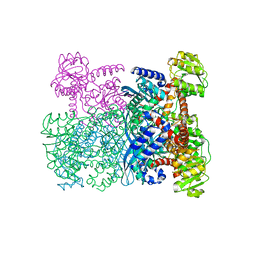 | | GLUTAMATE DEHYDROGENASE | | 分子名称: | GLUTAMATE DEHYDROGENASE | | 著者 | Britton, K.L, Baker, P.J, Stillman, T.J, Rice, D.W. | | 登録日 | 1996-04-03 | | 公開日 | 1997-03-12 | | 最終更新日 | 2024-02-07 | | 実験手法 | X-RAY DIFFRACTION (1.96 Å) | | 主引用文献 | The structure of Pyrococcus furiosus glutamate dehydrogenase reveals a key role for ion-pair networks in maintaining enzyme stability at extreme temperatures.
Structure, 3, 1995
|
|
1ZP5
 | | Crystal structure of the complex between MMP-8 and a N-hydroxyurea inhibitor | | 分子名称: | CALCIUM ION, N-{2-[(4'-CYANO-1,1'-BIPHENYL-4-YL)OXY]ETHYL}-N'-HYDROXY-N-METHYLUREA, Neutrophil collagenase, ... | | 著者 | Campestre, C, Agamennone, M, Tortorella, P, Preziuso, S, Biasone, A, Gavuzzo, E, Pochetti, G, Mazza, F, Tschesche, H, Gallina, C. | | 登録日 | 2005-05-16 | | 公開日 | 2005-12-06 | | 最終更新日 | 2023-08-23 | | 実験手法 | X-RAY DIFFRACTION (1.8 Å) | | 主引用文献 | N-Hydroxyurea as zinc binding group in matrix metalloproteinase inhibition: Mode of binding in a complex with MMP-8.
Bioorg.Med.Chem.Lett., 16, 2006
|
|
5M3U
 | | The X-ray structure of human V216F phosphoglycerate kinase 1 mutant | | 分子名称: | 3-PHOSPHOGLYCERIC ACID, ADENOSINE-5'-DIPHOSPHATE, MAGNESIUM ION, ... | | 著者 | Ilari, A, Fiorillo, A, Cipollone, A, Petrosino, M. | | 登録日 | 2016-10-17 | | 公開日 | 2017-12-20 | | 最終更新日 | 2024-01-17 | | 実験手法 | X-RAY DIFFRACTION (1.81 Å) | | 主引用文献 | The phosphoglycerate kinase 1 variants found in carcinoma cells display different catalytic activity and conformational stability compared to the native enzyme.
PLoS ONE, 13, 2018
|
|
5M1R
 | | X-ray structure of human G166D PGK-1 mutant | | 分子名称: | ADENOSINE-5'-DIPHOSPHATE, MAGNESIUM ION, Phosphoglycerate kinase 1 | | 著者 | Ilari, A, Cipollone, A, Fiorillo, A, Petrosino, M. | | 登録日 | 2016-10-10 | | 公開日 | 2017-12-20 | | 最終更新日 | 2024-01-17 | | 実験手法 | X-RAY DIFFRACTION (1.64 Å) | | 主引用文献 | The phosphoglycerate kinase 1 variants found in carcinoma cells display different catalytic activity and conformational stability compared to the native enzyme.
PLoS ONE, 13, 2018
|
|
5MXM
 | | The X-ray structure of human M190I phosphoglycerate kinase 1 mutant | | 分子名称: | 2-[BIS-(2-HYDROXY-ETHYL)-AMINO]-2-HYDROXYMETHYL-PROPANE-1,3-DIOL, 3-PHOSPHOGLYCERIC ACID, ADENOSINE-5'-DIPHOSPHATE, ... | | 著者 | Ilari, A, Fiorillo, A, Cipollone, A, Petrosino, M. | | 登録日 | 2017-01-23 | | 公開日 | 2018-02-14 | | 最終更新日 | 2024-05-08 | | 実験手法 | X-RAY DIFFRACTION (2.05 Å) | | 主引用文献 | The phosphoglycerate kinase 1 variants found in carcinoma cells display different catalytic activity and conformational stability compared to the native enzyme.
PLoS ONE, 13, 2018
|
|
5M6Z
 | | The X-ray structure of human M189I PGK-1 mutant in partially closed conformation | | 分子名称: | 3-PHOSPHOGLYCERIC ACID, ADENOSINE-5'-DIPHOSPHATE, MAGNESIUM ION, ... | | 著者 | Ilari, A, Fiorillo, A, Petrosino, M, Cipollone, A. | | 登録日 | 2016-10-26 | | 公開日 | 2017-11-29 | | 最終更新日 | 2024-01-17 | | 実験手法 | X-RAY DIFFRACTION (1.67 Å) | | 主引用文献 | The phosphoglycerate kinase 1 variants found in carcinoma cells display different catalytic activity and conformational stability compared to the native enzyme.
PLoS ONE, 13, 2018
|
|
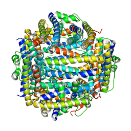
1K89
 | | K89L MUTANT OF GLUTAMATE DEHYDROGENASE | | 分子名称: | GLUTAMATE DEHYDROGENASE | | 著者 | Stillman, T.J, Migueis, A.M.B, Wang, X.G, Baker, P.J, Britton, K.L, Engel, P.C, Rice, D.W. | | 登録日 | 1998-06-05 | | 公開日 | 1999-01-27 | | 最終更新日 | 2024-04-03 | | 実験手法 | X-RAY DIFFRACTION (2.05 Å) | | 主引用文献 | Insights into the mechanism of domain closure and substrate specificity of glutamate dehydrogenase from Clostridium symbiosum.
J.Mol.Biol., 285, 1999
|
|
1AUP
 | | GLUTAMATE DEHYDROGENASE | | 分子名称: | NAD-SPECIFIC GLUTAMATE DEHYDROGENASE | | 著者 | Baker, P.J, Waugh, M.L, Stillman, T.J, Turnbull, A.P, Rice, D.W. | | 登録日 | 1997-09-01 | | 公開日 | 1998-03-18 | | 最終更新日 | 2024-04-03 | | 実験手法 | X-RAY DIFFRACTION (2.5 Å) | | 主引用文献 | Determinants of substrate specificity in the superfamily of amino acid dehydrogenases.
Biochemistry, 36, 1997
|
|
1BGV
 | | GLUTAMATE DEHYDROGENASE | | 分子名称: | GLUTAMATE DEHYDROGENASE, GLUTAMIC ACID | | 著者 | Stillman, T.J, Baker, P.J, Britton, K.L, Rice, D.W. | | 登録日 | 1998-06-01 | | 公開日 | 1998-10-14 | | 最終更新日 | 2024-02-07 | | 実験手法 | X-RAY DIFFRACTION (1.9 Å) | | 主引用文献 | Conformational flexibility in glutamate dehydrogenase. Role of water in substrate recognition and catalysis.
J.Mol.Biol., 234, 1993
|
|


