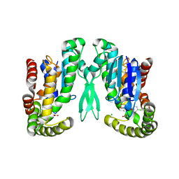
8YTE
 
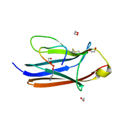 | | Crystal Structure of TrkA D5 domain in complex with macrocyclic peptide | | 分子名称: | 1,2-ETHANEDIOL, AMINOMETHYLAMIDE, High affinity nerve growth factor receptor, ... | | 著者 | Yamada, T, Mihara, K, Ueda, T, Yamauchi, D, Shimizu, M, Ando, A, Mayumi, K, Nakata, Z, Mikamiyama, H. | | 登録日 | 2024-03-25 | | 公開日 | 2024-07-10 | | 最終更新日 | 2024-07-24 | | 実験手法 | X-RAY DIFFRACTION (2.26 Å) | | 主引用文献 | Discovery and Hit to Lead Optimization of Macrocyclic Peptides as Novel Tropomyosin Receptor Kinase A Antagonists.
J.Med.Chem., 67, 2024
|
|
8YTD
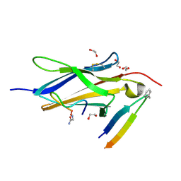 | | Crystal Structure of TrkA D5 domain in complex with two different macrocyclic peptides | | 分子名称: | 1,2-ETHANEDIOL, High affinity nerve growth factor receptor, Macrocyclic Peptide | | 著者 | Yamada, T, Mihara, K, Ueda, T, Yamauchi, D, Shimizu, M, Ando, A, Mayumi, K, Nakata, Z, Mikamiyama, H. | | 登録日 | 2024-03-25 | | 公開日 | 2024-07-10 | | 最終更新日 | 2024-07-24 | | 実験手法 | X-RAY DIFFRACTION (2.34 Å) | | 主引用文献 | Discovery and Hit to Lead Optimization of Macrocyclic Peptides as Novel Tropomyosin Receptor Kinase A Antagonists.
J.Med.Chem., 67, 2024
|
|
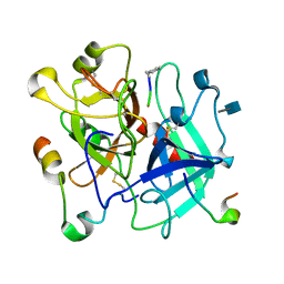
2RKB
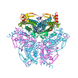
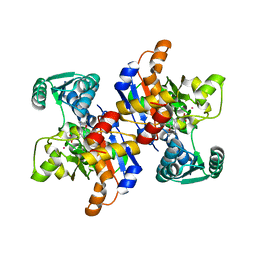 | | Serine dehydratase like-1 from human cancer cells | | 分子名称: | POTASSIUM ION, PYRIDOXAL-5'-PHOSPHATE, Serine dehydratase-like | | 著者 | Yamada, T, Komoto, J, Kasuya, T, Mori, H, Ogawa, H, Takusagawa, F. | | 登録日 | 2007-10-16 | | 公開日 | 2008-04-01 | | 最終更新日 | 2017-10-25 | | 実験手法 | X-RAY DIFFRACTION (2.8 Å) | | 主引用文献 | A catalytic mechanism that explains a low catalytic activity of serine dehydratase like-1 from human cancer cells: Crystal structure and site-directed mutagenesis studies.
Biochim.Biophys.Acta, 1780, 2008
|
|
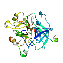
2H5L
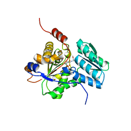
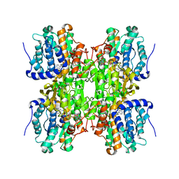 | | S-Adenosylhomocysteine hydrolase containing NAD and 3-deaza-D-eritadenine | | 分子名称: | (2R,3R)-4-(4-AMINO-1H-IMIDAZO[4,5-C]PYRIDIN-1-YL)-2,3-DIHYDROXYBUTANOIC ACID, Adenosylhomocysteinase, NICOTINAMIDE-ADENINE-DINUCLEOTIDE | | 著者 | Yamada, T, Komoto, J, Takusagawa, F. | | 登録日 | 2006-05-26 | | 公開日 | 2007-04-10 | | 最終更新日 | 2024-02-14 | | 実験手法 | X-RAY DIFFRACTION (2.8 Å) | | 主引用文献 | Structure and function of eritadenine and its 3-deaza analogues: Potent inhibitors of S-adenosylhomocysteine hydrolase and hypocholesterolemic agents.
Biochem.Pharm., 73, 2007
|
|
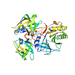
6KK8
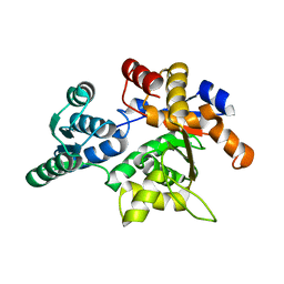
 | | XN joint refinement of manganese catalase from Thermus Thermophilus HB27 | | 分子名称: | 1,2-ETHANEDIOL, MANGANESE (III) ION, OXYGEN ATOM, ... | | 著者 | Yamada, T, Yano, N, Kusaka, K. | | 登録日 | 2019-07-24 | | 公開日 | 2019-09-04 | | 最終更新日 | 2024-04-03 | | 実験手法 | NEUTRON DIFFRACTION (1.37 Å), X-RAY DIFFRACTION | | 主引用文献 | Single-crystal time-of-flight neutron Laue methods: application to manganese catalase from Thermus thermophilus HB27
J.Appl.Crystallogr., 2019
|
|
1PWH
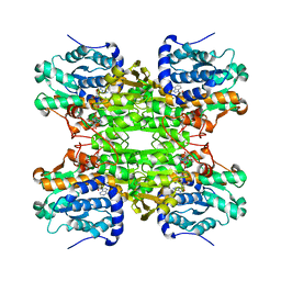
 | | Rat Liver L-Serine Dehydratase- Complex with PYRIDOXYL-(O-METHYL-SERINE)-5-MONOPHOSPHATE | | 分子名称: | L-serine dehydratase, N-({3-HYDROXY-2-METHYL-5-[(PHOSPHONOOXY)METHYL]PYRIDIN-4-YL}METHYL)-O-METHYL-L-SERINE, POTASSIUM ION | | 著者 | Yamada, T, Komoto, J, Takata, Y, Ogawa, H, Takusagawa, F. | | 登録日 | 2003-07-01 | | 公開日 | 2003-12-02 | | 最終更新日 | 2023-08-16 | | 実験手法 | X-RAY DIFFRACTION (2.6 Å) | | 主引用文献 | Crystal structure of serine dehydratase from rat liver.
Biochemistry, 42, 2003
|
|
1PWE
 | | Rat Liver L-Serine Dehydratase Apo Enzyme | | 分子名称: | L-serine dehydratase | | 著者 | Yamada, T, Komoto, J, Takata, Y, Ogawa, H, Takusagawa, F. | | 登録日 | 2003-07-01 | | 公開日 | 2003-12-02 | | 最終更新日 | 2024-02-14 | | 実験手法 | X-RAY DIFFRACTION (2.8 Å) | | 主引用文献 | Crystal structure of serine dehydratase from rat liver.
Biochemistry, 42, 2003
|
|
1XWF
 | | K185N mutated S-adenosylhomocysteine hydrolase | | 分子名称: | ADENOSINE, Adenosylhomocysteinase, NICOTINAMIDE-ADENINE-DINUCLEOTIDE | | 著者 | Yamada, T, Takata, Y, Komoto, J, Gomi, T, Ogawa, H, Fujioka, M, Takusagawa, F. | | 登録日 | 2004-11-01 | | 公開日 | 2005-09-20 | | 最終更新日 | 2024-02-14 | | 実験手法 | X-RAY DIFFRACTION (2.8 Å) | | 主引用文献 | Catalytic mechanism of S-adenosylhomocysteine hydrolase: Roles of His 54, Asp130, Glu155, Lys185, and Aspl89.
Int.J.Biochem.Cell Biol., 37, 2005
|
|
1Z9H
 | | Microsomal prostaglandin E synthase type-2 | | 分子名称: | ACETATE ION, CHLORIDE ION, INDOMETHACIN, ... | | 著者 | Yamada, T, Komoto, J, Watanabe, K, Ohmiya, Y, Takusagawa, F. | | 登録日 | 2005-04-02 | | 公開日 | 2005-05-17 | | 最終更新日 | 2024-02-14 | | 実験手法 | X-RAY DIFFRACTION (2.6 Å) | | 主引用文献 | Crystal Structure and Possible Catalytic Mechanism of Microsomal Prostaglandin E Synthase Type 2 (mPGES-2).
J.Mol.Biol., 348, 2005
|
|
3VXF
 | | X/N Joint refinement of Human alpha-thrombin-Bivalirudin complex PD5 | | 分子名称: | 2-acetamido-2-deoxy-beta-D-glucopyranose, BIVALIRUDIN, Thrombin heavy chain, ... | | 著者 | Yamada, T, Kurihara, K, Masumi, K, Tamada, T, Tomoyori, K, Ohnishi, Y, Tanaka, I, Kuroki, R, Niimura, N. | | 登録日 | 2012-09-12 | | 公開日 | 2013-09-04 | | 最終更新日 | 2020-07-29 | | 実験手法 | NEUTRON DIFFRACTION (1.602 Å), X-RAY DIFFRACTION | | 主引用文献 | Neutron and X-ray crystallographic analysis of the human alpha-thrombin-bivalirudin complex at pD 5.0: protonation states and hydration structure of the enzyme-product complex
Biochim.Biophys.Acta, 1834, 2013
|
|
3VXE
 | | Human alpha-thrombin-Bivalirudin complex at PD5.0 | | 分子名称: | 2-acetamido-2-deoxy-beta-D-glucopyranose, BIVALIRUDIN, Thrombin heavy chain, ... | | 著者 | Yamada, T, Kurihara, K, Masumi, K, Tamada, T, Tomoyori, K, Ohnishi, Y, Tanaka, I, Kuroki, R, Niimura, N. | | 登録日 | 2012-09-12 | | 公開日 | 2013-09-04 | | 最終更新日 | 2023-11-08 | | 実験手法 | X-RAY DIFFRACTION (1.25 Å) | | 主引用文献 | Neutron and X-ray crystallographic analysis of the human alpha-thrombin-bivalirudin complex at pD 5.0: protonation states and hydration structure of the enzyme-product complex
Biochim.Biophys.Acta, 1834, 2013
|
|
2F2F
 | | Crystal structure of cytolethal distending toxin (CDT) from Actinobacillus actinomycetemcomitans | | 分子名称: | Cytolethal distending toxin A, Cytolethal distending toxin B, cytolethal distending toxin C | | 著者 | Yamada, T, Komoto, J, Saiki, K, Konishi, K, Takusagawa, F. | | 登録日 | 2005-11-16 | | 公開日 | 2006-03-28 | | 最終更新日 | 2023-08-23 | | 実験手法 | X-RAY DIFFRACTION (2.4 Å) | | 主引用文献 | Variation of loop sequence alters stability of cytolethal distending toxin (CDT): crystal structure of CDT from Actinobacillus actinomycetemcomitans
Protein Sci., 15, 2006
|
|
7YVQ
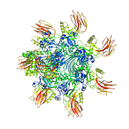 | | Complex structure of Clostridioides difficile binary toxin folded CDTa-bound CDTb-pore (short). | | 分子名称: | ADP-ribosylating binary toxin binding subunit CdtB, ADP-ribosylating binary toxin enzymatic subunit CdtA, CALCIUM ION | | 著者 | Yamada, T, Kawamoto, A, Yoshida, T, Sato, Y, Kato, T, Tsuge, H. | | 登録日 | 2022-08-19 | | 公開日 | 2022-10-26 | | 最終更新日 | 2024-07-03 | | 実験手法 | ELECTRON MICROSCOPY (3.18 Å) | | 主引用文献 | Cryo-EM structures of the translocational binary toxin complex CDTa-bound CDTb-pore from Clostridioides difficile.
Nat Commun, 13, 2022
|
|
7YVS
 | | Complex structure of Clostridioides difficile binary toxin unfolded CDTa-bound CDTb-pore (short). | | 分子名称: | ADP-ribosylating binary toxin binding subunit CdtB, ADP-ribosylating binary toxin enzymatic subunit CdtA, CALCIUM ION | | 著者 | Yamada, T, Kawamoto, A, Yoshida, T, Sato, Y, Kato, T, Tsuge, H. | | 登録日 | 2022-08-19 | | 公開日 | 2022-10-26 | | 最終更新日 | 2024-07-03 | | 実験手法 | ELECTRON MICROSCOPY (2.8 Å) | | 主引用文献 | Cryo-EM structures of the translocational binary toxin complex CDTa-bound CDTb-pore from Clostridioides difficile.
Nat Commun, 13, 2022
|
|
7YVW
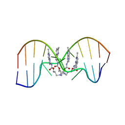 | | NMR determination of the 2:1 binding motif structure involving cytosine flipping out for the recognition of the CGG/CGG triad DNA | | 分子名称: | 3-[3-[(7-methyl-1,8-naphthyridin-2-yl)carbamoyloxy]propylamino]propyl ~{N}-(7-methyl-1,8-naphthyridin-2-yl)carbamate, DNA (5'-D(*CP*AP*TP*TP*CP*GP*GP*TP*TP*AP*G)-3'), DNA (5'-D(*CP*TP*AP*AP*CP*GP*GP*AP*AP*TP*G)-3') | | 著者 | Furuita, K, Yamada, T, Sakurabayashi, S, Nomura, M, Kojima, C, Nakatani, K. | | 登録日 | 2022-08-19 | | 公開日 | 2023-06-14 | | 最終更新日 | 2024-05-15 | | 実験手法 | SOLUTION NMR | | 主引用文献 | NMR determination of the 2:1 binding complex of naphthyridine carbamate dimer (NCD) and CGG/CGG triad in double-stranded DNA.
Nucleic Acids Res., 50, 2022
|
|
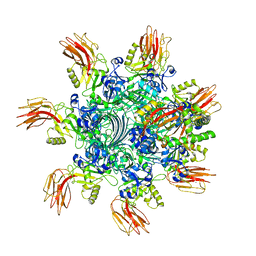
7VNN
 | | Complex structure of Clostridioides difficile enzymatic component (CDTa) and binding component (CDTb) pore with long stem | | 分子名称: | ADP-ribosylating binary toxin binding subunit CdtB, CALCIUM ION, CdtA | | 著者 | Yamada, T, Kawamoto, A, Yoshida, T, Sato, Y, Kato, T, Tsuge, H. | | 登録日 | 2021-10-11 | | 公開日 | 2022-10-26 | | 最終更新日 | 2024-06-19 | | 実験手法 | ELECTRON MICROSCOPY (2.64 Å) | | 主引用文献 | Cryo-EM structures of the translocational binary toxin complex CDTa-bound CDTb-pore from Clostridioides difficile.
Nat Commun, 13, 2022
|
|
7VNJ
 | | Complex structure of Clostridioides difficile enzymatic component (CDTa) and binding component (CDTb) pore with short stem | | 分子名称: | ADP-ribosylating binary toxin binding subunit CdtB, ADP-ribosyltransferase enzymatic component, CALCIUM ION | | 著者 | Yamada, T, Kawamoto, A, Yoshida, T, Sato, Y, Kato, T, Tsuge, H. | | 登録日 | 2021-10-11 | | 公開日 | 2022-10-26 | | 最終更新日 | 2024-06-19 | | 実験手法 | ELECTRON MICROSCOPY (2.56 Å) | | 主引用文献 | Cryo-EM structures of the translocational binary toxin complex CDTa-bound CDTb-pore from Clostridioides difficile.
Nat Commun, 13, 2022
|
|
6KLO
 | | Complex structure of Iota toxin enzymatic component (Ia) and binding component (Ib) pore with short stem | | 分子名称: | CALCIUM ION, Iota toxin component Ia, Iota toxin component Ib | | 著者 | Yoshida, T, Yamada, T, Kawamoto, A, Mitsuoka, K, Iwasaki, K, Tsuge, H. | | 登録日 | 2019-07-30 | | 公開日 | 2020-01-15 | | 最終更新日 | 2024-03-27 | | 実験手法 | ELECTRON MICROSCOPY (2.8 Å) | | 主引用文献 | Cryo-EM structures reveal translocational unfolding in the clostridial binary iota toxin complex.
Nat.Struct.Mol.Biol., 27, 2020
|
|
2PBJ
 | | GSH-heme bound microsomal prostaglandin E synthase | | 分子名称: | CHLORIDE ION, GLUTATHIONE, PROTOPORPHYRIN IX CONTAINING FE, ... | | 著者 | Takusagawa, F, Yamada, T. | | 登録日 | 2007-03-28 | | 公開日 | 2008-02-12 | | 最終更新日 | 2024-02-21 | | 実験手法 | X-RAY DIFFRACTION (2.8 Å) | | 主引用文献 | PGH2 degradation pathway catalyzed by GSH-heme complex bound microsomal prostaglandin E2 synthase type 2: the first example of a dual-function enzyme.
Biochemistry, 46, 2007
|
|
6KLW
 | | Complex structure of Iota toxin enzymatic component (Ia) and binding component (Ib) pore with long stem | | 分子名称: | CALCIUM ION, Iota toxin component Ia, Iota toxin component Ib | | 著者 | Yoshida, T, Yamada, T, Kawamoto, A, Mitsuoka, K, Iwasaki, K, Tsuge, H. | | 登録日 | 2019-07-30 | | 公開日 | 2020-01-15 | | 最終更新日 | 2024-03-27 | | 実験手法 | ELECTRON MICROSCOPY (2.9 Å) | | 主引用文献 | Cryo-EM structures reveal translocational unfolding in the clostridial binary iota toxin complex.
Nat.Struct.Mol.Biol., 27, 2020
|
|
6KLX
 | | Pore structure of Iota toxin binding component (Ib) | | 分子名称: | CALCIUM ION, Iota toxin component Ib | | 著者 | Yoshida, T, Yamada, T, Kawamoto, A, Mitsuoka, K, Iwasaki, K, Tsuge, H. | | 登録日 | 2019-07-30 | | 公開日 | 2020-01-15 | | 最終更新日 | 2024-03-27 | | 実験手法 | ELECTRON MICROSCOPY (2.9 Å) | | 主引用文献 | Cryo-EM structures reveal translocational unfolding in the clostridial binary iota toxin complex.
Nat.Struct.Mol.Biol., 27, 2020
|
|
1LZ5
 | | STRUCTURAL AND FUNCTIONAL ANALYSES OF THE ARG-GLY-ASP SEQUENCE INTRODUCED INTO HUMAN LYSOZYME | | 分子名称: | CHLORIDE ION, HUMAN LYSOZYME | | 著者 | Matsushima, M, Inaka, K, Yamada, T, Sekiguchi, K, Kikuchi, M. | | 登録日 | 1993-02-03 | | 公開日 | 1993-10-31 | | 最終更新日 | 2017-11-29 | | 実験手法 | X-RAY DIFFRACTION (1.8 Å) | | 主引用文献 | Structural and functional analyses of the Arg-Gly-Asp sequence introduced into human lysozyme.
J.Biol.Chem., 268, 1993
|
|
1LZ6
 | | STRUCTURAL AND FUNCTIONAL ANALYSES OF THE ARG-GLY-ASP SEQUENCE INTRODUCED INTO HUMAN LYSOZYME | | 分子名称: | CHLORIDE ION, HUMAN LYSOZYME | | 著者 | Matsushima, M, Inaka, K, Yamada, T, Sekiguchi, K, Kikuchi, M. | | 登録日 | 1993-02-03 | | 公開日 | 1993-10-31 | | 最終更新日 | 2017-11-29 | | 実験手法 | X-RAY DIFFRACTION (1.8 Å) | | 主引用文献 | Structural and functional analyses of the Arg-Gly-Asp sequence introduced into human lysozyme.
J.Biol.Chem., 268, 1993
|
|
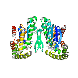
1F3P
 | | FERREDOXIN REDUCTASE (BPHA4)-NADH COMPLEX | | 分子名称: | FERREDOXIN REDUCTASE, FLAVIN-ADENINE DINUCLEOTIDE, NICOTINAMIDE-ADENINE-DINUCLEOTIDE | | 著者 | Senda, T, Yamada, T, Sakurai, N, Kubota, M, Nishizaki, T, Masai, E, Fukuda, M, Mitsuidagger, Y. | | 登録日 | 2000-06-06 | | 公開日 | 2001-06-06 | | 最終更新日 | 2024-03-13 | | 実験手法 | X-RAY DIFFRACTION (2.4 Å) | | 主引用文献 | Crystal structure of NADH-dependent ferredoxin reductase component in biphenyl dioxygenase.
J.Mol.Biol., 304, 2000
|
|
3U2J
 | | Neutron crystal structure of human Transthyretin | | 分子名称: | Transthyretin | | 著者 | Yokoyama, T, Mizuguchi, M, Nabeshima, Y, Kusaka, K, Yamada, T, Hosoya, T, Ohhara, T, Kurihara, K, Tomoyori, K, Tanaka, I, Niimura, N. | | 登録日 | 2011-10-03 | | 公開日 | 2012-02-22 | | 最終更新日 | 2023-11-01 | | 実験手法 | NEUTRON DIFFRACTION (2 Å) | | 主引用文献 | Hydrogen-bond network and pH sensitivity in transthyretin: Neutron crystal structure of human transthyretin
J.Struct.Biol., 177, 2012
|
|


