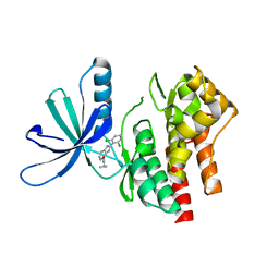
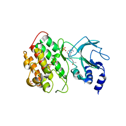
1BPT
 
 | | CREVICE-FORMING MUTANTS OF BPTI: CRYSTAL STRUCTURES OF F22A, Y23A, N43G, AND F45A | | 分子名称: | BOVINE PANCREATIC TRYPSIN INHIBITOR, PHOSPHATE ION | | 著者 | Housset, D, Wlodawer, A, Tao, F, Fuchs, J, Woodward, C. | | 登録日 | 1991-12-11 | | 公開日 | 1993-01-15 | | 最終更新日 | 2024-11-13 | | 実験手法 | X-RAY DIFFRACTION (2 Å) | | 主引用文献 | Crevice-forming mutants in the rigid core of bovine pancreatic trypsin inhibitor: crystal structures of F22A, Y23A, N43G, and F45A.
Protein Sci., 2, 1993
|
|
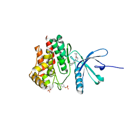
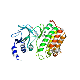
1FAN
 | | CREVICE-FORMING MUTANTS IN THE RIGID CORE OF BOVINE PANCREATIC TRYPSIN INHIBITOR: CRYSTAL STRUCTURES OF F22A, Y23A, N43G, AND F45A | | 分子名称: | BOVINE PANCREATIC TRYPSIN INHIBITOR | | 著者 | Danishefsky, A.T, Wlodawer, A, Kim, K.-S, Tao, F, Woodward, C. | | 登録日 | 1992-08-21 | | 公開日 | 1993-10-31 | | 最終更新日 | 2024-11-20 | | 実験手法 | X-RAY DIFFRACTION (2 Å) | | 主引用文献 | Crevice-forming mutants in the rigid core of bovine pancreatic trypsin inhibitor: crystal structures of F22A, Y23A, N43G, and F45A.
Protein Sci., 2, 1993
|
|
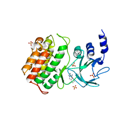
1BTI
 | | CREVICE-FORMING MUTANTS IN THE RIGID CORE OF BOVINE PANCREATIC TRYPSIN INHIBITOR: CRYSTAL STRUCTURES OF F22A, Y23A, N43G, AND F45A | | 分子名称: | BOVINE PANCREATIC TRYPSIN INHIBITOR | | 著者 | Housset, D, Tao, F, Kim, K.-S, Fuchs, J, Woodward, C, Wlodawer, A. | | 登録日 | 1991-07-11 | | 公開日 | 1993-10-31 | | 最終更新日 | 2024-11-06 | | 実験手法 | X-RAY DIFFRACTION (2.2 Å) | | 主引用文献 | Crevice-forming mutants in the rigid core of bovine pancreatic trypsin inhibitor: crystal structures of F22A, Y23A, N43G, and F45A.
Protein Sci., 2, 1993
|
|
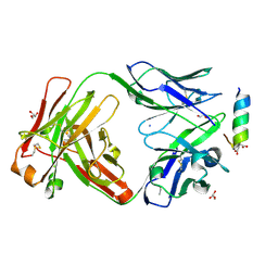
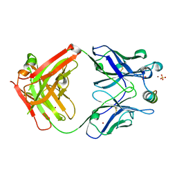
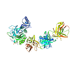
6VVU
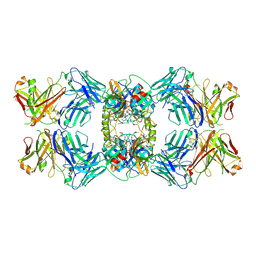 | | Anti-Tryptase fab E104.v1 bound to tryptase | | 分子名称: | CALCIUM ION, Fab E104.v1 heavy chain, Fab E104.v1 light chain, ... | | 著者 | Ultsch, M, Koerber, J.T. | | 登録日 | 2020-02-18 | | 公開日 | 2020-12-30 | | 最終更新日 | 2024-11-06 | | 実験手法 | X-RAY DIFFRACTION (3 Å) | | 主引用文献 | Bivalent antibody pliers inhibit beta-tryptase by an allosteric mechanism dependent on the IgG hinge.
Nat Commun, 11, 2020
|
|
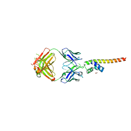
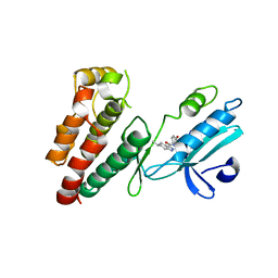
7L6K
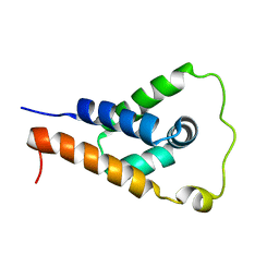 | | ApoL1 N-terminal domain | | 分子名称: | Apolipoprotein L1 | | 著者 | Holliday, M.J, Ultsch, M, Moran, P, Fairbrother, W.J, Kirchhofer, D. | | 登録日 | 2020-12-23 | | 公開日 | 2021-08-04 | | 最終更新日 | 2024-05-15 | | 実験手法 | SOLUTION NMR | | 主引用文献 | Structures of the ApoL1 and ApoL2 N-terminal domains reveal a non-classical four-helix bundle motif.
Commun Biol, 4, 2021
|
|
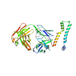
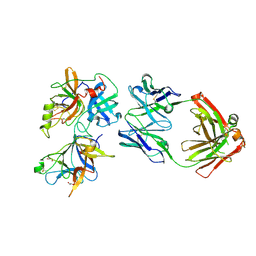
7LFA
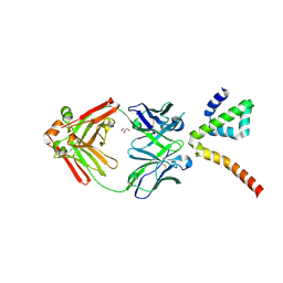 | | Fab 3B6 bound to ApoL1 NTD | | 分子名称: | Apolipoprotein L1, CHLORIDE ION, Fab 3B6 heavy chain, ... | | 著者 | Ultsch, M, Kirchhofer, D. | | 登録日 | 2021-01-15 | | 公開日 | 2021-08-04 | | 最終更新日 | 2024-11-06 | | 実験手法 | X-RAY DIFFRACTION (1.857 Å) | | 主引用文献 | Structures of the ApoL1 and ApoL2 N-terminal domains reveal a non-classical four-helix bundle motif.
Commun Biol, 4, 2021
|
|
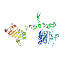
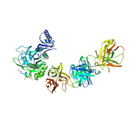
7LF8
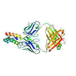 | | Fab 6D12 bound to ApoL2 NTD | | 分子名称: | Apolipoprotein L2, Fab 6D12 heavy chain, Fab 6D12 light chain, ... | | 著者 | Ultsch, M, Kirchhofer, D. | | 登録日 | 2021-01-15 | | 公開日 | 2021-08-04 | | 最終更新日 | 2024-10-09 | | 実験手法 | X-RAY DIFFRACTION (2.15 Å) | | 主引用文献 | Structures of the ApoL1 and ApoL2 N-terminal domains reveal a non-classical four-helix bundle motif.
Commun Biol, 4, 2021
|
|
7LFD
 | | Fab 7D6 bound to ApoL1 BH3 like peptide | | 分子名称: | AMMONIUM ION, Apolipoprotein L1 BH3 like peptide, CITRATE ANION, ... | | 著者 | Ultsch, M, Kirchhofer, D. | | 登録日 | 2021-01-16 | | 公開日 | 2021-08-04 | | 最終更新日 | 2024-10-30 | | 実験手法 | X-RAY DIFFRACTION (2.157 Å) | | 主引用文献 | Structures of the ApoL1 and ApoL2 N-terminal domains reveal a non-classical four-helix bundle motif.
Commun Biol, 4, 2021
|
|
7LFB
 | | Fab 7D6 bound to ApoL1 NTD | | 分子名称: | 2-(N-MORPHOLINO)-ETHANESULFONIC ACID, Apolipoprotein L1, Fab 7D6 heavy chain, ... | | 著者 | Ultsch, M, Kirchhofer, D. | | 登録日 | 2021-01-16 | | 公開日 | 2021-08-04 | | 最終更新日 | 2024-11-13 | | 実験手法 | X-RAY DIFFRACTION (1.913 Å) | | 主引用文献 | Structures of the ApoL1 and ApoL2 N-terminal domains reveal a non-classical four-helix bundle motif.
Commun Biol, 4, 2021
|
|
7LF7
 | | Fab 6D12 bound to ApoL1 NTD | | 分子名称: | Apolipoprotein L1, ETHANOL, Fab 6D12 heavy chain, ... | | 著者 | Ultsch, M, Kirchhofer, D. | | 登録日 | 2021-01-15 | | 公開日 | 2021-08-04 | | 最終更新日 | 2023-10-18 | | 実験手法 | X-RAY DIFFRACTION (2.026 Å) | | 主引用文献 | Structures of the ApoL1 and ApoL2 N-terminal domains reveal a non-classical four-helix bundle motif.
Commun Biol, 4, 2021
|
|
3U2P
 | | Crystal structure of N-terminal three extracellular domains of ErbB4/Her4 | | 分子名称: | 2-acetamido-2-deoxy-beta-D-glucopyranose, Receptor tyrosine-protein kinase erbB-4 | | 著者 | Liu, P, Bouyain, S, Elgenbrot, C, Leahy, D.J. | | 登録日 | 2011-10-04 | | 公開日 | 2011-11-09 | | 最終更新日 | 2024-11-20 | | 実験手法 | X-RAY DIFFRACTION (2.57 Å) | | 主引用文献 | The ErbB4 extracellular region retains a tethered-like conformation in the absence of the tether.
Protein Sci., 21, 2012
|
|
6MV5
 | |
6NW2
 | | Structure of human RIPK1 kinase domain in complex with compound 11 | | 分子名称: | (5R)-5-methyl-N-[(3S)-5-methyl-4-oxo-2,3,4,5-tetrahydro-1,5-benzoxazepin-3-yl]-4,5,6,7-tetrahydro-2H-indazole-3-carboxamide, Receptor-interacting serine/threonine-protein kinase 1 | | 著者 | Fong, R, Lupardus, P.J. | | 登録日 | 2019-02-05 | | 公開日 | 2019-05-01 | | 最終更新日 | 2024-03-13 | | 実験手法 | X-RAY DIFFRACTION (2 Å) | | 主引用文献 | Potent and selective inhibitors of receptor-interacting protein kinase 1 that lack an aromatic back pocket group.
Bioorg.Med.Chem.Lett., 29, 2019
|
|
6O1F
 | |
6U3I
 | |
6U2F
 | |
5WAL
 | |
5WEV
 | |
5T8Q
 | |
5T8O
 | | Crystal structure of murine NF-kappaB inducing kinase (NIK) bound to Imidazobenzoxepin Compound 3 | | 分子名称: | 10-(3-methyl-3-oxidanyl-but-1-ynyl)-5,6-dihydroimidazo[1,2-d][1,4]benzoxazepine-2-carboxamide, Mitogen-activated protein kinase kinase kinase 14, SULFATE ION | | 著者 | Smith, M.A, McEwan, P, Hymowitz, S.G. | | 登録日 | 2016-09-08 | | 公開日 | 2017-01-11 | | 最終更新日 | 2024-05-08 | | 実験手法 | X-RAY DIFFRACTION (2.41 Å) | | 主引用文献 | Structure-Based Design of Tricyclic NF-kappa B Inducing Kinase (NIK) Inhibitors That Have High Selectivity over Phosphoinositide-3-kinase (PI3K).
J. Med. Chem., 60, 2017
|
|
5T8P
 | | Crystal structure of murine NF-kappaB inducing kinase (NIK) bound to benzoxepin compound 2 | | 分子名称: | 6,7-dihydrothieno[4,5]oxepino[1,2-~{c}]pyridine-2-carboxamide, Mitogen-activated protein kinase kinase kinase 14, SULFATE ION | | 著者 | Smith, M.A, McEwan, P.A. | | 登録日 | 2016-09-08 | | 公開日 | 2017-01-11 | | 最終更新日 | 2024-05-08 | | 実験手法 | X-RAY DIFFRACTION (2.32 Å) | | 主引用文献 | Structure-Based Design of Tricyclic NF-kappa B Inducing Kinase (NIK) Inhibitors That Have High Selectivity over Phosphoinositide-3-kinase (PI3K).
J. Med. Chem., 60, 2017
|
|
1FLT
 | |
6BQD
 | | TAF1-BD2 bromodomain in complex with (E)-3-(6-(but-2-en-1-yl)-7-oxo-6,7-dihydro-1H-pyrrolo[2,3-c]pyridin-4-yl)-N,N-dimethylbenzamide | | 分子名称: | 3-{6-[(2E)-but-2-en-1-yl]-7-oxo-6,7-dihydro-1H-pyrrolo[2,3-c]pyridin-4-yl}-N,N-dimethylbenzamide, Transcription initiation factor TFIID subunit 1 | | 著者 | Murray, J.M, Tang, Y. | | 登録日 | 2017-11-27 | | 公開日 | 2019-02-20 | | 最終更新日 | 2023-10-04 | | 実験手法 | X-RAY DIFFRACTION (2.136 Å) | | 主引用文献 | Water molecules in protein-ligand interfaces. Evaluation of software tools and SAR comparison.
J. Comput. Aided Mol. Des., 33, 2019
|
|
6E4Y
 | |
6E4Z
 | |


