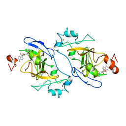
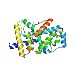
8X5J
 
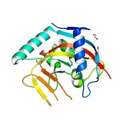 | | The Crystal Structure of PARP5A from Biortus. | | 分子名称: | 1,2-ETHANEDIOL, L(+)-TARTARIC ACID, Poly [ADP-ribose] polymerase, ... | | 著者 | Wang, F, Cheng, W, Yuan, Z, Qi, J, Wu, B. | | 登録日 | 2023-11-17 | | 公開日 | 2023-12-27 | | 実験手法 | X-RAY DIFFRACTION (1.7 Å) | | 主引用文献 | The Crystal Structure of PARP5A from Biortus.
To Be Published
|
|
8XFL
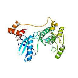 | |
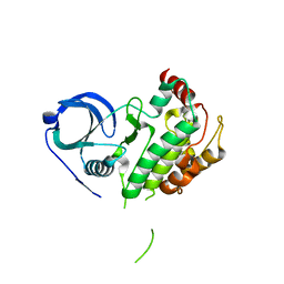
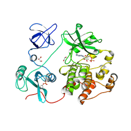
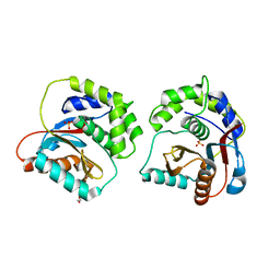
8X5L
 | | The Crystal Structure of PRKACA from Biortus. | | 分子名称: | (2S)-2-(4-chlorophenyl)-2-hydroxy-2-[4-(1H-pyrazol-4-yl)phenyl]ethanaminium, SODIUM ION, cAMP-dependent protein kinase catalytic subunit alpha | | 著者 | Wang, F, Cheng, W, Lv, Z, Lin, D, Pan, W. | | 登録日 | 2023-11-17 | | 公開日 | 2023-12-27 | | 実験手法 | X-RAY DIFFRACTION (2.75 Å) | | 主引用文献 | The Crystal Structure of PRKACA from Biortus.
To Be Published
|
|
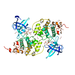
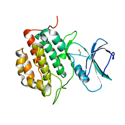
8XU4
 | | The Crystal Structure of MAPK2 from Biortus. | | 分子名称: | MALONIC ACID, MAP kinase-activated protein kinase 2 | | 著者 | Wang, F, Cheng, W, Yuan, Z, Qi, J, Shen, Z. | | 登録日 | 2024-01-12 | | 公開日 | 2024-01-24 | | 実験手法 | X-RAY DIFFRACTION (3.4 Å) | | 主引用文献 | The Crystal Structure of MAPK2 from Biortus.
To Be Published
|
|
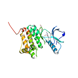
8XN6
 | | The Crystal Structure of GSK3b from Biortus. | | 分子名称: | 1,2-ETHANEDIOL, DI(HYDROXYETHYL)ETHER, Glycogen synthase kinase-3 beta, ... | | 著者 | Wang, F, Cheng, W, Yuan, Z, Qi, J, Wu, B. | | 登録日 | 2023-12-29 | | 公開日 | 2024-01-24 | | 実験手法 | X-RAY DIFFRACTION (2.4 Å) | | 主引用文献 | The Crystal Structure of GSK3b from Biortus.
To Be Published
|
|
8XPT
 | | The Crystal Structure of EHMT1 from Biortus. | | 分子名称: | Histone-lysine N-methyltransferase EHMT1, S-ADENOSYL-L-HOMOCYSTEINE, SULFATE ION, ... | | 著者 | Wang, F, Cheng, W, Yuan, Z, Lin, D, Bao, C. | | 登録日 | 2024-01-04 | | 公開日 | 2024-01-24 | | 実験手法 | X-RAY DIFFRACTION (3.35 Å) | | 主引用文献 | The Crystal Structure of EHMT1 from Biortus.
To Be Published
|
|
8XN8
 | | The Crystal Structure of SRC from Biortus. | | 分子名称: | 1,2-ETHANEDIOL, ADENOSINE-5'-DIPHOSPHATE, GLYCEROL, ... | | 著者 | Wang, F, Cheng, W, Yuan, Z, Lin, D, Bao, C. | | 登録日 | 2023-12-29 | | 公開日 | 2024-01-24 | | 実験手法 | X-RAY DIFFRACTION (1.95 Å) | | 主引用文献 | The Crystal Structure of SRC from Biortus.
To Be Published
|
|
8XPZ
 | | The Crystal Structure of TTBK1 from Biortus. | | 分子名称: | 1,2-ETHANEDIOL, Tau-tubulin kinase 1 | | 著者 | Wang, F, Cheng, W, Yuan, Z, Lin, D, Ni, C. | | 登録日 | 2024-01-04 | | 公開日 | 2024-01-24 | | 実験手法 | X-RAY DIFFRACTION (2.6 Å) | | 主引用文献 | The Crystal Structure of TTBK1 from Biortus.
To Be Published
|
|
8XPV
 | | The Crystal Structure of EphA2 from Biortus. | | 分子名称: | 1,2-ETHANEDIOL, 1-(3,3-dimethylbutyl)-3-{2-fluoro-4-methyl-5-[7-methyl-2-(methylamino)pyrido[2,3-d]pyrimidin-6-yl]phenyl}urea, Ephrin type-A receptor 2, ... | | 著者 | Wang, F, Cheng, W, Lv, Z, Meng, Q, Xu, Y. | | 登録日 | 2024-01-04 | | 公開日 | 2024-01-24 | | 実験手法 | X-RAY DIFFRACTION (1.55 Å) | | 主引用文献 | The Crystal Structure of EphA2 from Biortus.
To Be Published
|
|
8XU5
 | |
8XI7
 | | The Crystal Structure of UCHL1 from Biortus. | | 分子名称: | 1,2-ETHANEDIOL, SULFATE ION, Ubiquitin carboxyl-terminal hydrolase isozyme L1 | | 著者 | Wang, F, Cheng, W, Yuan, Z, Qi, J, Shen, Z. | | 登録日 | 2023-12-19 | | 公開日 | 2024-03-06 | | 実験手法 | X-RAY DIFFRACTION (1.95 Å) | | 主引用文献 | The Crystal Structure of UCHL1 from Biortus.
To Be Published
|
|
8XP5
 | | The Crystal Structure of p53/BCL-xL fusion complex from Biortus. | | 分子名称: | Bcl-2-like protein 1,Cellular tumor antigen p53, ZINC ION | | 著者 | Wang, F, Cheng, W, Yuan, Z, Lin, D, Bao, C. | | 登録日 | 2024-01-03 | | 公開日 | 2024-03-06 | | 実験手法 | X-RAY DIFFRACTION (2.55 Å) | | 主引用文献 | The Crystal Structure of p53/BCL-xL fusion complex from Biortus.
To Be Published
|
|
8XPG
 | | The Crystal Structure of polo box domain of Plk4 from Biortus. | | 分子名称: | SULFATE ION, Serine/threonine-protein kinase PLK4 | | 著者 | Wang, F, Cheng, W, Lv, Z, Meng, Q, Zhang, B. | | 登録日 | 2024-01-03 | | 公開日 | 2024-03-06 | | 実験手法 | X-RAY DIFFRACTION (2.7 Å) | | 主引用文献 | The Crystal Structure of polo box domain of Plk4 from Biortus.
To Be Published
|
|
8XOV
 | | The Crystal Structure of N-terminal kinase domain of human RSK-1 from Biortus. | | 分子名称: | 1,2-ETHANEDIOL, PHOSPHOAMINOPHOSPHONIC ACID-ADENYLATE ESTER, Ribosomal protein S6 kinase alpha-1 | | 著者 | Wang, F, Cheng, W, Lv, Z, Meng, Q, Zhang, B. | | 登録日 | 2024-01-02 | | 公開日 | 2024-03-06 | | 実験手法 | X-RAY DIFFRACTION (2.55 Å) | | 主引用文献 | The Crystal Structure of N-terminal kinase domain of human RSK-1 from Biortus.
To Be Published
|
|
8XEY
 | | The Crystal Structure of C-terminal kinase domain of RSK2 from Biortus | | 分子名称: | 1,2-ETHANEDIOL, DI(HYDROXYETHYL)ETHER, Ribosomal protein S6 kinase alpha-3 | | 著者 | Wang, F, Cheng, W, Lv, Z, Meng, Q, Zhang, B. | | 登録日 | 2023-12-13 | | 公開日 | 2024-03-06 | | 実験手法 | X-RAY DIFFRACTION (2.65 Å) | | 主引用文献 | The Crystal Structure of C-terminal kinase domain of RSK2 from Biortus
To Be Published
|
|
8XQK
 | | The Crystal Structure of Apaf from Biortus. | | 分子名称: | Apoptotic protease-activating factor 1, PHOSPHATE ION | | 著者 | Wang, F, Cheng, W, Lv, Z, Ju, C, Ni, C. | | 登録日 | 2024-01-05 | | 公開日 | 2024-03-06 | | 実験手法 | X-RAY DIFFRACTION (2.85 Å) | | 主引用文献 | The Crystal Structure of Apaf from Biortus.
To Be Published
|
|
8XQD
 | |
8XLD
 | | Structure of the GFP:GFP-nanobody complex from Biortus. | | 分子名称: | 1,2-ETHANEDIOL, Nanobody(Staygold-S2G10)-Nanobody(Staygold-S4F1), ZINC ION, ... | | 著者 | Wang, F, Cheng, W, Yuan, Z, Lin, D, Bao, C. | | 登録日 | 2023-12-25 | | 公開日 | 2024-03-06 | | 実験手法 | X-RAY DIFFRACTION (2.1 Å) | | 主引用文献 | Structure of the GFP:GFP-nanobody complex from Biortus.
To Be Published
|
|
8XI8
 | |
8XPX
 | | The Crystal Structure of PARP12 from Biortus. | | 分子名称: | 1,2-ETHANEDIOL, ACETATE ION, DI(HYDROXYETHYL)ETHER, ... | | 著者 | Wang, F, Cheng, W, Lv, Z, Qi, J, Shen, Z. | | 登録日 | 2024-01-04 | | 公開日 | 2024-03-06 | | 実験手法 | X-RAY DIFFRACTION (1.75 Å) | | 主引用文献 | The Crystal Structure of PARP12 from Biortus.
To Be Published
|
|
4HPV
 | | Crystal structure of S-Adenosylmethionine synthetase from Sulfolobus solfataricus | | 分子名称: | S-adenosylmethionine synthase | | 著者 | Wang, F, Hurley, K.A, Helmich, K.E, Singh, S, Bingman, C.A, Thorson, J.S, Phillips Jr, G.N, Enzyme Discovery for Natural Product Biosynthesis (NatPro) | | 登録日 | 2012-10-24 | | 公開日 | 2012-11-14 | | 最終更新日 | 2017-11-15 | | 実験手法 | X-RAY DIFFRACTION (2.214 Å) | | 主引用文献 | Understanding molecular recognition of promiscuity of thermophilic methionine adenosyltransferase sMAT from Sulfolobus solfataricus.
Febs J., 281, 2014
|
|
6EF8
 | | Cryo-EM of the OmcS nanowires from Geobacter sulfurreducens | | 分子名称: | C-type cytochrome OmcS, HEME C | | 著者 | Wang, F, Gu, Y, Egelman, E.H, Malvankar, N.S. | | 登録日 | 2018-08-16 | | 公開日 | 2019-04-10 | | 最終更新日 | 2019-11-27 | | 実験手法 | ELECTRON MICROSCOPY (3.7 Å) | | 主引用文献 | Structure of Microbial Nanowires Reveals Stacked Hemes that Transport Electrons over Micrometers.
Cell, 177, 2019
|
|
4L2Z
 | | Crystal structure of S-Adenosylmethionine synthetase from Sulfolobus solfataricus complexed with SAE and PPi | | 分子名称: | DIPHOSPHATE, MAGNESIUM ION, PHOSPHATE ION, ... | | 著者 | Wang, F, Hurley, K.A, Helmich, K.E, Singh, S, Bingman, C.A, Thorson, J.S, Phillips Jr, G.N, Enzyme Discovery for Natural Product Biosynthesis (NatPro) | | 登録日 | 2013-06-05 | | 公開日 | 2013-06-19 | | 最終更新日 | 2023-12-06 | | 実験手法 | X-RAY DIFFRACTION (2.494 Å) | | 主引用文献 | Understanding molecular recognition of promiscuity of thermophilic methionine adenosyltransferase sMAT from Sulfolobus solfataricus.
Febs J., 281, 2014
|
|
5EU8
 | | Structure of FIPV main protease in complex with dual inhibitors | | 分子名称: | 1,2-ETHANEDIOL, N-[(5-METHYLISOXAZOL-3-YL)CARBONYL]ALANYL-L-VALYL-N~1~-((1R,2Z)-4-(BENZYLOXY)-4-OXO-1-{[(3R)-2-OXOPYRROLIDIN-3-YL]METHYL}BUT-2-ENYL)-L-LEUCINAMIDE, ZINC ION, ... | | 著者 | Wang, F, Chen, C, Liu, X, Yang, K, Xu, X, Yang, H. | | 登録日 | 2015-11-18 | | 公開日 | 2015-12-30 | | 最終更新日 | 2023-11-15 | | 実験手法 | X-RAY DIFFRACTION (2.447 Å) | | 主引用文献 | Crystal Structure of Feline Infectious Peritonitis Virus Main Protease in Complex with Synergetic Dual Inhibitors
J.Virol., 90, 2015
|
|
2K86
 | | Solution Structure of FOXO3a Forkhead domain | | 分子名称: | Forkhead box protein O3 | | 著者 | Wang, F, Marshall, C.B, Li, G, Plevin, M.J, Ikura, M. | | 登録日 | 2008-09-02 | | 公開日 | 2008-10-14 | | 最終更新日 | 2024-05-01 | | 実験手法 | SOLUTION NMR | | 主引用文献 | Biochemical and structural characterization of an intramolecular interaction in FOXO3a and its binding with p53.
J.Mol.Biol., 384, 2008
|
|


