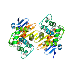
3EW9
 
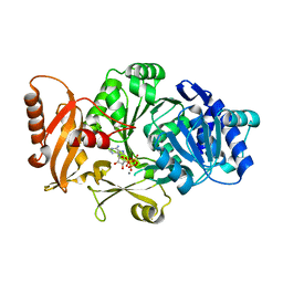
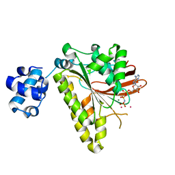 | | RADA recombinase from METHANOCOCCUS MARIPALUDIS in complex with AMPPNP and potassium ions | | 分子名称: | DNA repair and recombination protein radA, MAGNESIUM ION, PHOSPHOAMINOPHOSPHONIC ACID-ADENYLATE ESTER, ... | | 著者 | Li, Y, He, Y, Luo, Y. | | 登録日 | 2008-10-14 | | 公開日 | 2009-05-05 | | 最終更新日 | 2023-09-06 | | 実験手法 | X-RAY DIFFRACTION (2.4 Å) | | 主引用文献 | Conservation of a conformational switch in RadA recombinase from Methanococcus maripaludis.
Acta Crystallogr.,Sect.D, 65, 2009
|
|
6PWC
 | | A complex structure of arrestin-2 bound to neurotensin receptor 1 | | 分子名称: | Beta-arrestin-1, Fab30 heavy chain, Fab30 light chain, ... | | 著者 | Yin, W, Li, Z, Jin, M, Yin, Y.-L, de Waal, P.W, Pal, K, Gao, X, He, Y, Gao, J, Wang, X, Zhang, Y, Zhou, H, Melcher, K, Jiang, Y, Cong, Y, Zhou, X.E, Yu, X, Xu, H.E. | | 登録日 | 2019-07-22 | | 公開日 | 2019-12-04 | | 最終更新日 | 2025-06-04 | | 実験手法 | ELECTRON MICROSCOPY (4.9 Å) | | 主引用文献 | A complex structure of arrestin-2 bound to a G protein-coupled receptor.
Cell Res., 29, 2019
|
|
3DHV
 | |
7NRJ
 | |
7O5T
 | |
7O5N
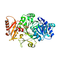
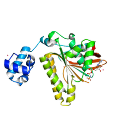
 | | Crystal Structure of a Class D carbapenemase complexed with Avibactam | | 分子名称: | (2S,5R)-1-formyl-5-[(sulfooxy)amino]piperidine-2-carboxamide, 1-BUTANOL, Beta-lactamase, ... | | 著者 | Zhou, Q, Zhang, Z, He, Y, Jin, Y. | | 登録日 | 2021-04-09 | | 公開日 | 2022-04-20 | | 最終更新日 | 2024-11-13 | | 実験手法 | X-RAY DIFFRACTION (1.6 Å) | | 主引用文献 | An Ion-Pair Induced Intermediate Complex Captured in Class D Carbapenemase Reveals Chloride Ion as a Janus Effector Modulating Activity
Acs Cent.Sci., 2023
|
|
8QNZ
 | | Crystal Structure of a Class D Carbapenemase Complexed with Hydrolyzed Imipenem | | 分子名称: | (2R)-2-[(2S,3R)-1,3-bis(oxidanyl)-1-oxidanylidene-butan-2-yl]-4-(2-methanimidamidoethylsulfanyl)-2,3-dihydro-1H-pyrrole -5-carboxylic acid, 1-BUTANOL, BROMIDE ION, ... | | 著者 | Zhou, Q, He, Y, Jin, Y. | | 登録日 | 2023-09-27 | | 公開日 | 2023-11-08 | | 最終更新日 | 2024-03-13 | | 実験手法 | X-RAY DIFFRACTION (1.53 Å) | | 主引用文献 | An Ion-Pair Induced Intermediate Complex Captured in Class D Carbapenemase Reveals Chloride Ion as a Janus Effector Modulating Activity.
Acs Cent.Sci., 9, 2023
|
|
7O9N
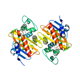 | |
3FCC
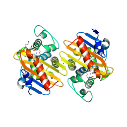
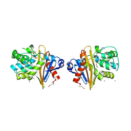
 | | CRYSTAL STRUCTURE OF DLTA PROTEIN IN COMPLEX WITH ATP and MAGNESIUM | | 分子名称: | ADENOSINE-5'-TRIPHOSPHATE, D-alanine--poly(phosphoribitol) ligase subunit 1, MAGNESIUM ION | | 著者 | Osman, K.T, Du, L, He, Y, Luo, Y. | | 登録日 | 2008-11-21 | | 公開日 | 2009-04-14 | | 最終更新日 | 2023-09-06 | | 実験手法 | X-RAY DIFFRACTION (2.32 Å) | | 主引用文献 | Crystal structure of Bacillus cereus D-alanyl carrier protein ligase (DltA) in complex with ATP.
J.Mol.Biol., 388, 2009
|
|
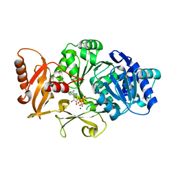
3FCE
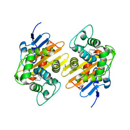
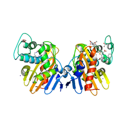
 | | Crystal Structure of Bacillus cereus D-alanyl Carrier Protein Ligase DltA in Complex with ATP: Implications for Adenylation Mechanism | | 分子名称: | ADENOSINE-5'-TRIPHOSPHATE, CALCIUM ION, D-alanine--poly(phosphoribitol) ligase subunit 1 | | 著者 | Osman, K.T, Du, L, He, Y, Luo, Y. | | 登録日 | 2008-11-21 | | 公開日 | 2009-04-14 | | 最終更新日 | 2023-09-06 | | 実験手法 | X-RAY DIFFRACTION (1.9 Å) | | 主引用文献 | Crystal structure of Bacillus cereus D-alanyl carrier protein ligase (DltA) in complex with ATP.
J.Mol.Biol., 388, 2009
|
|
7Q14
 | |
7O5Q
 | | Crystal Structure of a Class D Carbapenemase Complexed with Hydrolyzed Oxacillin | | 分子名称: | (2R,4S)-2-[(1R)-2-butoxy-1-[(5-methyl-3-phenyl-1,2-oxazol-4-yl)carbonylamino]-2-oxidanylidene-ethyl]-5,5-dimethyl-1,3-thiazolidin-3-ium-4-carboxylic acid, (2R,4S)-2-[(R)-carboxy{[(5-methyl-3-phenyl-1,2-oxazol-4-yl)carbonyl]amino}methyl]-5,5-dimethyl-1,3-thiazolidine-4-carbo xylic acid, 1-BUTANOL, ... | | 著者 | Zhou, Q, He, Y, Jin, Y. | | 登録日 | 2021-04-09 | | 公開日 | 2022-04-20 | | 最終更新日 | 2024-01-31 | | 実験手法 | X-RAY DIFFRACTION (1.85 Å) | | 主引用文献 | Crystal Structure of a Class D Carbapenemase Complexed with Hydrolyzed Oxacillin
To Be Published
|
|
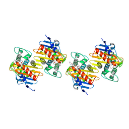
2B21
 | | RADA Recombinase in complex with AMPPNP at pH 6.0 | | 分子名称: | DNA repair and recombination protein radA, MAGNESIUM ION, PHOSPHOAMINOPHOSPHONIC ACID-ADENYLATE ESTER, ... | | 著者 | Qian, X, He, Y, Wu, Y, Luo, Y. | | 登録日 | 2005-09-16 | | 公開日 | 2006-08-29 | | 最終更新日 | 2023-08-23 | | 実験手法 | X-RAY DIFFRACTION (2.4 Å) | | 主引用文献 | Crystal Structure of Methanococcus Voltae Rada at an Acidic pH
To be Published
|
|
8FFZ
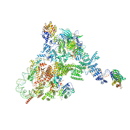 | | TFIIIA-TFIIIC-Brf1-TBP complex bound to 5S rRNA gene | | 分子名称: | DNA (151-MER), Transcription factor IIIA, Transcription factor IIIB 70 kDa subunit,TATA-box-binding protein, ... | | 著者 | Talyzina, A, He, Y. | | 登録日 | 2022-12-11 | | 公開日 | 2023-06-21 | | 最終更新日 | 2023-08-16 | | 実験手法 | ELECTRON MICROSCOPY (3.8 Å) | | 主引用文献 | Structural basis of TFIIIC-dependent RNA polymerase III transcription initiation.
Mol.Cell, 83, 2023
|
|
6O9L
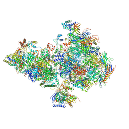 | | Human holo-PIC in the closed state | | 分子名称: | CDK-activating kinase assembly factor MAT1, Cyclin-H, Cyclin-dependent kinase 7, ... | | 著者 | Yan, C.L, Dodd, T, He, Y, Tainer, J.A, Tsutakawa, S.E, Ivanov, I. | | 登録日 | 2019-03-14 | | 公開日 | 2019-05-29 | | 最終更新日 | 2024-03-13 | | 実験手法 | ELECTRON MICROSCOPY (7.2 Å) | | 主引用文献 | Transcription preinitiation complex structure and dynamics provide insight into genetic diseases.
Nat.Struct.Mol.Biol., 26, 2019
|
|
1JNB
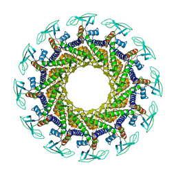 | | CONNECTOR PROTEIN FROM BACTERIOPHAGE PHI29 | | 分子名称: | UPPER COLLAR PROTEIN | | 著者 | Simpson, A.A, Leiman, P.G, Tao, Y, He, Y, Badasso, M.O, Jardine, P.J, Anderson, D.L, Rossmann, M.G. | | 登録日 | 2001-07-23 | | 公開日 | 2001-08-15 | | 最終更新日 | 2024-04-03 | | 実験手法 | X-RAY DIFFRACTION (3.2 Å) | | 主引用文献 | Structure determination of the head-tail connector of bacteriophage phi29.
Acta Crystallogr.,Sect.D, 57, 2001
|
|
7PEP
 | | Crystal Structure of a Class D Carbapenemase Complexed with Hydrolyzed Imipenem | | 分子名称: | (2R,4S)-2-[(1S,2R)-1-carboxy-2-hydroxypropyl]-4-[(2-{[(Z)-iminomethyl]amino}ethyl)sulfanyl]-3,4-dihydro-2H-pyrrole-5-ca rboxylic acid, 1-BUTANOL, BROMIDE ION, ... | | 著者 | Zhou, Q, He, Y, Jin, Y. | | 登録日 | 2021-08-11 | | 公開日 | 2022-08-24 | | 最終更新日 | 2024-03-06 | | 実験手法 | X-RAY DIFFRACTION (1.7 Å) | | 主引用文献 | An Ion-Pair Induced Intermediate Complex Captured in Class D Carbapenemase Reveals Chloride Ion as a Janus Effector Modulating Activity
Acs Cent.Sci., 2023
|
|
7PEI
 | |
7PSF
 | |
7PSE
 | | Crystal Structure of a Class D Carbapenemase_K73ALY Complexed with Oxacillin | | 分子名称: | (2R,4S)-5,5-dimethyl-2-[(1R)-1-{[(5-methyl-3-phenyl-1,2-oxazol-4-yl)carbonyl]amino}-2-oxoethyl]-1,3-thiazolidine-4-carb oxylic acid, 1-BUTANOL, Beta-lactamase, ... | | 著者 | Zhou, Q, He, Y, Jin, Y. | | 登録日 | 2021-09-23 | | 公開日 | 2022-10-05 | | 最終更新日 | 2024-10-16 | | 実験手法 | X-RAY DIFFRACTION (2.32 Å) | | 主引用文献 | An Ion-Pair Induced Intermediate Complex Captured in Class D Carbapenemase Reveals Chloride Ion as a Janus Effector Modulating Activity
Acs Cent.Sci., 2023
|
|
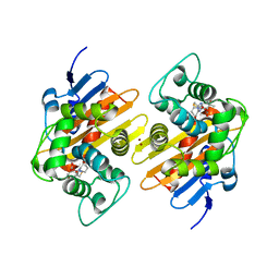
1S17
 | | Identification of Novel Potent Bicyclic Peptide Deformylase Inhibitors | | 分子名称: | 2-(3,4-DIHYDRO-3-OXO-2H-BENZO[B][1,4]THIAZIN-2-YL)-N-HYDROXYACETAMIDE, GLYCEROL, NICKEL (II) ION, ... | | 著者 | Molteni, V, He, X, Nabakka, J, Yang, K, Kreusch, A, Gordon, P, Bursulaya, B, Ryder, N.S, Goldberg, R, He, Y. | | 登録日 | 2004-01-05 | | 公開日 | 2004-03-30 | | 最終更新日 | 2023-08-23 | | 実験手法 | X-RAY DIFFRACTION (1.95 Å) | | 主引用文献 | Identification of novel potent bicyclic peptide deformylase inhibitors
Bioorg.Med.Chem.Lett., 14, 2004
|
|
7PGO
 | | Crystal Structure of a Class D Carbapenemase_R250A | | 分子名称: | 1-BUTANOL, BROMIDE ION, Beta-lactamase | | 著者 | Zhou, Q, He, Y, Jin, Y. | | 登録日 | 2021-08-15 | | 公開日 | 2022-08-24 | | 最終更新日 | 2024-03-06 | | 実験手法 | X-RAY DIFFRACTION (1.85 Å) | | 主引用文献 | An Ion-Pair Induced Intermediate Complex Captured in Class D Carbapenemase Reveals Chloride Ion as a Janus Effector Modulating Activity
Acs Cent.Sci., 2023
|
|
7PFN
 | |
7PEH
 | | Crystal Structure of a Class D Carbapenemase | | 分子名称: | 1-BUTANOL, Beta-lactamase | | 著者 | Zhou, Q, He, Y, Jin, Y. | | 登録日 | 2021-08-10 | | 公開日 | 2022-08-24 | | 最終更新日 | 2024-03-06 | | 実験手法 | X-RAY DIFFRACTION (1.92 Å) | | 主引用文献 | An Ion-Pair Induced Intermediate Complex Captured in Class D Carbapenemase Reveals Chloride Ion as a Janus Effector Modulating Activity
Acs Cent.Sci., 2023
|
|
3KO1
 | | Cystal structure of thermosome from Acidianus tengchongensis strain S5 | | 分子名称: | ADENOSINE-5'-DIPHOSPHATE, Chaperonin | | 著者 | Huo, Y, Zhang, K, Hu, Z, Wang, L, Zhai, Y, Zhou, Q, Lander, G, He, Y, Zhu, J, Xu, W, Dong, Z, Sun, F. | | 登録日 | 2009-11-12 | | 公開日 | 2010-11-03 | | 最終更新日 | 2023-11-01 | | 実験手法 | X-RAY DIFFRACTION (3.7 Å) | | 主引用文献 | Crystal structure of group II chaperonin in the open state.
Structure, 18, 2010
|
|


