7NE0
 
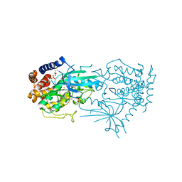
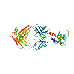
 | | Structure of the ternary complex between Netrin-1, Repulsive-Guidance Molecule-B (RGMB) and Neogenin | | 分子名称: | 1,3,4,6-tetra-O-sulfo-beta-D-fructofuranose-(2-1)-2,3,4,6-tetra-O-sulfonato-alpha-D-glucopyranose, 2-acetamido-2-deoxy-beta-D-glucopyranose, CALCIUM ION, ... | | 著者 | Robinson, R.A, Griffiths, S.C, van de Haar, L.L, Malinauskas, T, van Battum, E.Y, Zelina, P, Schwab, R.A, Karia, D, Malinauskaite, L, Brignani, S, van den Munkhof, M, Dudukcu, O, De Ruiter, A.A, Van den Heuvel, D.M.A, Bishop, B, Elegheert, J, Aricescu, A.R, Pasterkamp, R.J, Siebold, C. | | 登録日 | 2021-02-02 | | 公開日 | 2021-03-31 | | 最終更新日 | 2024-01-31 | | 実験手法 | X-RAY DIFFRACTION (3.25 Å) | | 主引用文献 | Simultaneous binding of Guidance Cues NET1 and RGM blocks extracellular NEO1 signaling.
Cell, 184, 2021
|
|
5E2U
 | |
5E2T
 | |
5E2V
 | |
5NGB
 | |
5E2W
 | |
4MA3
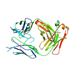
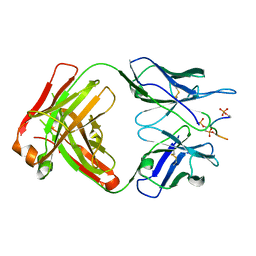
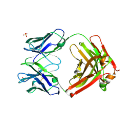
 | | Crystal structure of anti-hinge rabbit antibody C2095 | | 分子名称: | ACETATE ION, C2095 heavy chain, C2095 light chain, ... | | 著者 | Malia, T, Teplyakov, A, Gilliland, G.L. | | 登録日 | 2013-08-15 | | 公開日 | 2014-03-26 | | 最終更新日 | 2023-09-20 | | 実験手法 | X-RAY DIFFRACTION (2 Å) | | 主引用文献 | Structure and specificity of an antibody targeting a proteolytically cleaved IgG hinge.
Proteins, 82, 2014
|
|
8UDC
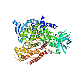
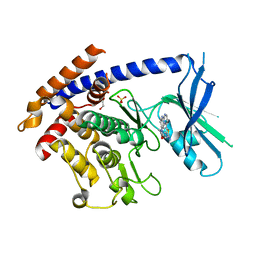 | | Crystal structure of TcPINK1 in complex with CYC116 | | 分子名称: | (4P)-4-(2-amino-4-methyl-1,3-thiazol-5-yl)-N-[4-(morpholin-4-yl)phenyl]pyrimidin-2-amine, DI(HYDROXYETHYL)ETHER, SULFATE ION, ... | | 著者 | Veyron, S, Rasool, S, Trempe, J.F. | | 登録日 | 2023-09-28 | | 公開日 | 2024-04-17 | | 実験手法 | X-RAY DIFFRACTION (3.1 Å) | | 主引用文献 | Structural Characterization of a small-molecule inhibitor of PINK1, a precursor tool compound for the study of Parkinson's disease
To Be Published
|
|
8UCT
 | | Crystal structure of TcPINK1 in complex with PRT | | 分子名称: | 2,3-DIHYDROXY-1,4-DITHIOBUTANE, 2-{[(1R,2S)-2-aminocyclohexyl]amino}-4-{[3-(2H-1,2,3-triazol-2-yl)phenyl]amino}pyrimidine-5-carboxamide, SULFATE ION, ... | | 著者 | Veyron, S, Rasool, S, Trempe, J.F. | | 登録日 | 2023-09-27 | | 公開日 | 2024-05-08 | | 実験手法 | X-RAY DIFFRACTION (2.93 Å) | | 主引用文献 | Characterization of a new family of PINK1 inhibitors
To Be Published
|
|
3L5W
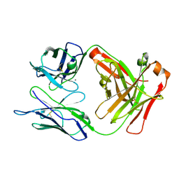
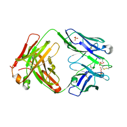
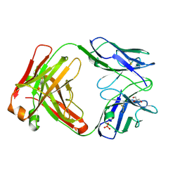
 | | Crystal structure of the complex between IL-13 and C836 FAB | | 分子名称: | C836 HEAVY CHAIN, C836 LIGHT CHAIN, GLYCEROL, ... | | 著者 | Teplyakov, A, Obmolova, G, Malia, T, Gilliland, G.L. | | 登録日 | 2009-12-22 | | 公開日 | 2010-04-14 | | 最終更新日 | 2023-09-06 | | 実験手法 | X-RAY DIFFRACTION (2 Å) | | 主引用文献 | Human framework adaptation of a mouse anti-human IL-13 antibody.
J.Mol.Biol., 398, 2010
|
|
3L7F
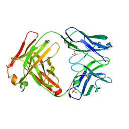
 | | Structure of IL-13 antibody H2L6, A humanized variant OF C836 | | 分子名称: | CALCIUM ION, H2L6 HEAVY CHAIN, H2L6 LIGHT CHAIN, ... | | 著者 | Teplyakov, A, Obmolova, G, Malia, T, Gilliland, G.L. | | 登録日 | 2009-12-28 | | 公開日 | 2010-11-10 | | 最終更新日 | 2023-09-06 | | 実験手法 | X-RAY DIFFRACTION (2.6 Å) | | 主引用文献 | Human framework adaptation of a mouse anti-human IL-13 antibody.
J.Mol.Biol., 398, 2010
|
|
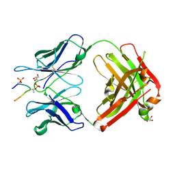
5I1E
 | | CRYSTAL STRUCTURE OF HUMAN GERMLINE ANTIBODY IGHV3-53/IGKV1-39 | | 分子名称: | FAB HEAVY CHAIN, FAB LIGHT CHAIN, GLYCEROL, ... | | 著者 | Teplyakov, A, Obmolova, G, Malia, T, Luo, J, Gilliland, G. | | 登録日 | 2016-02-05 | | 公開日 | 2016-06-08 | | 最終更新日 | 2016-08-03 | | 実験手法 | X-RAY DIFFRACTION (2.7 Å) | | 主引用文献 | Structural diversity in a human antibody germline library.
Mabs, 8, 2016
|
|
5I1H
 | | CRYSTAL STRUCTURE OF HUMAN GERMLINE ANTIBODY IGHV3-53/IGKV3-20 | | 分子名称: | FAB HEAVY CHAIN, FAB LIGHT CHAIN, SULFATE ION | | 著者 | Teplyakov, A, Obmolova, G, Malia, T, Luo, J, Gilliland, G. | | 登録日 | 2016-02-05 | | 公開日 | 2016-06-08 | | 最終更新日 | 2023-09-27 | | 実験手法 | X-RAY DIFFRACTION (2.222 Å) | | 主引用文献 | Structural diversity in a human antibody germline library.
Mabs, 8, 2016
|
|
5I18
 | | CRYSTAL STRUCTURE OF HUMAN GERMLINE ANTIBODY IGHV1-69/IGKV4-1 | | 分子名称: | FAB HEAVY CHAIN, FAB LIGHT CHAIN, GLYCEROL | | 著者 | Teplyakov, A, Obmolova, G, Malia, T, Luo, J, Gilliland, G. | | 登録日 | 2016-02-05 | | 公開日 | 2016-02-24 | | 最終更新日 | 2023-09-27 | | 実験手法 | X-RAY DIFFRACTION (1.92 Å) | | 主引用文献 | Structural diversity in a human antibody germline library.
Mabs, 8, 2016
|
|
5I1K
 | | CRYSTAL STRUCTURE OF HUMAN GERMLINE ANTIBODY IGHV5-51/IGKV3-20 | | 分子名称: | 2-[N-CYCLOHEXYLAMINO]ETHANE SULFONIC ACID, FAB HEAVY CHAIN, FAB LIGHT CHAIN, ... | | 著者 | Teplyakov, A, Obmolova, G, Malia, T, Luo, J, Gilliland, G. | | 登録日 | 2016-02-05 | | 公開日 | 2016-06-08 | | 最終更新日 | 2016-08-03 | | 実験手法 | X-RAY DIFFRACTION (1.65 Å) | | 主引用文献 | Structural diversity in a human antibody germline library.
Mabs, 8, 2016
|
|
5I16
 | | CRYSTAL STRUCTURE OF HUMAN GERMLINE ANTIBODY IGHV1-69/IGKV3-11 | | 分子名称: | 2-(N-MORPHOLINO)-ETHANESULFONIC ACID, FAB HEAVY CHAIN, FAB LIGHT CHAIN, ... | | 著者 | Teplyakov, A, Obmolova, G, Malia, T, Luo, J, Gilliland, G. | | 登録日 | 2016-02-05 | | 公開日 | 2016-06-08 | | 最終更新日 | 2023-09-27 | | 実験手法 | X-RAY DIFFRACTION (1.9 Å) | | 主引用文献 | Structural diversity in a human antibody germline library.
Mabs, 8, 2016
|
|
5I1C
 | | CRYSTAL STRUCTURE OF HUMAN GERMLINE ANTIBODY IGHV3-23/IGKV3-20 | | 分子名称: | 2-(N-MORPHOLINO)-ETHANESULFONIC ACID, CHLORIDE ION, FAB HEAVY CHAIN, ... | | 著者 | Teplyakov, A, Obmolova, G, Malia, T, Luo, J, Gilliland, G. | | 登録日 | 2016-02-05 | | 公開日 | 2016-06-08 | | 最終更新日 | 2023-09-27 | | 実験手法 | X-RAY DIFFRACTION (2.25 Å) | | 主引用文献 | Structural diversity in a human antibody germline library.
Mabs, 8, 2016
|
|
5I1A
 | | CRYSTAL STRUCTURE OF HUMAN GERMLINE ANTIBODY IGHV3-23/IGKV3-11 | | 分子名称: | ACETATE ION, CNTO388 HEAVY CHAIN, CNTO388 LIGHT CHAIN, ... | | 著者 | Teplyakov, A, Obmolova, G, Malia, T, Luo, J, Gilliland, G. | | 登録日 | 2016-02-05 | | 公開日 | 2016-06-08 | | 最終更新日 | 2023-09-27 | | 実験手法 | X-RAY DIFFRACTION (2 Å) | | 主引用文献 | Structural diversity in a human antibody germline library.
Mabs, 8, 2016
|
|
5I1G
 | | CRYSTAL STRUCTURE OF HUMAN GERMLINE ANTIBODY IGHV3-53/IGKV3-11 | | 分子名称: | FAB HEAVY CHAIN, FAB LIGHT CHAIN, SULFATE ION | | 著者 | Teplyakov, A, Obmolova, G, Malia, T, Luo, J, Gilliland, G. | | 登録日 | 2016-02-05 | | 公開日 | 2016-06-08 | | 最終更新日 | 2016-08-03 | | 実験手法 | X-RAY DIFFRACTION (2.3 Å) | | 主引用文献 | Structural diversity in a human antibody germline library.
Mabs, 8, 2016
|
|
5I19
 | | CRYSTAL STRUCTURE OF HUMAN GERMLINE ANTIBODY IGHV3-23/IGKV1-39 | | 分子名称: | FAB HEAVY CHAIN, FAB LIGHT CHAIN | | 著者 | Teplyakov, A, Obmolova, G, Malia, T, Luo, J, Gilliland, G. | | 登録日 | 2016-02-05 | | 公開日 | 2016-06-08 | | 最終更新日 | 2023-09-27 | | 実験手法 | X-RAY DIFFRACTION (2.8 Å) | | 主引用文献 | Structural diversity in a human antibody germline library.
Mabs, 8, 2016
|
|
5I1J
 | | CRYSTAL STRUCTURE OF HUMAN GERMLINE ANTIBODY IGHV5-51/IGKV3-11 | | 分子名称: | FAB HEAVY CHAIN, FAB LIGHT CHAIN, GLYCEROL | | 著者 | Teplyakov, A, Obmolova, G, Malia, T, Luo, J, Gilliland, G. | | 登録日 | 2016-02-05 | | 公開日 | 2016-06-08 | | 最終更新日 | 2016-08-03 | | 実験手法 | X-RAY DIFFRACTION (2.5 Å) | | 主引用文献 | Structural diversity in a human antibody germline library.
Mabs, 8, 2016
|
|
3L7E
 | | Crystal structure of ANTI-IL-13 antibody C836 | | 分子名称: | ACETATE ION, C836 HEAVY CHAIN, C836 LIGHT CHAIN, ... | | 著者 | Teplyakov, A, Obmolova, G, Malia, T, Gilliland, G.L. | | 登録日 | 2009-12-28 | | 公開日 | 2010-11-10 | | 最終更新日 | 2023-09-06 | | 実験手法 | X-RAY DIFFRACTION (2.5 Å) | | 主引用文献 | Antigen recognition by antibody C836 through adjustment of VL/VH packing
Acta Crystallogr.,Sect.F, 67, 2011
|
|
2WNO
 | | X-ray Structure of CUB_C domain from TSG-6 | | 分子名称: | CALCIUM ION, COBALT (II) ION, TUMOR NECROSIS FACTOR-INDUCIBLE GENE 6 PROTEIN | | 著者 | Briggs, D.C, Day, A.J. | | 登録日 | 2009-07-13 | | 公開日 | 2010-09-01 | | 最終更新日 | 2023-12-13 | | 実験手法 | X-RAY DIFFRACTION (2.3 Å) | | 主引用文献 | Metal Ion-Dependent Heavy Chain Transfer Activity of Tsg-6 Mediates Assembly of the Cumulus-Oocyte Matrix.
J.Biol.Chem., 290, 2015
|
|
1YQA
 | |
5I17
 | | CRYSTAL STRUCTURE OF HUMAN GERMLINE ANTIBODY IGHV1-69/IGKV3-20 | | 分子名称: | FAB HEAVY CHAIN, FAB LIGHT CHAIN, SULFATE ION | | 著者 | Teplyakov, A, Obmolova, G, Malia, T, Luo, J, Gilliland, G. | | 登録日 | 2016-02-05 | | 公開日 | 2016-06-08 | | 最終更新日 | 2016-08-03 | | 実験手法 | X-RAY DIFFRACTION (3.3 Å) | | 主引用文献 | Structural diversity in a human antibody germline library.
Mabs, 8, 2016
|
|


