3H6Q
 
 | | Macrocypin, a beta-trefoil cysteine protease inhibitor | | 分子名称: | Macrocypin 1a | | 著者 | Renko, M, Sabotic, J, Brzin, J, Turk, D. | | 登録日 | 2009-04-23 | | 公開日 | 2009-10-20 | | 最終更新日 | 2024-02-21 | | 実験手法 | X-RAY DIFFRACTION (1.643 Å) | | 主引用文献 | Versatile loops in mycocypins inhibit three protease families.
J.Biol.Chem., 285, 2010
|
|
2LAK
 | | Solution NMR structure of the AHSA1-like protein RHE_CH02687 (1-152) from Rhizobium etli, Northeast Structural Genomics Consortium Target ReR242 | | 分子名称: | AHSA1-like protein RHE_CH02687 | | 著者 | Yang, Y, Ramelot, T.A, Cort, J.R, Wang, D, Ciccosanti, C, Janjua, H, Nair, R, Rost, B, Acton, T.B, Xiao, R, Everett, J.K, Montelione, G.T, Kennedy, M.A, Northeast Structural Genomics Consortium (NESG) | | 登録日 | 2011-03-16 | | 公開日 | 2011-04-13 | | 最終更新日 | 2024-05-15 | | 実験手法 | SOLUTION NMR | | 主引用文献 | Solution NMR structure of the AHSA1-like protein RHE_CH02687 (1-152) from Rhizobium etli, Northeast Structural Genomics Consortium Target ReR242
To be Published
|
|
3VBS
 | | Crystal structure of human Enterovirus 71 | | 分子名称: | Genome Polyprotein, capsid protein VP1, capsid protein VP2, ... | | 著者 | Wang, X, Peng, W, Ren, J, Hu, Z, Xu, J, Lou, Z, Li, X, Yin, W, Shen, X, Porta, C, Walter, T.S, Evans, G, Axford, D, Owen, R, Rowlands, D.J, Wang, J, Stuart, D.I, Fry, E.E, Rao, Z. | | 登録日 | 2012-01-02 | | 公開日 | 2012-02-29 | | 最終更新日 | 2023-09-13 | | 実験手法 | X-RAY DIFFRACTION (3 Å) | | 主引用文献 | A sensor-adaptor mechanism for enterovirus uncoating from structures of EV71.
Nat.Struct.Mol.Biol., 19, 2012
|
|
6RRH
 | | GOLGI ALPHA-MANNOSIDASE II | | 分子名称: | 1,2-ETHANEDIOL, Alpha-mannosidase 2, ZINC ION | | 著者 | Armstrong, Z, Lahav, D, Johnson, R, Kuo, C.L, Beenakker, T.J.M, de Boer, C, Wong, C.S, van Rijssel, E.R, Debets, M, Geurink, P.P, Ovaa, H, van der Stelt, M, Codee, J.D.C, Aerts, J.M.F.G, Wu, L, Overkleeft, H.S, Davies, G.J. | | 登録日 | 2019-05-18 | | 公開日 | 2020-07-08 | | 最終更新日 | 2024-01-24 | | 実験手法 | X-RAY DIFFRACTION (2.07 Å) | | 主引用文献 | Manno- epi -cyclophellitols Enable Activity-Based Protein Profiling of Human alpha-Mannosidases and Discovery of New Golgi Mannosidase II Inhibitors.
J.Am.Chem.Soc., 142, 2020
|
|
3VCE
 | | Thaumatin by LB based Hanging Drop Vapour Diffusion after 18.1 MGy X-Ray dose at ESRF ID29 beamline (Best Case) | | 分子名称: | GLYCEROL, Thaumatin I | | 著者 | Belmonte, L, Pechkova, E, Scudieri, D, Nicolini, C. | | 登録日 | 2012-01-04 | | 公開日 | 2012-11-21 | | 最終更新日 | 2023-09-13 | | 実験手法 | X-RAY DIFFRACTION (2.3 Å) | | 主引用文献 | Langmuir-blodgett nanotemplate and radiation resistance in protein crystals: state of the art.
Crit Rev Eukaryot Gene Expr, 22, 2012
|
|
3VCJ
 | | Thaumatin by LB Hanging Drop Vapour Diffusion after 9.05 MGy X-Ray dose at ESRF ID29 beamline (Best Case) | | 分子名称: | GLYCEROL, Thaumatin I | | 著者 | Belmonte, L, Pechkova, E, Scudieri, D, Nicolini, C. | | 登録日 | 2012-01-04 | | 公開日 | 2012-11-21 | | 最終更新日 | 2023-09-13 | | 実験手法 | X-RAY DIFFRACTION (2.3 Å) | | 主引用文献 | Langmuir-blodgett nanotemplate and radiation resistance in protein crystals: state of the art.
Crit Rev Eukaryot Gene Expr, 22, 2012
|
|
2L06
 | | Solution NMR structure of the PBS linker polypeptide domain (fragment 254-400) of phycobilisome linker protein ApcE from Synechocystis sp. PCC 6803. Northeast Structural Genomics Consortium Target SgR209C | | 分子名称: | Phycobilisome LCM core-membrane linker polypeptide | | 著者 | Ramelot, T.A, Yang, Y, Cort, J.R, Hamilton, K, Ciccosanti, C, Lee, D, Acton, T.B, Xiao, R, Everett, J.K, Montelione, G.T, Kennedy, M.A, Northeast Structural Genomics Consortium (NESG) | | 登録日 | 2010-06-30 | | 公開日 | 2010-08-25 | | 最終更新日 | 2024-05-15 | | 実験手法 | SOLUTION NMR | | 主引用文献 | Solution NMR structure of the PBS linker polypeptide domain of phycobilisome
linker protein apcE from Synechocystis sp. Northeast Structural Genomics Consortium
Target SgR209C
To be Published
|
|
2KZV
 | | Solution NMR structure of CV_0373(175-257) protein from Chromobacterium violaceum, Northeast Structural Genomics Consortium Target CvR118A | | 分子名称: | Uncharacterized protein | | 著者 | Yang, Y, Ramelot, T.A, Wang, D, Ciccosanti, C, Mao, L, Janjua, H, Acton, T.B, Xiao, R, Everett, J.K, Montelione, G.T, Kennedy, M.A, Northeast Structural Genomics Consortium (NESG) | | 登録日 | 2010-06-25 | | 公開日 | 2010-08-25 | | 最終更新日 | 2024-05-15 | | 実験手法 | SOLUTION NMR | | 主引用文献 | Solution NMR structure of CV_0373(175-257) protein from Chromobacterium violaceum, Northeast Structural Genomics Consortium Target CvR118A.
To be Published
|
|
2LME
 | | Solid-state NMR structure of the membrane anchor domain of the trimeric autotransporter YadA | | 分子名称: | Adhesin yadA | | 著者 | Shahid, S.A, Bardiaux, B, Franks, W.T, Habeck, M, Linke, D, van Rossum, B. | | 登録日 | 2011-11-30 | | 公開日 | 2012-11-07 | | 最終更新日 | 2024-05-15 | | 実験手法 | SOLID-STATE NMR | | 主引用文献 | Membrane-protein structure determination by solid-state NMR spectroscopy of microcrystals.
Nat.Methods, 9, 2012
|
|
7YL7
 | | Structure of hIAPP-TF-type3 | | 分子名称: | Islet amyloid polypeptide | | 著者 | Li, D, Zhang, X. | | 登録日 | 2022-07-25 | | 公開日 | 2022-12-28 | | 最終更新日 | 2024-05-08 | | 実験手法 | ELECTRON MICROSCOPY (3.3 Å) | | 主引用文献 | A new polymorphism of human amylin fibrils with similar protofilaments and a conserved core.
Iscience, 25, 2022
|
|
3GOP
 | |
3HAR
 | |
3D9B
 | | Symmetric structure of E. coli AcrB | | 分子名称: | Acriflavine resistance protein B, NICKEL (II) ION | | 著者 | Veesler, D, Blangy, S, Cambillau, C, Sciara, G. | | 登録日 | 2008-05-27 | | 公開日 | 2008-07-01 | | 最終更新日 | 2023-08-30 | | 実験手法 | X-RAY DIFFRACTION (3.42 Å) | | 主引用文献 | There is a baby in the bath water: AcrB contamination is a major problem in membrane-protein crystallization.
Acta Crystallogr.,Sect.F, 64, 2008
|
|
3VB8
 | | Crystal Structure of Engineered Protein, Northeast Structural Genomics Consortium Target OR43 | | 分子名称: | Engineered protein, SULFATE ION | | 著者 | Seetharaman, J, Su, M, Procko, E, Baker, D, Ciccosanti, C, Sahdev, S, Xiao, R, Everett, J.K, Acton, T.B, Montelione, G.T, Hunt, J.F, Tong, L, Northeast Structural Genomics Consortium (NESG) | | 登録日 | 2011-12-31 | | 公開日 | 2012-06-06 | | 最終更新日 | 2023-09-13 | | 実験手法 | X-RAY DIFFRACTION (2.9 Å) | | 主引用文献 | Computational design of a protein-based enzyme inhibitor.
J.Mol.Biol., 425, 2013
|
|
3HC4
 | | BHA10 IgG1 Fab quadruple mutant variant - antibody directed at human LTBR | | 分子名称: | ACETATE ION, IMMUNOGLOBULIN IGG1 FAB, HEAVY CHAIN, ... | | 著者 | Arndt, J.W, Jordan, J.L, Lugovskoy, A, Wang, D. | | 登録日 | 2009-05-05 | | 公開日 | 2009-08-04 | | 最終更新日 | 2023-09-06 | | 実験手法 | X-RAY DIFFRACTION (1.62 Å) | | 主引用文献 | Structural understanding of stabilization patterns in engineered bispecific Ig-like antibody molecules
Proteins, 77, 2009
|
|
3VCD
 | | Computationally Designed Self-assembling Octahedral Cage protein, O333, Crystallized in space group R32 | | 分子名称: | CHLORIDE ION, Propanediol utilization polyhedral body protein PduT, SULFATE ION | | 著者 | Sawaya, M.R, King, N.P, Sheffler, W, Baker, D, Yeates, T.O. | | 登録日 | 2012-01-03 | | 公開日 | 2012-06-06 | | 最終更新日 | 2024-02-28 | | 実験手法 | X-RAY DIFFRACTION (2.35 Å) | | 主引用文献 | Computational design of self-assembling protein nanomaterials with atomic level accuracy.
Science, 336, 2012
|
|
3VGP
 | | Crystal structure of the C-terminal globular domain of oligosaccharyltransferase (AF_0329) from Archaeoglobus fulgidus | | 分子名称: | Transmembrane oligosaccharyl transferase, putative | | 著者 | Matsumoto, S, Igura, M, Nyirenda, J, Yuzawa, S, Noda, N.N, Inagaki, F, Kohda, D. | | 登録日 | 2011-08-18 | | 公開日 | 2012-07-04 | | 実験手法 | X-RAY DIFFRACTION (1.75 Å) | | 主引用文献 | Crystal Structure of the C-Terminal Globular Domain of Oligosaccharyltransferase from Archaeoglobus fulgidus at 1.75 A Resolution
Biochemistry, 51, 2012
|
|
7YM0
 | | Lysoplasmalogen-specific phospholipase D (LyPls-PLD) with Ca2+ | | 分子名称: | CALCIUM ION, Lysoplasmalogenase | | 著者 | Yasutake, Y, Sakasegawa, S, Sugimori, D, Murayama, K. | | 登録日 | 2022-07-27 | | 公開日 | 2023-01-04 | | 実験手法 | X-RAY DIFFRACTION (2.91 Å) | | 主引用文献 | Structural basis for the substrate specificity switching of lysoplasmalogen-specific phospholipase D from Thermocrispum sp. RD004668.
Biosci.Biotechnol.Biochem., 87, 2022
|
|
1CIZ
 | | X-RAY STRUCTURE OF HUMAN STROMELYSIN CATALYTIC DOMAIN COMPLEXES WITH NON-PEPTIDE INHIBITORS: IMPLICATION FOR INHIBITOR SELECTIVITY | | 分子名称: | 3-(1H-INDOL-3-YL)-2-[4-(4-PHENYL-PIPERIDIN-1-YL)-BENZENESULFONYLAMINO]-PROPIONIC ACID, CALCIUM ION, PROTEIN (STROMELYSIN-1), ... | | 著者 | Pavlovsky, A.G, Williams, M.G, Ye, Q.-Z, Ortwine, D.F, Purchase II, C.F, White, A.D, Dhanaraj, V, Roth, B.D, Johnson, L.L, Hupe, D, Humblet, C, Blundell, T.L. | | 登録日 | 1999-04-06 | | 公開日 | 1999-09-01 | | 最終更新日 | 2023-08-09 | | 実験手法 | X-RAY DIFFRACTION (1.64 Å) | | 主引用文献 | X-ray structure of human stromelysin catalytic domain complexed with nonpeptide inhibitors: implications for inhibitor selectivity.
Protein Sci., 8, 1999
|
|
3GRB
 | | Crystal structure of the F87M/L110M mutant of human transthyretin at pH 6.5 | | 分子名称: | ACETATE ION, GLYCEROL, Transthyretin, ... | | 著者 | Palmieri, L.C, Freire, J.B.B, Foguel, D, Lima, L.M.T.R. | | 登録日 | 2009-03-25 | | 公開日 | 2010-04-07 | | 最終更新日 | 2023-09-06 | | 実験手法 | X-RAY DIFFRACTION (1.75 Å) | | 主引用文献 | Novel Zn2+-binding sites in human transthyretin: implications for amyloidogenesis and retinol-binding protein recognition.
J.Biol.Chem., 285, 2010
|
|
3VJH
 | | Human PPAR GAMMA ligand binding domain in complex with JKPL35 | | 分子名称: | (2S)-2-[4-methoxy-3-({[4-(trifluoromethyl)benzoyl]amino}methyl)benzyl]pentanoic acid, Peroxisome proliferator-activated receptor gamma | | 著者 | Tomioka, D, Kuwabara, N, Hashimoto, H, Sato, M, Shimizu, T. | | 登録日 | 2011-10-20 | | 公開日 | 2012-08-29 | | 最終更新日 | 2023-11-08 | | 実験手法 | X-RAY DIFFRACTION (2.22 Å) | | 主引用文献 | Peroxisome proliferator-activated receptors (PPARs) have multiple binding points that accommodate ligands in various conformations: phenylpropanoic acid-type PPAR ligands bind to PPAR in different conformations, depending on the subtype.
J.Med.Chem., 55, 2012
|
|
7YYO
 | | Cryo-EM structure of an a-carboxysome RuBisCO enzyme at 2.9 A resolution | | 分子名称: | 2-CARBOXYARABINITOL-1,5-DIPHOSPHATE, MAGNESIUM ION, Ribulose bisphosphate carboxylase large chain, ... | | 著者 | Mann, D, Evans, S.L, Bergeron, J.R.C. | | 登録日 | 2022-02-18 | | 公開日 | 2023-01-25 | | 最終更新日 | 2023-06-14 | | 実験手法 | ELECTRON MICROSCOPY (2.87 Å) | | 主引用文献 | Single-particle cryo-EM analysis of the shell architecture and internal organization of an intact alpha-carboxysome.
Structure, 31, 2023
|
|
2L8O
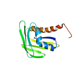 | | Solution structure of Chr148 from Cytophaga hutchinsonii, Northeast Structural Genomics Consortium Target Chr148 | | 分子名称: | Activator of Hsp90 ATPase homologue 1-like C-terminal domain-containing protein | | 著者 | Liu, Y, Lee, D, Ciccosanti, C, Nair, L, Rost, B, Acton, T, Xiao, R, Everett, J, Montelione, G, Prestegard, J, Northeast Structural Genomics Consortium (NESG) | | 登録日 | 2011-01-21 | | 公開日 | 2011-03-02 | | 最終更新日 | 2024-05-15 | | 実験手法 | SOLUTION NMR | | 主引用文献 | Solution structure of Chr148 from Cytophaga hutchinsonii. Northeast Structural Genomics Consortium Target Chr148
To be Published
|
|
3GRO
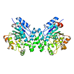 | | Human palmitoyl-protein thioesterase 1 | | 分子名称: | Palmitoyl-protein thioesterase 1, UNKNOWN ATOM OR ION | | 著者 | Dobrovetsky, E, Seitova, A, Tong, Y, Tempel, W, Dong, A, Arrowsmith, C.H, Edwards, A.M, Bountra, C, Weigelt, J, Bochkarev, A, Cossar, D, Park, H, Structural Genomics Consortium (SGC) | | 登録日 | 2009-03-26 | | 公開日 | 2009-04-07 | | 最終更新日 | 2023-11-22 | | 実験手法 | X-RAY DIFFRACTION (2.53 Å) | | 主引用文献 | Human palmitoyl-protein thioesterase 1
To be Published
|
|
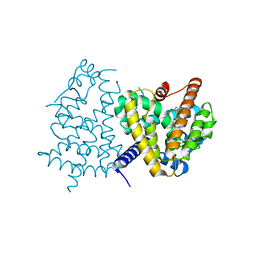
6RKA
 | | Inter-dimeric interface controls function and stability of S-methionine adenosyltransferase from U. urealiticum | | 分子名称: | ADENOSINE-5'-TRIPHOSPHATE, Methionine adenosyltransferase, PHOSPHATE ION, ... | | 著者 | Shahar, A, Zarivach, R, Bershtein, S, Kleiner, D, Shmulevich, F. | | 登録日 | 2019-04-30 | | 公開日 | 2019-09-25 | | 最終更新日 | 2024-01-24 | | 実験手法 | X-RAY DIFFRACTION (2.5 Å) | | 主引用文献 | The interdimeric interface controls function and stability of Ureaplasma urealiticum methionine S-adenosyltransferase.
J.Mol.Biol., 431, 2019
|
|


