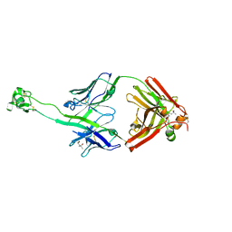
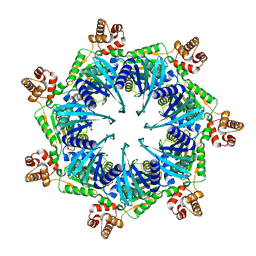
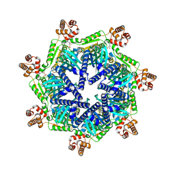
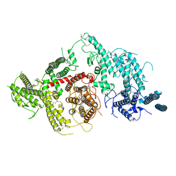
6JIV
 
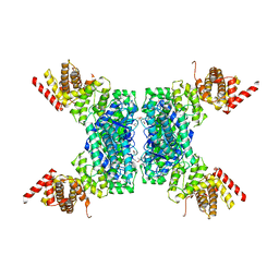 | | SspE crystal structure | | 分子名称: | SspE protein | | 著者 | Bing, Y.Z, Yang, H.G. | | 登録日 | 2019-02-23 | | 公開日 | 2020-03-25 | | 最終更新日 | 2020-07-08 | | 実験手法 | X-RAY DIFFRACTION (3.31 Å) | | 主引用文献 | SspABCD-SspE is a phosphorothioation-sensing bacterial defence system with broad anti-phage activities.
Nat Microbiol, 5, 2020
|
|
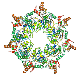
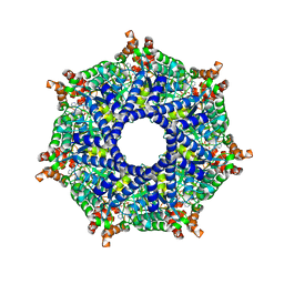
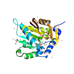
7KD7
 | |
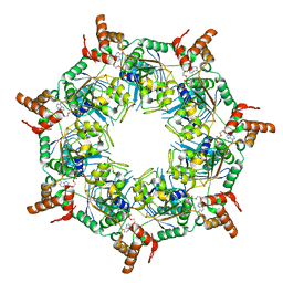
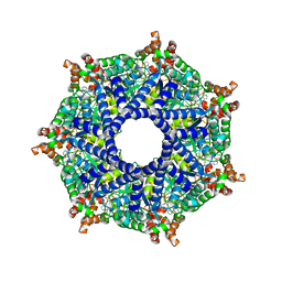
6JUF
 | | SspB crystal structure | | 分子名称: | SspB protein | | 著者 | Liqiong, L, Yubing, Z. | | 登録日 | 2019-04-13 | | 公開日 | 2020-03-25 | | 最終更新日 | 2024-03-27 | | 実験手法 | X-RAY DIFFRACTION (1.587 Å) | | 主引用文献 | SspABCD-SspE is a phosphorothioation-sensing bacterial defence system with broad anti-phage activities.
Nat Microbiol, 5, 2020
|
|
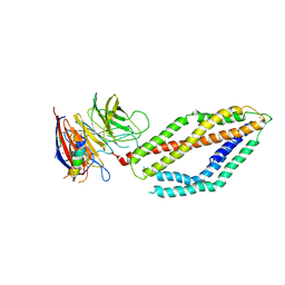
7MFU
 | | Crystal structure of synthetic nanobody (Sb14+Sb68) complexes with SARS-CoV-2 receptor binding domain | | 分子名称: | 1,2-ETHANEDIOL, GLYCEROL, Spike protein S1, ... | | 著者 | Jiang, J, Ahmad, J, Natarajan, K, Boyd, L.F, Margulies, D.H. | | 登録日 | 2021-04-11 | | 公開日 | 2021-06-02 | | 最終更新日 | 2023-10-18 | | 実験手法 | X-RAY DIFFRACTION (1.7 Å) | | 主引用文献 | Structures of synthetic nanobody-SARS-CoV-2 receptor-binding domain complexes reveal distinct sites of interaction.
J.Biol.Chem., 297, 2021
|
|
7MFV
 | | Crystal structure of synthetic nanobody (Sb16) | | 分子名称: | 1,2-ETHANEDIOL, Synthetic Nanobody #16 (Sb16) | | 著者 | Jiang, J, Ahmad, J, Natarajan, K, Boyd, L.F, Margulies, D.H. | | 登録日 | 2021-04-11 | | 公開日 | 2021-06-02 | | 最終更新日 | 2023-10-18 | | 実験手法 | X-RAY DIFFRACTION (1.9 Å) | | 主引用文献 | Structures of synthetic nanobody-SARS-CoV-2 receptor-binding domain complexes reveal distinct sites of interaction.
J.Biol.Chem., 297, 2021
|
|
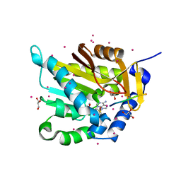
6OO0
 | |
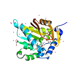
6OPA
 | |
6PW6
 | | The HIV-1 Envelope Glycoprotein Clone BG505 SOSIP.664 in Complex with Three Copies of the Bovine Broadly Neutralizing Antibody, NC-Cow1, Fragment Antigen Binding Domain | | 分子名称: | 2-acetamido-2-deoxy-beta-D-glucopyranose, 2-acetamido-2-deoxy-beta-D-glucopyranose-(1-4)-2-acetamido-2-deoxy-beta-D-glucopyranose, Broadly Neutralizing Antibody NC-Cow1 Heavy Chain, ... | | 著者 | Berndsen, Z.T, Ward, A.B. | | 登録日 | 2019-07-22 | | 公開日 | 2020-06-24 | | 最終更新日 | 2020-07-29 | | 実験手法 | ELECTRON MICROSCOPY (4.5 Å) | | 主引用文献 | Structural basis of broad HIV neutralization by a vaccine-induced cow antibody.
Sci Adv, 6, 2020
|
|
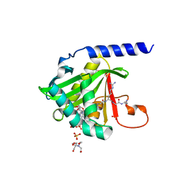
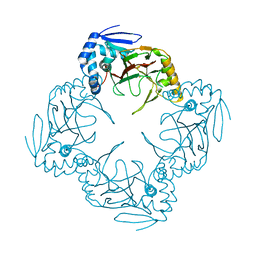
7U1M
 | | Crystal structure of NTMT1 in complex with compound YD206 | | 分子名称: | (1R,3S,4R)-1-azabicyclo[2.2.2]octan-3-yl {2-[2-(4-fluorophenyl)-1,3-thiazol-4-yl]propan-2-yl}carbamate, N-terminal Xaa-Pro-Lys N-methyltransferase 1, S-ADENOSYL-L-HOMOCYSTEINE | | 著者 | Yadav, R, Noinaj, N. | | 登録日 | 2022-02-21 | | 公開日 | 2022-12-14 | | 最終更新日 | 2023-10-25 | | 実験手法 | X-RAY DIFFRACTION (3.17 Å) | | 主引用文献 | Venglustat Inhibits Protein N-Terminal Methyltransferase 1 in a Substrate-Competitive Manner.
J.Med.Chem., 65, 2022
|
|
1KM8
 | | The Structure of a Cytotoxic Ribonuclease From the Oocyte of Rana Catesbeiana (Bullfrog) | | 分子名称: | PHOSPHATE ION, RIBONUCLEASE, OOCYTES | | 著者 | Chern, S.-S, Musayev, F.N, Amiraslanov, I.R, Liao, Y.-D, Liaw, Y.-C. | | 登録日 | 2001-12-14 | | 公開日 | 2003-09-09 | | 最終更新日 | 2023-08-16 | | 実験手法 | X-RAY DIFFRACTION (1.9 Å) | | 主引用文献 | The Structure of a Cytotoxic Ribonuclease From the Oocyte of Rana Catesbeiana (Bullfrog)
To be Published
|
|
1KM9
 | | The Structure of a Cytotoxic Ribonuclease From the Oocyte of Rana Catesbeiana (Bullfrog) | | 分子名称: | PHOSPHATE ION, RIBONUCLEASE, OOCYTES | | 著者 | Chern, S.-S, Musayev, F.N, Amiraslanov, I.R, Liao, Y.-D, Liaw, Y.-C. | | 登録日 | 2001-12-14 | | 公開日 | 2003-09-09 | | 最終更新日 | 2023-08-16 | | 実験手法 | X-RAY DIFFRACTION (1.96 Å) | | 主引用文献 | The Structure of a Cytotoxic Ribonuclease From the Oocyte of Rana Catesbeiana (Bullfrog)
To be Published
|
|
8TBY
 | | Apo Bcs1, unsymmetrized | | 分子名称: | Mitochondrial chaperone BCS1 | | 著者 | Zhan, J, Xia, D. | | 登録日 | 2023-06-29 | | 公開日 | 2024-06-05 | | 最終更新日 | 2024-06-12 | | 実験手法 | ELECTRON MICROSCOPY (4.4 Å) | | 主引用文献 | Conformations of Bcs1L undergoing ATP hydrolysis suggest a concerted translocation mechanism for folded iron-sulfur protein substrate.
Nat Commun, 15, 2024
|
|
8T5U
 | | ATP-1 state of Bcs1 (C7 symmetrized) | | 分子名称: | ADENOSINE-5'-TRIPHOSPHATE, MAGNESIUM ION, Mitochondrial chaperone BCS1 | | 著者 | Zhan, J, Xia, D. | | 登録日 | 2023-06-14 | | 公開日 | 2024-06-05 | | 最終更新日 | 2024-06-12 | | 実験手法 | ELECTRON MICROSCOPY (3.13 Å) | | 主引用文献 | Conformations of Bcs1L undergoing ATP hydrolysis suggest a concerted translocation mechanism for folded iron-sulfur protein substrate.
Nat Commun, 15, 2024
|
|
8TI0
 | | ATP-1 state of Bcs1 (unsymmetrized) | | 分子名称: | ADENOSINE-5'-TRIPHOSPHATE, MAGNESIUM ION, Mitochondrial chaperone BCS1 | | 著者 | Zhan, J, Xia, D. | | 登録日 | 2023-07-18 | | 公開日 | 2024-06-05 | | 最終更新日 | 2024-06-12 | | 実験手法 | ELECTRON MICROSCOPY (3.77 Å) | | 主引用文献 | Conformations of Bcs1L undergoing ATP hydrolysis suggest a concerted translocation mechanism for folded iron-sulfur protein substrate.
Nat Commun, 15, 2024
|
|
8T14
 | | ADP-bound Bcs1 (C7 symmetrized) | | 分子名称: | ADENOSINE-5'-DIPHOSPHATE, Mitochondrial chaperone BCS1 | | 著者 | Zhan, J, Xia, D. | | 登録日 | 2023-06-01 | | 公開日 | 2024-06-05 | | 最終更新日 | 2024-06-12 | | 実験手法 | ELECTRON MICROSCOPY (3.18 Å) | | 主引用文献 | Conformations of Bcs1L undergoing ATP hydrolysis suggest a concerted translocation mechanism for folded iron-sulfur protein substrate.
Nat Commun, 15, 2024
|
|
8T7U
 | | ADP-bound Bcs1 (unsymmetrized) | | 分子名称: | ADENOSINE-5'-DIPHOSPHATE, Mitochondrial chaperone BCS1 | | 著者 | Zhan, J, Xia, D. | | 登録日 | 2023-06-21 | | 公開日 | 2024-06-05 | | 最終更新日 | 2024-06-12 | | 実験手法 | ELECTRON MICROSCOPY (3.74 Å) | | 主引用文献 | Conformations of Bcs1L undergoing ATP hydrolysis suggest a concerted translocation mechanism for folded iron-sulfur protein substrate.
Nat Commun, 15, 2024
|
|
8TP1
 | | ATP-2 state of Bcs1 (C7 symmetrized) | | 分子名称: | ADENOSINE-5'-TRIPHOSPHATE, MAGNESIUM ION, Mitochondrial chaperone BCS1 | | 著者 | Zhan, J, Xia, D. | | 登録日 | 2023-08-04 | | 公開日 | 2024-06-05 | | 最終更新日 | 2024-06-12 | | 実験手法 | ELECTRON MICROSCOPY (3.02 Å) | | 主引用文献 | Conformations of Bcs1L undergoing ATP hydrolysis suggest a concerted translocation mechanism for folded iron-sulfur protein substrate.
Nat Commun, 15, 2024
|
|
8TPL
 | | ATP-2 state of Bcs1 (unsymmetrized) | | 分子名称: | ADENOSINE-5'-TRIPHOSPHATE, MAGNESIUM ION, Mitochondrial chaperone BCS1 | | 著者 | Zhan, J, Xia, D. | | 登録日 | 2023-08-04 | | 公開日 | 2024-06-05 | | 最終更新日 | 2024-06-12 | | 実験手法 | ELECTRON MICROSCOPY (3.46 Å) | | 主引用文献 | Conformations of Bcs1L undergoing ATP hydrolysis suggest a concerted translocation mechanism for folded iron-sulfur protein substrate.
Nat Commun, 15, 2024
|
|
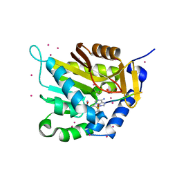
6MPV
 | |
5E2B
 | | Crystal structure of NTMT1 in complex with N-terminally methylated PPKRIA peptide | | 分子名称: | GLYCEROL, N-terminal Xaa-Pro-Lys N-methyltransferase 1, RCC1, ... | | 著者 | Dong, C, Tempel, W, Bountra, C, Arrowsmith, C.H, Edwards, A.M, Min, J, Structural Genomics Consortium (SGC) | | 登録日 | 2015-09-30 | | 公開日 | 2015-10-28 | | 最終更新日 | 2015-12-02 | | 実験手法 | X-RAY DIFFRACTION (1.95 Å) | | 主引用文献 | Structural basis for substrate recognition by the human N-terminal methyltransferase 1.
Genes Dev., 29, 2015
|
|
5E1B
 | | Crystal structure of NRMT1 in complex with SPKRIA peptide | | 分子名称: | GLYCEROL, N-terminal Xaa-Pro-Lys N-methyltransferase 1, RCC1, ... | | 著者 | Dong, C, Tempel, W, Bountra, C, Arrowsmith, C.H, Edwards, A.M, Min, J, Structural Genomics Consortium (SGC) | | 登録日 | 2015-09-29 | | 公開日 | 2015-10-28 | | 最終更新日 | 2023-09-27 | | 実験手法 | X-RAY DIFFRACTION (1.65 Å) | | 主引用文献 | Structural basis for substrate recognition by the human N-terminal methyltransferase 1.
Genes Dev., 29, 2015
|
|
5E1D
 | | NTMT1 in complex with YPKRIA peptide | | 分子名称: | GLYCEROL, N-terminal Xaa-Pro-Lys N-methyltransferase 1, RCC1, ... | | 著者 | Dong, C, Tempel, W, Bountra, C, Arrowsmith, C.H, Edwards, A.M, Min, J, Structural Genomics Consortium (SGC) | | 登録日 | 2015-09-29 | | 公開日 | 2015-10-28 | | 最終更新日 | 2024-03-06 | | 実験手法 | X-RAY DIFFRACTION (1.45 Å) | | 主引用文献 | Structural basis for substrate recognition by the human N-terminal methyltransferase 1.
Genes Dev., 29, 2015
|
|
5E1O
 | | Crystal structure of NTMT1 in complex with RPKRIA peptide | | 分子名称: | GLYCEROL, N-terminal Xaa-Pro-Lys N-methyltransferase 1, RCC1, ... | | 著者 | Dong, C, Tempel, W, Bountra, C, Arrowsmith, C.H, Edwards, A.M, Min, J, Structural Genomics Consortium (SGC) | | 登録日 | 2015-09-29 | | 公開日 | 2015-10-28 | | 最終更新日 | 2024-03-06 | | 実験手法 | X-RAY DIFFRACTION (2 Å) | | 主引用文献 | Structural basis for substrate recognition by the human N-terminal methyltransferase 1.
Genes Dev., 29, 2015
|
|
7JGD
 | |
1JR7
 | |


