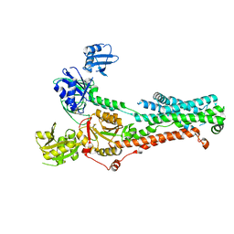
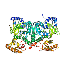
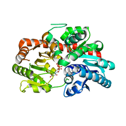
8WG5
 
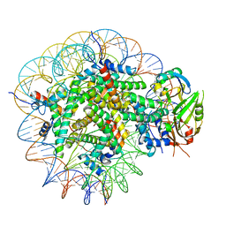 | | Cryo-EM structure of USP16 bound to H2AK119Ub nucleosome | | 分子名称: | DNA (147-MER), Histone H2A type 1-B/E, Histone H2B type 1-K, ... | | 著者 | Ai, H.S, He, Z.Z, Deng, Z.H, Liu, L. | | 登録日 | 2023-09-20 | | 公開日 | 2023-12-27 | | 最終更新日 | 2024-07-10 | | 実験手法 | ELECTRON MICROSCOPY (3.05 Å) | | 主引用文献 | Structural and mechanistic basis for nucleosomal H2AK119 deubiquitination by single-subunit deubiquitinase USP16.
Nat.Struct.Mol.Biol., 2024
|
|
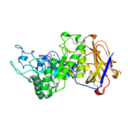
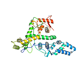
3VAX
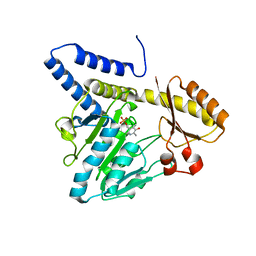 | | Crystal structure of DndA from streptomyces lividans | | 分子名称: | PYRIDOXAL-5'-PHOSPHATE, Putative uncharacterized protein dndA | | 著者 | Zhang, Z, Chen, F, Lin, K, Wu, G. | | 登録日 | 2011-12-30 | | 公開日 | 2013-01-02 | | 最終更新日 | 2024-03-20 | | 実験手法 | X-RAY DIFFRACTION (2.4 Å) | | 主引用文献 | Crystal structure of the cysteine desulfurase DndA from Streptomyces lividans which is involved in DNA phosphorothioation
Plos One, 7, 2012
|
|
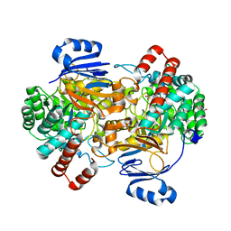
7SI6
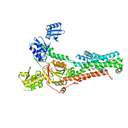 | | Structure of ATP7B in state 1 | | 分子名称: | MAGNESIUM ION, P-type Cu(+) transporter, TETRAFLUOROALUMINATE ION | | 著者 | Bitter, R.M, Oh, S.C, Hite, R.K, Yuan, P. | | 登録日 | 2021-10-12 | | 公開日 | 2022-03-16 | | 実験手法 | ELECTRON MICROSCOPY (3.32 Å) | | 主引用文献 | Structure of the Wilson disease copper transporter ATP7B.
Sci Adv, 8, 2022
|
|
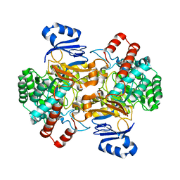
7SI7
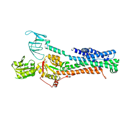 | | Structure of ATP7B in state 2 | | 分子名称: | MAGNESIUM ION, P-type Cu(+) transporter, TETRAFLUOROALUMINATE ION | | 著者 | Bitter, R.M, Oh, S.C, Hite, R.K, Yuan, P. | | 登録日 | 2021-10-12 | | 公開日 | 2022-03-16 | | 最終更新日 | 2024-06-05 | | 実験手法 | ELECTRON MICROSCOPY (3.49 Å) | | 主引用文献 | Structure of the Wilson disease copper transporter ATP7B.
Sci Adv, 8, 2022
|
|
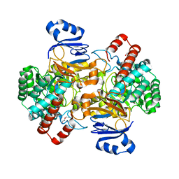
7SI3
 | | Consensus structure of ATP7B | | 分子名称: | MAGNESIUM ION, P-type Cu(+) transporter, TETRAFLUOROALUMINATE ION | | 著者 | Bitter, R.M, Oh, S.C, Hite, R.K, Yuan, P. | | 登録日 | 2021-10-12 | | 公開日 | 2022-03-16 | | 実験手法 | ELECTRON MICROSCOPY (3.19 Å) | | 主引用文献 | Structure of the Wilson disease copper transporter ATP7B.
Sci Adv, 8, 2022
|
|
5HBZ
 | |
5HC1
 | |
4DS8
 | | Complex structure of abscisic acid receptor PYL3-(+)-ABA-HAB1 in the presence of Mn2+ | | 分子名称: | (2Z,4E)-5-[(1S)-1-hydroxy-2,6,6-trimethyl-4-oxocyclohex-2-en-1-yl]-3-methylpenta-2,4-dienoic acid, Abscisic acid receptor PYL3, GLYCEROL, ... | | 著者 | Zhang, X, Zhang, Q, Wang, G, Chen, Z. | | 登録日 | 2012-02-18 | | 公開日 | 2012-06-06 | | 最終更新日 | 2023-11-08 | | 実験手法 | X-RAY DIFFRACTION (2.21 Å) | | 主引用文献 | Complex Structures of the Abscisic Acid Receptor PYL3/RCAR13 Reveal a Unique Regulatory Mechanism
Structure, 20, 2012
|
|
5EYI
 | | Structure of PRRSV apo-NSP11 at 2.16A | | 分子名称: | 2-(2-METHOXYETHOXY)ETHANOL, CHLORIDE ION, Non-structural protein 11, ... | | 著者 | Zhang, M.F, Chen, Z. | | 登録日 | 2015-11-25 | | 公開日 | 2016-10-12 | | 最終更新日 | 2024-03-20 | | 実験手法 | X-RAY DIFFRACTION (2.16 Å) | | 主引用文献 | Structural Biology of the Arterivirus nsp11 Endoribonucleases.
J. Virol., 91, 2017
|
|
7EYO
 | | Crystal structure of leech hyaluronidase | | 分子名称: | 2-acetamido-2-deoxy-beta-D-glucopyranose-(1-4)-2-acetamido-2-deoxy-beta-D-glucopyranose, GLYCEROL, Hyaluronoglucuronidase | | 著者 | Huang, H, Hou, X.D, Rao, Y.J, Kang, Z. | | 登録日 | 2021-05-31 | | 公開日 | 2022-05-25 | | 最終更新日 | 2023-11-29 | | 実験手法 | X-RAY DIFFRACTION (1.85 Å) | | 主引用文献 | Structure and cleavage pattern of a hyaluronate 3-glycanohydrolase in the glycoside hydrolase 79 family.
Carbohydr Polym, 277, 2022
|
|
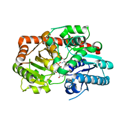
6SJ2
 | | Amidohydrolase, AHS with 3-HAA | | 分子名称: | 3-HYDROXYANTHRANILIC ACID, Amidohydrolase, GLYCEROL, ... | | 著者 | Naismith, J.H, Song, H. | | 登録日 | 2019-08-12 | | 公開日 | 2020-01-15 | | 最終更新日 | 2024-05-15 | | 実験手法 | X-RAY DIFFRACTION (1.25 Å) | | 主引用文献 | The Biosynthesis of the Benzoxazole in Nataxazole Proceeds via an Unstable Ester and has Synthetic Utility.
Angew.Chem.Int.Ed.Engl., 59, 2020
|
|
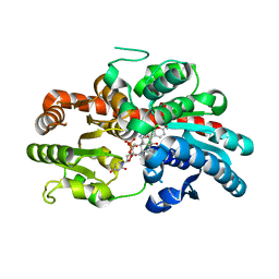
6SJ1
 | | Amidohydrolase, AHS | | 分子名称: | Amidohydrolase, ZINC ION | | 著者 | Naismith, J.H, Song, H. | | 登録日 | 2019-08-12 | | 公開日 | 2020-01-15 | | 最終更新日 | 2024-01-24 | | 実験手法 | X-RAY DIFFRACTION (2.06 Å) | | 主引用文献 | The Biosynthesis of the Benzoxazole in Nataxazole Proceeds via an Unstable Ester and has Synthetic Utility.
Angew.Chem.Int.Ed.Engl., 59, 2020
|
|
6SJ4
 | | Amidohydrolase, AHS with substrate analog | | 分子名称: | 1,2-ETHANEDIOL, 3-(3-hydroxyphenyl)carbonyloxybenzoic acid, Amidohydrolase, ... | | 著者 | Naismith, J.H, Song, H. | | 登録日 | 2019-08-12 | | 公開日 | 2020-01-15 | | 最終更新日 | 2024-01-24 | | 実験手法 | X-RAY DIFFRACTION (1.81 Å) | | 主引用文献 | The Biosynthesis of the Benzoxazole in Nataxazole Proceeds via an Unstable Ester and has Synthetic Utility.
Angew.Chem.Int.Ed.Engl., 59, 2020
|
|
6SIW
 | | PaaK family AMP-ligase with AMP | | 分子名称: | 1,2-ETHANEDIOL, 2-(2-(2-(2-(2-(2-ETHOXYETHOXY)ETHOXY)ETHOXY)ETHOXY)ETHOXY)ETHANOL, ADENOSINE MONOPHOSPHATE, ... | | 著者 | Naismith, J.H, Song, H. | | 登録日 | 2019-08-12 | | 公開日 | 2020-01-15 | | 最終更新日 | 2024-05-15 | | 実験手法 | X-RAY DIFFRACTION (1.96 Å) | | 主引用文献 | The Biosynthesis of the Benzoxazole in Nataxazole Proceeds via an Unstable Ester and has Synthetic Utility.
Angew.Chem.Int.Ed.Engl., 59, 2020
|
|
6SIZ
 | | PaaK family AMP-ligase with ANP and substrate | | 分子名称: | 1,2-ETHANEDIOL, 3-HYDROXYANTHRANILIC ACID, AMP-dependent synthetase and ligase, ... | | 著者 | Naismith, J.H, Song, H. | | 登録日 | 2019-08-12 | | 公開日 | 2020-01-15 | | 最終更新日 | 2024-01-24 | | 実験手法 | X-RAY DIFFRACTION (1.77 Å) | | 主引用文献 | The Biosynthesis of the Benzoxazole in Nataxazole Proceeds via an Unstable Ester and has Synthetic Utility.
Angew.Chem.Int.Ed.Engl., 59, 2020
|
|
6SJ3
 | | Amidohydrolase, AHS with 3-HBA | | 分子名称: | 1,2-ETHANEDIOL, 3-HYDROXYBENZOIC ACID, Amidohydrolase, ... | | 著者 | Naismith, J.H, Song, H. | | 登録日 | 2019-08-12 | | 公開日 | 2020-01-15 | | 最終更新日 | 2024-01-24 | | 実験手法 | X-RAY DIFFRACTION (1.17 Å) | | 主引用文献 | The Biosynthesis of the Benzoxazole in Nataxazole Proceeds via an Unstable Ester and has Synthetic Utility.
Angew.Chem.Int.Ed.Engl., 59, 2020
|
|
7ES4
 | | the crystral structure of DndH-C-domain | | 分子名称: | DNA phosphorothioation-dependent restriction protein DptH | | 著者 | Wu, D. | | 登録日 | 2021-05-08 | | 公開日 | 2022-05-11 | | 最終更新日 | 2022-12-21 | | 実験手法 | X-RAY DIFFRACTION (2.3 Å) | | 主引用文献 | The functional coupling between restriction and DNA phosphorothioate modification systems underlying the DndFGH restriction complex
Nat Catal, 2022
|
|
7EXX
 | | The structure of DndG | | 分子名称: | DNA phosphorothioation-dependent restriction protein DptG, SODIUM ION | | 著者 | Wu, D, Wang, L. | | 登録日 | 2021-05-28 | | 公開日 | 2022-06-01 | | 最終更新日 | 2022-12-21 | | 実験手法 | X-RAY DIFFRACTION (2.5 Å) | | 主引用文献 | The functional coupling between restriction and DNA phosphorothioate modification systems underlying the DndFGH restriction complex
Nat Catal, 2022
|
|
8HVR
 | |
8IND
 | | Crystal structure of UGT74AN3-UDP-RES | | 分子名称: | 2-AMINO-2-HYDROXYMETHYL-PROPANE-1,3-DIOL, 5-[(1R,2S,4R,6R,7R,10S,11S,14S,16R)-14-hydroxy-7,11-dimethyl-3-oxapentacyclo[8.8.0.02,4.02,7.011,16]octadecan-6-yl]pyran-2-one, Glycosyltransferase, ... | | 著者 | Huang, W. | | 登録日 | 2023-03-09 | | 公開日 | 2024-01-24 | | 実験手法 | X-RAY DIFFRACTION (1.85 Å) | | 主引用文献 | Substrate Promiscuity, Crystal Structure, and Application of a Plant UDP-Glycosyltransferase UGT74AN3
Acs Catalysis, 14, 2024
|
|
8INV
 | | Crystal structure of UGT74AN3-UDP-BUF | | 分子名称: | 2-AMINO-2-HYDROXYMETHYL-PROPANE-1,3-DIOL, Glycosyltransferase, URIDINE-5'-DIPHOSPHATE, ... | | 著者 | Long, F, Huang, W. | | 登録日 | 2023-03-10 | | 公開日 | 2024-01-24 | | 実験手法 | X-RAY DIFFRACTION (1.85 Å) | | 主引用文献 | Substrate Promiscuity, Crystal Structure, and Application of a Plant UDP-Glycosyltransferase UGT74AN3
Acs Catalysis, 14, 2024
|
|
8INA
 | | Crystal structure of UGT74AN3-UDP | | 分子名称: | GLYCEROL, Glycosyltransferase, URIDINE-5'-DIPHOSPHATE | | 著者 | Long, F, Huang, W. | | 登録日 | 2023-03-09 | | 公開日 | 2024-01-24 | | 実験手法 | X-RAY DIFFRACTION (1.86 Å) | | 主引用文献 | Substrate Promiscuity, Crystal Structure, and Application of a Plant UDP-Glycosyltransferase UGT74AN3
Acs Catalysis, 14, 2024
|
|
8INO
 | | Crystal structure of UGT74AN3 in complex UDP and PER | | 分子名称: | 2-AMINO-2-HYDROXYMETHYL-PROPANE-1,3-DIOL, 3-[(3S,5S,8S,9S,10R,13R,14S,17R)-10,13-dimethyl-3,5,14-tris(oxidanyl)-2,3,4,6,7,8,9,11,12,15,16,17-dodecahydro-1H-cyclopenta[a]phenanthren-17-yl]-2H-furan-5-one, Glycosyltransferase, ... | | 著者 | Long, F, Huang, W. | | 登録日 | 2023-03-10 | | 公開日 | 2024-01-24 | | 実験手法 | X-RAY DIFFRACTION (2.3 Å) | | 主引用文献 | Substrate Promiscuity, Crystal Structure, and Application of a Plant UDP-Glycosyltransferase UGT74AN3
Acs Catalysis, 14, 2024
|
|
7JVN
 | |
7JVM
 | |


