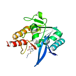
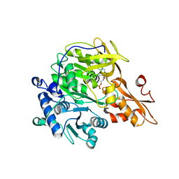
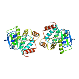
5YPN
 
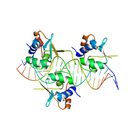
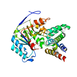
 | | Crystal structure of NDM-1 bound to hydrolyzed meropenem representing an EI2 complex | | 分子名称: | (2~{S},3~{R},4~{S})-2-[(2~{S},3~{R})-1,3-bis(oxidanyl)-1-oxidanylidene-butan-2-yl]-4-[(3~{S},5~{S})-5-(dimethylcarbamoy l)pyrrolidin-3-yl]sulfanyl-3-methyl-3,4-dihydro-2~{H}-pyrrole-5-carboxylic acid, Metallo-beta-lactamase NDM-1, SULFATE ION, ... | | 著者 | Feng, H, Liu, W, Wang, D. | | 登録日 | 2017-11-02 | | 公開日 | 2018-02-21 | | 最終更新日 | 2023-11-22 | | 実験手法 | X-RAY DIFFRACTION (2.12 Å) | | 主引用文献 | The mechanism of NDM-1-catalyzed carbapenem hydrolysis is distinct from that of penicillin or cephalosporin hydrolysis.
Nat Commun, 8, 2017
|
|
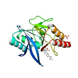
5YPL
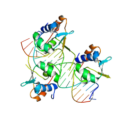
 | | Crystal structure of NDM-1 bound to hydrolyzed imipenem representing an EP complex | | 分子名称: | (2R,4S)-2-[(1S,2R)-1-carboxy-2-hydroxypropyl]-4-[(2-{[(Z)-iminomethyl]amino}ethyl)sulfanyl]-3,4-dihydro-2H-pyrrole-5-ca rboxylic acid, CHLORIDE ION, Metallo-beta-lactamase NDM-1, ... | | 著者 | Feng, H, Wang, D, Liu, W. | | 登録日 | 2017-11-02 | | 公開日 | 2018-02-21 | | 最終更新日 | 2023-11-22 | | 実験手法 | X-RAY DIFFRACTION (1.8 Å) | | 主引用文献 | The mechanism of NDM-1-catalyzed carbapenem hydrolysis is distinct from that of penicillin or cephalosporin hydrolysis.
Nat Commun, 8, 2017
|
|
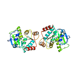
5YPM
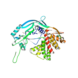
 | | Crystal structure of NDM-1 bound to hydrolyzed meropenem representing an EI1 complex | | 分子名称: | (2S,3R)-2-[(2S,3R)-1,3-bis(oxidanyl)-1-oxidanylidene-butan-2-yl]-4-[(3S,5S)-5-(dimethylcarbamoyl)pyrrolidin-3-yl]sulfan yl-3-methyl-2,3-dihydro-1H-pyrrole-5-carboxylic acid, Metallo-beta-lactamase NDM-1, SULFATE ION, ... | | 著者 | Feng, H, Wang, D, Liu, W. | | 登録日 | 2017-11-02 | | 公開日 | 2018-02-21 | | 最終更新日 | 2023-11-22 | | 実験手法 | X-RAY DIFFRACTION (2.15 Å) | | 主引用文献 | The mechanism of NDM-1-catalyzed carbapenem hydrolysis is distinct from that of penicillin or cephalosporin hydrolysis.
Nat Commun, 8, 2017
|
|
7DCT
 | |
7DCJ
 | |
7DCS
 | |
7DCU
 | |
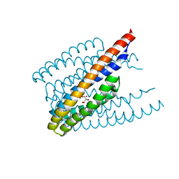
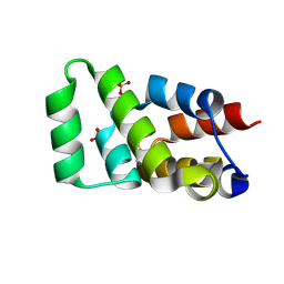
5B5R
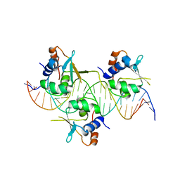
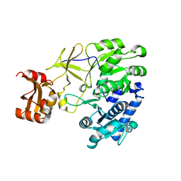
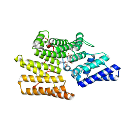
 | | Crystal structure of GSDMA3 | | 分子名称: | Gasdermin-A3 | | 著者 | Ding, J, Shao, F. | | 登録日 | 2016-05-14 | | 公開日 | 2016-06-15 | | 最終更新日 | 2017-09-27 | | 実験手法 | X-RAY DIFFRACTION (1.902 Å) | | 主引用文献 | Pore-forming activity and structural autoinhibition of the gasdermin family.
Nature, 535, 2016
|
|
3A9U
 | |
3A9V
 | |
6ITS
 | |
3AFK
 | |
4LJR
 | |
4LJL
 | |
4LJK
 | |
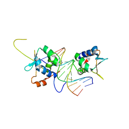
3WLC
 | | Crystal structure of dimeric GCaMP6m | | 分子名称: | CALCIUM ION, Myosin light chain kinase, Green fluorescent protein, ... | | 著者 | Ding, J, Luo, A.F, Hu, L.Y, Wang, D.C, Shao, F. | | 登録日 | 2013-11-08 | | 公開日 | 2014-01-22 | | 最終更新日 | 2023-12-06 | | 実験手法 | X-RAY DIFFRACTION (2.49 Å) | | 主引用文献 | Structural basis of the ultrasensitive calcium indicator GCaMP6.
Sci China Life Sci, 57, 2014
|
|
3WLD
 | | Crystal structure of monomeric GCaMP6m | | 分子名称: | CALCIUM ION, Myosin light chain kinase, Green fluorescent protein, ... | | 著者 | Ding, J, Luo, A.F, Hu, L.Y, Wang, D.C, Shao, F. | | 登録日 | 2013-11-08 | | 公開日 | 2014-01-22 | | 最終更新日 | 2023-12-06 | | 実験手法 | X-RAY DIFFRACTION (2.7 Å) | | 主引用文献 | Structural basis of the ultrasensitive calcium indicator GCaMP6.
Sci China Life Sci, 57, 2014
|
|
6AC5
 | |
3M3E
 | |
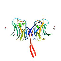
5Z2C
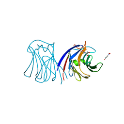
 | | Crystal structure of ALPK-1 N-terminal domain in complex with ADP-heptose | | 分子名称: | Alpha-protein kinase 1, [[(2R,3S,4R,5R)-5-(6-aminopurin-9-yl)-3,4-bis(oxidanyl)oxolan-2-yl]methoxy-oxidanyl-phosphoryl] [(2S,3S,4S,5S,6R)-6-[(1S)-1,2-bis(oxidanyl)ethyl]-3,4,5-tris(oxidanyl)oxan-2-yl] hydrogen phosphate | | 著者 | Ding, J, She, Y, Shao, F. | | 登録日 | 2018-01-02 | | 公開日 | 2018-08-22 | | 最終更新日 | 2024-03-27 | | 実験手法 | X-RAY DIFFRACTION (2.594 Å) | | 主引用文献 | Alpha-kinase 1 is a cytosolic innate immune receptor for bacterial ADP-heptose.
Nature, 561, 2018
|
|
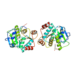
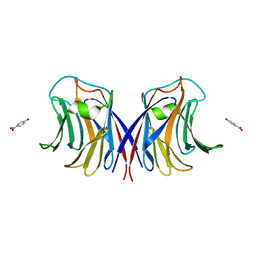
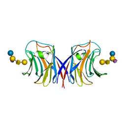
3M3C
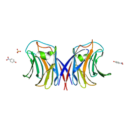 | | Crystal Structure of Agrocybe aegerita lectin AAL complexed with p-Nitrophenyl TF disaccharide | | 分子名称: | Anti-tumor lectin, P-NITROPHENOL, SULFATE ION, ... | | 著者 | Feng, L, Li, D, Wang, D. | | 登録日 | 2010-03-09 | | 公開日 | 2010-12-01 | | 最終更新日 | 2023-11-01 | | 実験手法 | X-RAY DIFFRACTION (2 Å) | | 主引用文献 | Structural insights into the recognition mechanism between an antitumor galectin AAL and the Thomsen-Friedenreich antigen
Faseb J., 24, 2010
|
|
3M3O
 | | Crystal Structure of Agrocybe aegerita lectin AAL mutant R85A complexed with p-Nitrophenyl TF disaccharide | | 分子名称: | 2-[BIS-(2-HYDROXY-ETHYL)-AMINO]-2-HYDROXYMETHYL-PROPANE-1,3-DIOL, Anti-tumor lectin, P-NITROPHENOL, ... | | 著者 | Feng, L, Li, D, Wang, D. | | 登録日 | 2010-03-09 | | 公開日 | 2010-12-01 | | 最終更新日 | 2023-11-01 | | 実験手法 | X-RAY DIFFRACTION (2.1 Å) | | 主引用文献 | Structural insights into the recognition mechanism between an antitumor galectin AAL and the Thomsen-Friedenreich antigen
Faseb J., 24, 2010
|
|
6ACI
 | |
6AC0
 | |
3M3Q
 | |


