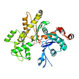
6JZP
 
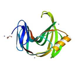 | |
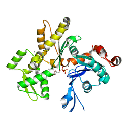
6K9X
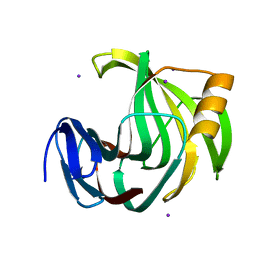
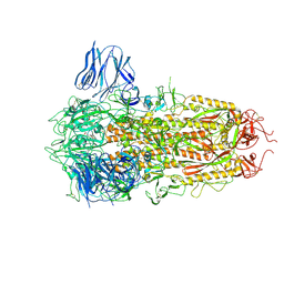
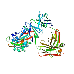
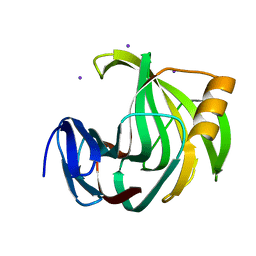 | | Crystal Structure Analysis of Protein | | 分子名称: | Endo-1,4-beta-xylanase 2, IODIDE ION, beta-D-xylopyranose-(1-4)-beta-D-xylopyranose-(1-4)-beta-D-xylopyranose | | 著者 | Li, C, Wan, Q. | | 登録日 | 2019-06-18 | | 公開日 | 2021-04-28 | | 最終更新日 | 2023-11-22 | | 実験手法 | X-RAY DIFFRACTION (1.2 Å) | | 主引用文献 | X-ray crystallographic studies of family 11 xylanase Michaelis and product complexes: implications for the catalytic mechanism
Acta Crystallographica Section D-Biological Crystallography, D70, 2014
|
|
6KVV
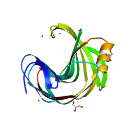 | |
6KWE
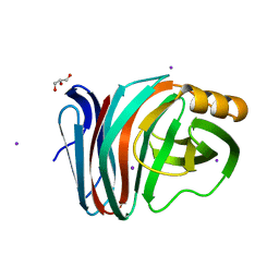 | |
6KWH
 | |
3RWA
 | |
3RWT
 | |
8YMJ
 | | Cryo-EM structure of Hepatitis B virus surface antigen subviral particle with D2 symmetry | | 分子名称: | Isoform S of Large envelope protein | | 著者 | Wang, T, Cao, L, Mu, A, Wang, Q, Rao, Z.H. | | 登録日 | 2024-03-09 | | 公開日 | 2024-09-18 | | 実験手法 | ELECTRON MICROSCOPY (6.6 Å) | | 主引用文献 | Inherent symmetry and flexibility in hepatitis B virus subviral particles
Science, 2024
|
|
8YMK
 | | Localized reconstruction of Hepatitis B virus surface antigen dimer in the subviral particle with D2 symmetry from dataset A0 | | 分子名称: | Isoform S of Large envelope protein | | 著者 | Wang, T, Cao, L, Mu, A, Wang, Q, Rao, Z.H. | | 登録日 | 2024-03-09 | | 公開日 | 2024-09-18 | | 実験手法 | ELECTRON MICROSCOPY (3.7 Å) | | 主引用文献 | Inherent symmetry and flexibility in hepatitis B virus subviral particles
Science, 2024
|
|
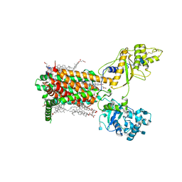
7RPK
 | | Cryo-EM structure of murine Dispatched in complex with Sonic hedgehog | | 分子名称: | (2S)-3-{[(S)-(2-aminoethoxy)(hydroxy)phosphoryl]oxy}-2-(hexanoyloxy)propyl hexanoate, 2-acetamido-2-deoxy-beta-D-glucopyranose, CALCIUM ION, ... | | 著者 | Asarnow, D, Wang, Q, Ding, K, Cheng, Y, Beachy, P.A. | | 登録日 | 2021-08-03 | | 公開日 | 2021-10-27 | | 最終更新日 | 2023-12-13 | | 実験手法 | ELECTRON MICROSCOPY (2.7 Å) | | 主引用文献 | Dispatched uses Na + flux to power release of lipid-modified Hedgehog.
Nature, 599, 2021
|
|
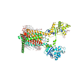
7RPJ
 | | Cryo-EM structure of murine Dispatched NNN mutant | | 分子名称: | 2-acetamido-2-deoxy-beta-D-glucopyranose, CHOLESTEROL HEMISUCCINATE, Protein dispatched homolog 1 | | 著者 | Asarnow, D, Wang, Q, Ding, K, Cheng, Y, Beachy, P.A. | | 登録日 | 2021-08-03 | | 公開日 | 2021-10-27 | | 最終更新日 | 2021-11-24 | | 実験手法 | ELECTRON MICROSCOPY (3.2 Å) | | 主引用文献 | Dispatched uses Na + flux to power release of lipid-modified Hedgehog.
Nature, 599, 2021
|
|
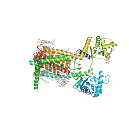
7RPH
 | | Cryo-EM structure of murine Dispatched 'R' conformation | | 分子名称: | 2-acetamido-2-deoxy-beta-D-glucopyranose, CHOLESTEROL HEMISUCCINATE, Lauryl Maltose Neopentyl Glycol, ... | | 著者 | Asarnow, D, Wang, Q, Ding, K, Cheng, Y, Beachy, P.A. | | 登録日 | 2021-08-03 | | 公開日 | 2021-10-27 | | 最終更新日 | 2023-12-13 | | 実験手法 | ELECTRON MICROSCOPY (2.5 Å) | | 主引用文献 | Dispatched uses Na + flux to power release of lipid-modified Hedgehog.
Nature, 599, 2021
|
|
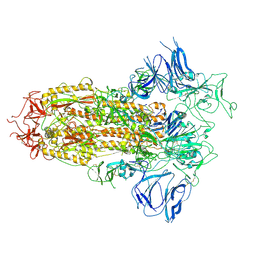
7RPI
 | | Cryo-EM structure of murine Dispatched 'T' conformation | | 分子名称: | 2-acetamido-2-deoxy-beta-D-glucopyranose, CHOLESTEROL HEMISUCCINATE, Lauryl Maltose Neopentyl Glycol, ... | | 著者 | Asarnow, D, Wang, Q, Ding, K, Cheng, Y, Beachy, P.A. | | 登録日 | 2021-08-03 | | 公開日 | 2021-10-27 | | 最終更新日 | 2023-12-13 | | 実験手法 | ELECTRON MICROSCOPY (2.5 Å) | | 主引用文献 | Dispatched uses Na + flux to power release of lipid-modified Hedgehog.
Nature, 599, 2021
|
|
7DTE
 | | SARS-CoV-2 RdRP catalytic complex with T33-1 RNA | | 分子名称: | Non-structural protein 7, Non-structural protein 8, RNA (33-MER), ... | | 著者 | Wang, Q, Gong, P. | | 登録日 | 2021-01-04 | | 公開日 | 2021-10-20 | | 最終更新日 | 2024-06-05 | | 実験手法 | ELECTRON MICROSCOPY (3 Å) | | 主引用文献 | Remdesivir overcomes the S861 roadblock in SARS-CoV-2 polymerase elongation complex.
Cell Rep, 37, 2021
|
|
8H3D
 | | Structure of apo SARS-CoV-2 spike protein with one RBD up | | 分子名称: | 2-acetamido-2-deoxy-beta-D-glucopyranose, Spike glycoprotein,Fibritin | | 著者 | Meng, F, Wang, Q, Xie, Y, Ni, X, Huang, N. | | 登録日 | 2022-10-08 | | 公開日 | 2023-03-22 | | 実験手法 | ELECTRON MICROSCOPY (3.27 Å) | | 主引用文献 | In Silico Discovery of Small Molecule Modulators Targeting the Achilles' Heel of SARS-CoV-2 Spike Protein.
Acs Cent.Sci., 9, 2023
|
|
8H3E
 | | Complex structure of a small molecule (SPC-14) bound SARS-CoV-2 spike protein, closed state | | 分子名称: | 2-acetamido-2-deoxy-beta-D-glucopyranose, 7-(6-nitro-2,3-dihydroindol-1-yl)-7-oxidanylidene-heptanoic acid, Spike glycoprotein,Fibritin | | 著者 | Meng, F, Wang, Q, Xie, Y, Ni, X, Huang, N. | | 登録日 | 2022-10-08 | | 公開日 | 2023-03-22 | | 実験手法 | ELECTRON MICROSCOPY (3.06 Å) | | 主引用文献 | In Silico Discovery of Small Molecule Modulators Targeting the Achilles' Heel of SARS-CoV-2 Spike Protein.
Acs Cent.Sci., 9, 2023
|
|
8HMH
 | | The closed state of RGLG2-VWA | | 分子名称: | E3 ubiquitin-protein ligase RGLG2, MAGNESIUM ION | | 著者 | Wang, Q. | | 登録日 | 2022-12-03 | | 公開日 | 2023-12-27 | | 最終更新日 | 2024-01-03 | | 実験手法 | X-RAY DIFFRACTION (2.56 Å) | | 主引用文献 | The regulation of RGLG2-VWA by Ca 2+ ions.
Biochim Biophys Acta Proteins Proteom, 1872, 2024
|
|
7Y9J
 | | Crystal structure of P450 BM3-TMK from Bacillus megaterium in complex with 5-nitro-1,2-benzisoxazole | | 分子名称: | 5-nitro-1,2-benzoxazole, Bifunctional cytochrome P450/NADPH--P450 reductase, PROTOPORPHYRIN IX CONTAINING FE | | 著者 | Wang, Q, Zhang, L.L, Liu, W.D, Huang, J.-W, Yang, Y, Chen, C.-C, Guo, R.-T. | | 登録日 | 2022-06-24 | | 公開日 | 2023-06-28 | | 最終更新日 | 2023-11-29 | | 実験手法 | X-RAY DIFFRACTION (1.83 Å) | | 主引用文献 | Engineering of a P450-based Kemp eliminase with a new mechanism
Chinese J Catal, 47, 2023
|
|
7Y9K
 | | Crystal structure of P450 BM3-TMK from Bacillus megaterium | | 分子名称: | Bifunctional cytochrome P450/NADPH--P450 reductase, PROTOPORPHYRIN IX CONTAINING FE | | 著者 | Wang, Q, Zhang, L.L, Liu, W.D, Huang, J.-W, Yang, Y, Chen, C.-C, Guo, R.-T. | | 登録日 | 2022-06-25 | | 公開日 | 2023-06-28 | | 最終更新日 | 2023-11-29 | | 実験手法 | X-RAY DIFFRACTION (2.23 Å) | | 主引用文献 | Engineering of a P450-based Kemp eliminase with a new mechanism
Chinese J Catal, 47, 2023
|
|
5DO2
 | | Complex structure of MERS-RBD bound with 4C2 antibody | | 分子名称: | 2-acetamido-2-deoxy-beta-D-glucopyranose, 4C2 heavy chain, 4C2 light chain, ... | | 著者 | Li, Y, Wan, Y, Liu, P, Zhao, J, Lu, G, Qi, J, Wang, Q, Lu, X, Wu, Y, Liu, W, Yuen, K.Y, Perlman, S, Gao, G.F, Yan, J. | | 登録日 | 2015-09-10 | | 公開日 | 2015-10-14 | | 最終更新日 | 2023-11-08 | | 実験手法 | X-RAY DIFFRACTION (2.409 Å) | | 主引用文献 | A humanized neutralizing antibody against MERS-CoV targeting the receptor-binding domain of the spike protein.
Cell Res., 25, 2015
|
|
7FIF
 | | Cryo-EM structure of the hedgehog release protein Disp from water bear (Hypsibius dujardini) | | 分子名称: | Protein dispatched-like protein 1 | | 著者 | Luo, Y, Wan, G, Wang, Q, Zhao, Y, Cong, Y, Li, D. | | 登録日 | 2021-07-31 | | 公開日 | 2021-09-08 | | 最終更新日 | 2024-06-12 | | 実験手法 | ELECTRON MICROSCOPY (6.5 Å) | | 主引用文献 | Architecture of Dispatched, a Transmembrane Protein Responsible for Hedgehog Release.
Front Mol Biosci, 8, 2021
|
|
3WP1
 | | Phosphorylation-dependent interaction between tumor suppressors Dlg and Lgl | | 分子名称: | Disks large homolog 4, Lethal(2) giant larvae protein homolog 2, SULFATE ION | | 著者 | Zhu, J, Shang, Y, Wan, Q, Xia, Y, Chen, J, Du, Q, Zhang, M. | | 登録日 | 2014-01-08 | | 公開日 | 2014-03-19 | | 最終更新日 | 2014-04-30 | | 実験手法 | X-RAY DIFFRACTION (2.804 Å) | | 主引用文献 | Phosphorylation-dependent interaction between tumor suppressors Dlg and Lgl
Cell Res., 24, 2014
|
|
3WP0
 | | Crystal structure of Dlg GK in complex with a phosphor-Lgl2 peptide | | 分子名称: | Disks large homolog 4, GLYCEROL, Lethal(2) giant larvae protein homolog 2 | | 著者 | Zhu, J, Shang, Y, Wan, Q, Xia, Y, Chen, J, Du, Q, Zhang, M. | | 登録日 | 2014-01-08 | | 公開日 | 2014-03-19 | | 最終更新日 | 2014-04-30 | | 実験手法 | X-RAY DIFFRACTION (2.039 Å) | | 主引用文献 | Phosphorylation-dependent interaction between tumor suppressors Dlg and Lgl
Cell Res., 24, 2014
|
|
2HF4
 | | Crystal structure of Monomeric Actin in its ATP-bound state | | 分子名称: | ADENOSINE-5'-TRIPHOSPHATE, Actin-5C, CALCIUM ION | | 著者 | Rould, M.A, Wan, Q, Joel, P.B, Lowey, S, Trybus, K.M. | | 登録日 | 2006-06-22 | | 公開日 | 2006-08-29 | | 最終更新日 | 2023-08-30 | | 実験手法 | X-RAY DIFFRACTION (1.8 Å) | | 主引用文献 | Crystal Structures of Expressed Non-polymerizable Monomeric Actin in the ADP and ATP States.
J.Biol.Chem., 281, 2006
|
|
2HF3
 | | Crystal structure of monomeric Actin in the ADP bound state | | 分子名称: | ADENOSINE-5'-DIPHOSPHATE, Actin-5C, CALCIUM ION | | 著者 | Rould, M.A, Wan, Q, Joel, P.B, Lowey, S, Trybus, K.M. | | 登録日 | 2006-06-22 | | 公開日 | 2006-08-29 | | 最終更新日 | 2023-08-30 | | 実験手法 | X-RAY DIFFRACTION (1.8 Å) | | 主引用文献 | Crystal Structures of Expressed Non-polymerizable Monomeric Actin in the ADP and ATP States.
J.Biol.Chem., 281, 2006
|
|


