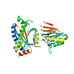
6ECB
 
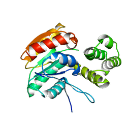 | |
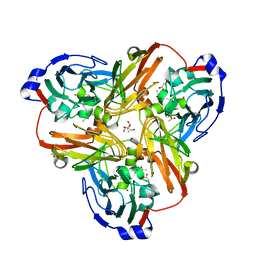
6JV2
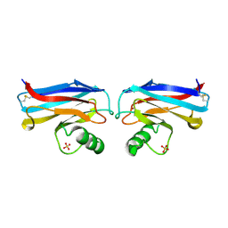
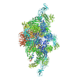 | | Structure of RyR2 (P/L-Ca2+/Ca2+-CaM dataset) | | 分子名称: | CALCIUM ION, Calmodulin-1, RyR2, ... | | 著者 | Gong, D.S, Chi, X.M, Zhou, G.W, Huang, G.X.Y, Lei, J.L, Yan, N. | | 登録日 | 2019-04-15 | | 公開日 | 2019-07-17 | | 最終更新日 | 2024-03-27 | | 実験手法 | ELECTRON MICROSCOPY (4.4 Å) | | 主引用文献 | Modulation of cardiac ryanodine receptor 2 by calmodulin.
Nature, 572, 2019
|
|
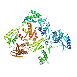
6TOF
 | | Crystal structure of human BCL6 BTB domain in complex with compound 4 | | 分子名称: | 1,2-ETHANEDIOL, 2-[(1,3-dimethyl-2-oxidanylidene-benzimidazol-5-yl)amino]pyridine-3-carbonitrile, ALA-TRP-VAL-ILE-PRO-ALA, ... | | 著者 | Shetty, K, Rodrigues, M.J, Le Bihan, Y.-V, van Montfort, R.L.M. | | 登録日 | 2019-12-11 | | 公開日 | 2020-04-22 | | 最終更新日 | 2024-01-24 | | 実験手法 | X-RAY DIFFRACTION (1.67 Å) | | 主引用文献 | AchievingIn VivoTarget Depletion through the Discovery and Optimization of Benzimidazolone BCL6 Degraders.
J.Med.Chem., 63, 2020
|
|
6TOK
 | | Crystal structure of human BCL6 BTB domain in complex with compound 23d | | 分子名称: | 1,2-ETHANEDIOL, 5-[[5-chloranyl-2-(3,5-dimethylpyrazol-1-yl)pyrimidin-4-yl]amino]-1-methyl-3-(3-methyl-3-oxidanyl-butyl)benzimidazol-2-one, ALA-TRP-VAL-ILE-PRO-ALA, ... | | 著者 | Rodrigues, M.J, Le Bihan, Y.-V, van Montfort, R.L.M. | | 登録日 | 2019-12-11 | | 公開日 | 2020-04-22 | | 最終更新日 | 2024-01-24 | | 実験手法 | X-RAY DIFFRACTION (1.43 Å) | | 主引用文献 | AchievingIn VivoTarget Depletion through the Discovery and Optimization of Benzimidazolone BCL6 Degraders.
J.Med.Chem., 63, 2020
|
|
6H3K
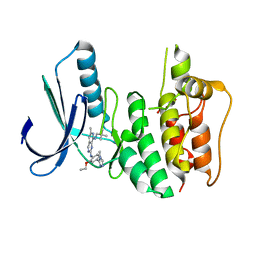 | | Introduction of a methyl group curbs metabolism of pyrido[3,4-d]pyrimidine MPS1 inhibitors and enables the discovery of the Phase 1 clinical candidate BOS172722. | | 分子名称: | 2-(2-(2-(2-(2-(2-ETHOXYETHOXY)ETHOXY)ETHOXY)ETHOXY)ETHOXY)ETHANOL, Dual specificity protein kinase TTK, ~{N}8-(2,2-dimethylpropyl)-~{N}2-[2-ethoxy-4-(4-methyl-1,2,4-triazol-3-yl)phenyl]-6-methyl-pyrido[3,4-d]pyrimidine-2,8-diamine | | 著者 | Woodward, H.L, Hoelder, S. | | 登録日 | 2018-07-19 | | 公開日 | 2018-09-19 | | 最終更新日 | 2024-05-15 | | 実験手法 | X-RAY DIFFRACTION (2.48 Å) | | 主引用文献 | Introduction of a Methyl Group Curbs Metabolism of Pyrido[3,4- d]pyrimidine Monopolar Spindle 1 (MPS1) Inhibitors and Enables the Discovery of the Phase 1 Clinical Candidate N2-(2-Ethoxy-4-(4-methyl-4 H-1,2,4-triazol-3-yl)phenyl)-6-methyl- N8-neopentylpyrido[3,4- d]pyrimidine-2,8-diamine (BOS172722).
J. Med. Chem., 61, 2018
|
|
6TON
 | | Crystal structure of human BCL6 BTB domain in complex with compound 25b | | 分子名称: | 1,2-ETHANEDIOL, 5-[[5-chloranyl-2-(2,2,6,6-tetramethylmorpholin-4-yl)pyrimidin-4-yl]amino]-1-methyl-3-(3-methyl-3-oxidanyl-butyl)benzimidazol-2-one, ALA-TRP-VAL-ILE-PRO-ALA, ... | | 著者 | Rodrigues, M.J, Le Bihan, Y.-V, van Montfort, R.L.M. | | 登録日 | 2019-12-11 | | 公開日 | 2020-04-22 | | 最終更新日 | 2024-01-24 | | 実験手法 | X-RAY DIFFRACTION (2.36 Å) | | 主引用文献 | AchievingIn VivoTarget Depletion through the Discovery and Optimization of Benzimidazolone BCL6 Degraders.
J.Med.Chem., 63, 2020
|
|
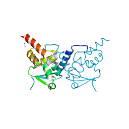
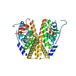
2AZU
 | |
1HV7
 | | PORCINE PANCREATIC ELASTASE COMPLEXED WITH GW311616A | | 分子名称: | ELASTASE 1, N-[2-(1-FORMYL-2-METHYL-PROPYL)-1-(4-PIPERIDIN-1-YL-BUT-2-ENOYL)-PYRROLIDIN-3-YL]-METHANESULFONAMIDE, SULFATE ION | | 著者 | Skarzynski, T, Singh, O.M. | | 登録日 | 2001-01-08 | | 公開日 | 2001-01-17 | | 最終更新日 | 2011-07-13 | | 実験手法 | X-RAY DIFFRACTION (1.7 Å) | | 主引用文献 | The discovery of a potent, intracellular, orally bioavailable, long duration inhibitor of human neutrophil elastase--GW311616A a development candidate
Bioorg.Med.Chem.Lett., 11, 2001
|
|
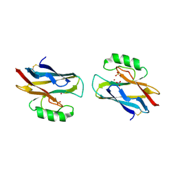
2ETS
 | |
2FHW
 | | Solution structure of human relaxin-3 | | 分子名称: | Relaxin 3 (Prorelaxin H3) (Insulin-like peptide INSL7) (Insulin-like peptide 7) | | 著者 | Rosengren, K.J, Craik, D.J. | | 登録日 | 2005-12-27 | | 公開日 | 2006-01-24 | | 最終更新日 | 2022-03-09 | | 実験手法 | SOLUTION NMR | | 主引用文献 | Solution structure and novel insights into the determinants of the receptor specificity of human relaxin-3.
J.Biol.Chem., 281, 2006
|
|
2FEA
 | |
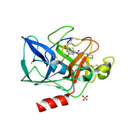
2F9Z
 | | Complex between the chemotaxis deamidase CheD and the chemotaxis phosphatase CheC from Thermotoga maritima | | 分子名称: | PROTEIN (chemotaxis methylation protein), chemotaxis protein CheC | | 著者 | Chao, X, Park, S.Y, Bilwes, A.M, Crane, B.R. | | 登録日 | 2005-12-06 | | 公開日 | 2006-06-06 | | 最終更新日 | 2023-08-30 | | 実験手法 | X-RAY DIFFRACTION (2.4 Å) | | 主引用文献 | A receptor-modifying deamidase in complex with a signaling phosphatase reveals reciprocal regulation.
Cell(Cambridge,Mass.), 124, 2006
|
|
2F46
 | |
2FJS
 | |
2FNA
 | |
2G36
 | |
2FNO
 | |
5W9C
 | |
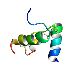
1A4C
 | | AZURIN MUTANT WITH MET 121 REPLACED BY HIS, PH 3.5 CRYSTAL FORM, DATA COLLECTED AT-180 DEGREES CELSIUS | | 分子名称: | AZURIN, COPPER (II) ION, NITRATE ION, ... | | 著者 | Messerschmidt, A, Prade, L. | | 登録日 | 1998-01-28 | | 公開日 | 1998-04-29 | | 最終更新日 | 2023-08-02 | | 実験手法 | X-RAY DIFFRACTION (2.45 Å) | | 主引用文献 | Rack-induced metal binding vs. flexibility: Met121His azurin crystal structures at different pH.
Proc.Natl.Acad.Sci.USA, 95, 1998
|
|
1A4B
 | |
1A4A
 | |
1D41
 | |
1SCF
 | | HUMAN RECOMBINANT STEM CELL FACTOR | | 分子名称: | CALCIUM ION, PENTAETHYLENE GLYCOL, STEM CELL FACTOR | | 著者 | Jiang, X, Gurel, O, Langley, K.E, Hendrickson, W.A. | | 登録日 | 1998-06-04 | | 公開日 | 2000-07-07 | | 最終更新日 | 2022-12-21 | | 実験手法 | X-RAY DIFFRACTION (2.2 Å) | | 主引用文献 | Structure of the active core of human stem cell factor and analysis of binding to its receptor kit.
EMBO J., 19, 2000
|
|
1TKX
 | | CRYSTAL STRUCTURE OF HIV-1 REVERSE TRANSCRIPTASE IN COMPLEX WITH GW490745 | | 分子名称: | 4-[(CYCLOPROPYLETHYNYL)OXY]-6-FLUORO-3-ISOPROPYLQUINOLIN-2(1H)-ONE, Pol polyprotein, Reverse transcriptase, ... | | 著者 | Ren, J, Hopkins, A.L, Stuart, D.I, Stammers, D.K. | | 登録日 | 2004-06-09 | | 公開日 | 2004-12-07 | | 最終更新日 | 2022-12-21 | | 実験手法 | X-RAY DIFFRACTION (2.85 Å) | | 主引用文献 | Design of non-nucleoside inhibitors of HIV-1 reverse transcriptase with improved drug resistance properties. 2.
J.Med.Chem., 47, 2004
|
|
1URI
 | | AZURIN MUTANT WITH MET 121 REPLACED BY GLN | | 分子名称: | AZURIN, COPPER (II) ION, SULFATE ION | | 著者 | Romero, A, Nar, H, Huber, R, Messerschmidt, A. | | 登録日 | 1996-11-14 | | 公開日 | 1997-04-01 | | 最終更新日 | 2018-04-18 | | 実験手法 | X-RAY DIFFRACTION (1.94 Å) | | 主引用文献 | X-ray analysis and spectroscopic characterization of M121Q azurin. A copper site model for stellacyanin.
J.Mol.Biol., 229, 1993
|
|


