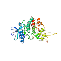
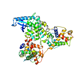
7DHN
 
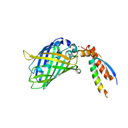
 | | The co-crystal structure of DYRK2 with a small molecule inhibitor 20 | | 分子名称: | 7-methoxy-2-methylsulfanyl-9-(piperidin-4-ylmethylsulfanyl)-[1,3]thiazolo[5,4-b]quinoline, Dual specificity tyrosine-phosphorylation-regulated kinase 2 | | 著者 | Wei, T, Xiao, J. | | 登録日 | 2020-11-16 | | 公開日 | 2021-11-17 | | 最終更新日 | 2023-11-29 | | 実験手法 | X-RAY DIFFRACTION (2.38 Å) | | 主引用文献 | Selective inhibition reveals the regulatory function of DYRK2 in protein synthesis and calcium entry.
Elife, 11, 2022
|
|
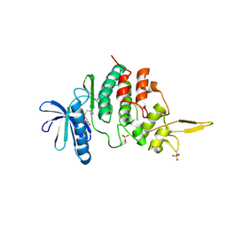
7DHO
 | | The co-crystal structure of DYRK2 with a small molecule inhibitor 14 | | 分子名称: | 2-methoxy-9-(piperidin-4-ylmethylsulfanyl)-7-propan-2-yloxy-acridine, Dual specificity tyrosine-phosphorylation-regulated kinase 2 | | 著者 | Wei, T, Xiao, J. | | 登録日 | 2020-11-16 | | 公開日 | 2021-11-17 | | 最終更新日 | 2023-11-29 | | 実験手法 | X-RAY DIFFRACTION (3.29 Å) | | 主引用文献 | Selective inhibition reveals the regulatory function of DYRK2 in protein synthesis and calcium entry.
Elife, 11, 2022
|
|
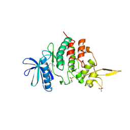
7DHH
 | | The co-crystal structure of DYRK2 with a small molecule inhibitor 19 | | 分子名称: | Dual specificity tyrosine-phosphorylation-regulated kinase 2, [9-(azetidin-3-ylmethylsulfanyl)-2,7-dimethoxy-acridin-4-yl]methanol | | 著者 | Wei, T, Xiao, J. | | 登録日 | 2020-11-14 | | 公開日 | 2021-11-17 | | 最終更新日 | 2023-11-29 | | 実験手法 | X-RAY DIFFRACTION (2.486 Å) | | 主引用文献 | Selective inhibition reveals the regulatory function of DYRK2 in protein synthesis and calcium entry.
Elife, 11, 2022
|
|
7DJO
 | | The co-crystal structure of DYRK2 with a small molecule inhibitor 17 | | 分子名称: | Dual specificity tyrosine-phosphorylation-regulated kinase 2, [2,7-dimethoxy-9-[[(3S)-pyrrolidin-3-yl]methylsulfanyl]acridin-4-yl]methanol | | 著者 | Wei, T, Xiao, J. | | 登録日 | 2020-11-20 | | 公開日 | 2021-11-24 | | 最終更新日 | 2023-11-29 | | 実験手法 | X-RAY DIFFRACTION (2.499 Å) | | 主引用文献 | Selective inhibition reveals the regulatory function of DYRK2 in protein synthesis and calcium entry.
Elife, 11, 2022
|
|
7DL6
 | | The co-crystal structure of DYRK2 with a small molecule inhibitor 18 | | 分子名称: | Dual specificity tyrosine-phosphorylation-regulated kinase 2, [2,7-dimethoxy-9-[[(3R)-pyrrolidin-3-yl]methylsulfanyl]acridin-4-yl]methanol | | 著者 | Wei, T, Xiao, J. | | 登録日 | 2020-11-26 | | 公開日 | 2021-12-01 | | 最終更新日 | 2023-11-29 | | 実験手法 | X-RAY DIFFRACTION (2.648 Å) | | 主引用文献 | Selective inhibition reveals the regulatory function of DYRK2 in protein synthesis and calcium entry.
Elife, 11, 2022
|
|
7EYA
 | |
7EY0
 | |
7DH3
 | |
7DNR
 | |
7DXI
 | |
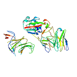
7EQ4
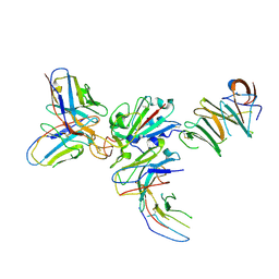
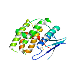
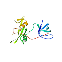
 | | Crystal Structure of the N-terminus of Nonstructural protein 1 from SARS-CoV-2 | | 分子名称: | Host translation inhibitor nsp1 | | 著者 | Liu, Y, Ke, Z, Hu, H, Zhao, K, Xiao, J, Xia, Y, Li, Y. | | 登録日 | 2021-04-29 | | 公開日 | 2021-06-09 | | 最終更新日 | 2023-11-29 | | 実験手法 | X-RAY DIFFRACTION (1.25 Å) | | 主引用文献 | Structural Basis and Function of the N Terminus of SARS-CoV-2 Nonstructural Protein 1.
Microbiol Spectr, 9, 2021
|
|
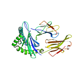
5F1I
 | | MHC with 9-mer peptide | | 分子名称: | 9-mer peptide, Beta2M, MHC class I DLA-88 | | 著者 | Liu, J, Chai, Y, QI, J, Gao, G.F. | | 登録日 | 2015-11-30 | | 公開日 | 2016-11-30 | | 最終更新日 | 2023-11-08 | | 実験手法 | X-RAY DIFFRACTION (2.904 Å) | | 主引用文献 | Diversified Anchoring Features the Peptide Presentation of DLA-88*50801: First Structural Insight into Domestic Dog MHC Class I
J Immunol., 197, 2016
|
|
5F1N
 | | MHC complexed to 11mer peptide | | 分子名称: | Beta-2-microglobulin, MHC class I antigen, Peptide from Cytochrome P450 family 1 subfamily B polypeptide 1 | | 著者 | Liu, J, Chai, Y, Qi, J, Gao, G.F. | | 登録日 | 2015-11-30 | | 公開日 | 2016-11-30 | | 最終更新日 | 2017-11-15 | | 実験手法 | X-RAY DIFFRACTION (2 Å) | | 主引用文献 | Diversified Anchoring Features the Peptide Presentation of DLA-88*50801: First Structural Insight into Domestic Dog MHC Class I
J Immunol., 197, 2016
|
|
7YOC
 | |
8X6B
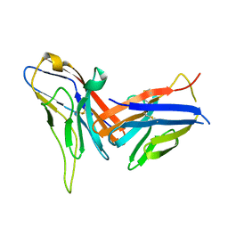 | | Crystal structure of immune receptor PVRIG in complex with ligand Nectin-2 | | 分子名称: | 2-acetamido-2-deoxy-beta-D-glucopyranose, Nectin-2, Transmembrane protein PVRIG | | 著者 | Hu, S.T, Han, P, Wang, H, Qi, J.X. | | 登録日 | 2023-11-21 | | 公開日 | 2024-04-24 | | 最終更新日 | 2024-05-08 | | 実験手法 | X-RAY DIFFRACTION (2 Å) | | 主引用文献 | Structural basis for the immune recognition and selectivity of the immune receptor PVRIG for ligand Nectin-2.
Structure, 2024
|
|
8WY4
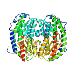
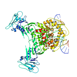
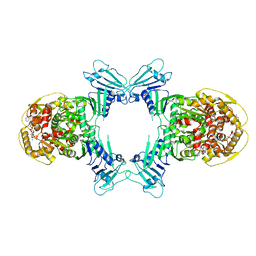 | | GajA tetramer with ATP | | 分子名称: | ADENOSINE-5'-TRIPHOSPHATE, Endonuclease GajA | | 著者 | Li, J, Wang, Z, Wang, L. | | 登録日 | 2023-10-30 | | 公開日 | 2024-02-28 | | 最終更新日 | 2024-05-22 | | 実験手法 | ELECTRON MICROSCOPY (2.81 Å) | | 主引用文献 | Structures and activation mechanism of the Gabija anti-phage system.
Nature, 629, 2024
|
|
8X51
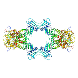
 | | Cryo-EM structure of Gabija GajA in complex with DNA(focused refinement) | | 分子名称: | CALCIUM ION, DNA (5'-D(*AP*AP*AP*AP*AP*TP*AP*AP*CP*CP*GP*GP*GP*TP*TP*AP*TP*TP*AP*AP*A)-3'), DNA (5'-D(*TP*TP*TP*AP*AP*TP*AP*AP*CP*CP*CP*GP*GP*TP*TP*AP*TP*TP*TP*TP*T)-3'), ... | | 著者 | Li, J, Wang, Z, Wang, L. | | 登録日 | 2023-11-16 | | 公開日 | 2024-02-28 | | 最終更新日 | 2024-05-22 | | 実験手法 | ELECTRON MICROSCOPY (2.92 Å) | | 主引用文献 | Structures and activation mechanism of the Gabija anti-phage system.
Nature, 629, 2024
|
|
8X5I
 | |
8WY5
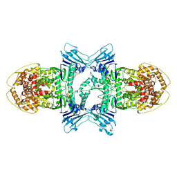
 | | Structure of Gabija GajA in complex with DNA | | 分子名称: | CALCIUM ION, DNA (5'-D(P*AP*AP*AP*AP*TP*AP*AP*CP*CP*GP*GP*GP*TP*TP*AP*TP*TP*AP*A)-3'), DNA (5'-D(P*TP*TP*AP*AP*TP*AP*AP*CP*CP*CP*GP*GP*TP*TP*AP*TP*TP*TP*T)-3'), ... | | 著者 | Li, J, Wang, Z, Wang, L. | | 登録日 | 2023-10-30 | | 公開日 | 2024-02-28 | | 最終更新日 | 2024-05-22 | | 実験手法 | ELECTRON MICROSCOPY (3.12 Å) | | 主引用文献 | Structures and activation mechanism of the Gabija anti-phage system.
Nature, 629, 2024
|
|
8X5N
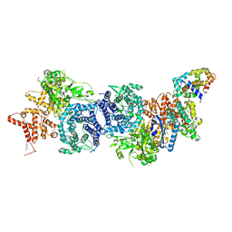
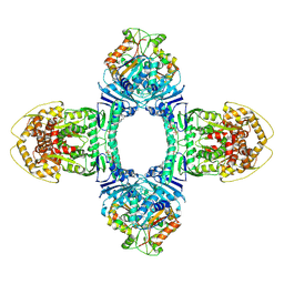 | | Tetramer Gabija with ATP | | 分子名称: | ADENOSINE-5'-TRIPHOSPHATE, Endonuclease GajA, Gabija protein GajB, ... | | 著者 | Li, J, Wang, Z, Wang, L. | | 登録日 | 2023-11-17 | | 公開日 | 2024-02-28 | | 最終更新日 | 2024-05-22 | | 実験手法 | ELECTRON MICROSCOPY (2.8 Å) | | 主引用文献 | Structures and activation mechanism of the Gabija anti-phage system.
Nature, 629, 2024
|
|
8XYB
 | |
8XY7
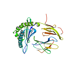
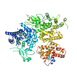 | | hPhK alpha-gamma subcomplex in active state | | 分子名称: | FARNESYL, Phosphorylase b kinase gamma catalytic chain, skeletal muscle/heart isoform, ... | | 著者 | Yang, X.K, Xiao, J.Y. | | 登録日 | 2024-01-19 | | 公開日 | 2024-04-03 | | 最終更新日 | 2024-04-10 | | 実験手法 | ELECTRON MICROSCOPY (2.9 Å) | | 主引用文献 | Architecture and activation of human muscle phosphorylase kinase.
Nat Commun, 15, 2024
|
|
7YQE
 | |
8XYA
 | |
7BWN
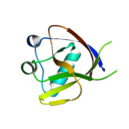
 | | Crystal Structure of a Designed Protein Heterocatenane | | 分子名称: | Cellular tumor antigen p53, Chimera of Green fluorescent protein and p53dim | | 著者 | Liu, Y.J, Duan, Z.L, Fang, J, Zhang, F, Xiao, J.Y, Zhang, W.B. | | 登録日 | 2020-04-15 | | 公開日 | 2020-06-17 | | 最終更新日 | 2023-11-29 | | 実験手法 | X-RAY DIFFRACTION (2.396 Å) | | 主引用文献 | Cellular Synthesis and X-ray Crystal Structure of a Designed Protein Heterocatenane.
Angew.Chem.Int.Ed.Engl., 59, 2020
|
|


