7KD7
 
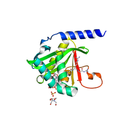 | |
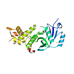
3BIY
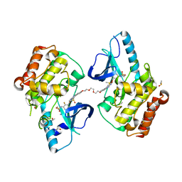
 | | Crystal structure of p300 histone acetyltransferase domain in complex with a bisubstrate inhibitor, Lys-CoA | | 分子名称: | BROMIDE ION, Histone acetyltransferase p300, [(2R,3S,4R,5R)-5-(6-amino-9H-purin-9-yl)-4-hydroxy-3-(phosphonooxy)tetrahydrofuran-2-yl]methyl (3R,20R)-20-carbamoyl-3-hydroxy-2,2-dimethyl-4,8,14,22-tetraoxo-12-thia-5,9,15,21-tetraazatricos-1-yl dihydrogen diphosphate | | 著者 | Liu, X, Wang, L, Zhao, K, Thompson, P.R, Hwang, Y, Marmorstein, R, Cole, P.A. | | 登録日 | 2007-12-02 | | 公開日 | 2008-02-12 | | 最終更新日 | 2024-02-21 | | 実験手法 | X-RAY DIFFRACTION (1.7 Å) | | 主引用文献 | The structural basis of protein acetylation by the p300/CBP transcriptional coactivator
Nature, 451, 2008
|
|
2R7G
 | |
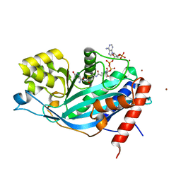
5ICV
 | | Crystal structure of human NatF (hNaa60) bound to a bisubstrate analogue | | 分子名称: | MET-LYS-ALA-VAL-LIG, N-alpha-acetyltransferase 60, [5-(6-amino-9H-purin-9-yl)-4-hydroxy-3-(phosphonooxy)furan-2-yl]methyl (3R)-4-{[3-({(E)-2-[(2,2-dihydroxyethyl)sulfanyl]ethenyl}amino)-3-oxopropyl]amino}-3-hydroxy-2,2-dimethyl-4-oxobutyl dihydrogen diphosphate | | 著者 | Stove, S.I, Magin, R.S, Marmorstein, R, Arnesen, T. | | 登録日 | 2016-02-23 | | 公開日 | 2016-06-22 | | 最終更新日 | 2024-01-10 | | 実験手法 | X-RAY DIFFRACTION (1.53 Å) | | 主引用文献 | Crystal Structure of the Golgi-Associated Human N alpha-Acetyltransferase 60 Reveals the Molecular Determinants for Substrate-Specific Acetylation.
Structure, 24, 2016
|
|
6UV5
 | |
5ICW
 | | Crystal structure of human NatF (hNaa60) homodimer bound to Coenzyme A | | 分子名称: | CHLORIDE ION, COENZYME A, N-alpha-acetyltransferase 60 | | 著者 | Stove, S.I, Magin, R.S, Marmorstein, R, Arnesen, T. | | 登録日 | 2016-02-23 | | 公開日 | 2016-06-22 | | 最終更新日 | 2024-01-10 | | 実験手法 | X-RAY DIFFRACTION (1.951 Å) | | 主引用文献 | Crystal Structure of the Golgi-Associated Human N alpha-Acetyltransferase 60 Reveals the Molecular Determinants for Substrate-Specific Acetylation.
Structure, 24, 2016
|
|
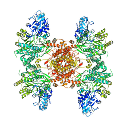
6UUW
 | | Structure of human ATP citrate lyase E599Q mutant in complex with Mg2+, citrate, ATP and CoA | | 分子名称: | (2S)-2-hydroxy-2-[2-oxo-2-(phosphonooxy)ethyl]butanedioic acid, ADENOSINE-5'-DIPHOSPHATE, ATP-citrate synthase, ... | | 著者 | Wei, X, Marmorstein, R. | | 登録日 | 2019-11-01 | | 公開日 | 2019-12-25 | | 最終更新日 | 2024-05-29 | | 実験手法 | ELECTRON MICROSCOPY (2.85 Å) | | 主引用文献 | Molecular basis for acetyl-CoA production by ATP-citrate lyase
Nat.Struct.Mol.Biol., 27, 2020
|
|
5J8C
 | |
5J8F
 | |
6UI9
 | |
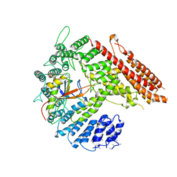
5JRQ
 | | BRAFV600E Kinase Domain In Complex with Chemically Linked Vemurafenib Inhibitor VEM-6-VEM | | 分子名称: | DIMETHYL SULFOXIDE, GLYCEROL, N-{2,4-difluoro-3-[5-(4-methoxyphenyl)-1H-pyrrolo[2,3-b]pyridine-3-carbonyl]phenyl}propane-1-sulfonamide, ... | | 著者 | Grasso, M.J, Marmorstein, R. | | 登録日 | 2016-05-06 | | 公開日 | 2016-09-14 | | 最終更新日 | 2023-10-18 | | 実験手法 | X-RAY DIFFRACTION (2.287 Å) | | 主引用文献 | Chemically Linked Vemurafenib Inhibitors Promote an Inactive BRAF(V600E) Conformation.
Acs Chem.Biol., 11, 2016
|
|
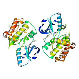
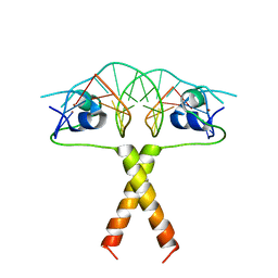
6VP9
 | | Cryo-EM structure of human NatB complex | | 分子名称: | CARBOXYMETHYL COENZYME *A, MDVFM peptide, N-alpha-acetyltransferase 20, ... | | 著者 | Deng, S, Marmorstein, R. | | 登録日 | 2020-02-02 | | 公開日 | 2020-09-23 | | 最終更新日 | 2024-03-06 | | 実験手法 | ELECTRON MICROSCOPY (3.46 Å) | | 主引用文献 | Molecular basis for N-terminal alpha-synuclein acetylation by human NatB.
Elife, 9, 2020
|
|
5JSM
 | |
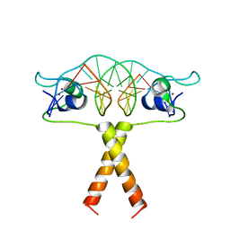
2B9D
 | | Crystal Structure of HPV E7 CR3 domain | | 分子名称: | E7 protein, ZINC ION | | 著者 | Liu, X, Clements, A, Zhao, K, Marmorstein, R. | | 登録日 | 2005-10-11 | | 公開日 | 2005-10-25 | | 最終更新日 | 2024-02-14 | | 実験手法 | X-RAY DIFFRACTION (1.6 Å) | | 主引用文献 | Structure of the human Papillomavirus E7 oncoprotein and its mechanism for inactivation of the retinoblastoma tumor suppressor.
J.Biol.Chem., 281, 2006
|
|
2ERE
 | |
2ER8
 | |
2ERG
 | |
7STX
 | |
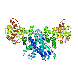
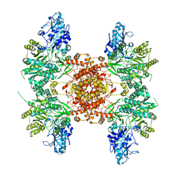
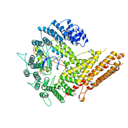
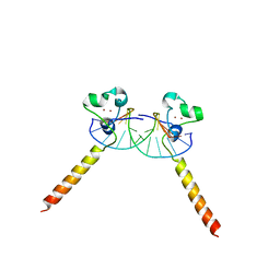
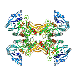
7SNI
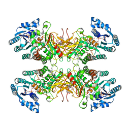 | | Structure of G6PD-D200N tetramer bound to NADP+ and G6P | | 分子名称: | 6-O-phosphono-beta-D-glucopyranose, Glucose-6-phosphate 1-dehydrogenase, NADP NICOTINAMIDE-ADENINE-DINUCLEOTIDE PHOSPHATE | | 著者 | Wei, X, Marmorstein, R. | | 登録日 | 2021-10-28 | | 公開日 | 2022-07-13 | | 最終更新日 | 2024-06-05 | | 実験手法 | ELECTRON MICROSCOPY (2.5 Å) | | 主引用文献 | Allosteric role of a structural NADP + molecule in glucose-6-phosphate dehydrogenase activity.
Proc.Natl.Acad.Sci.USA, 119, 2022
|
|
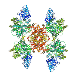
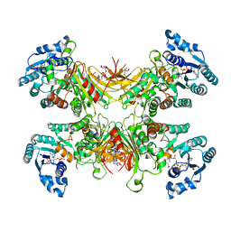
7SNG
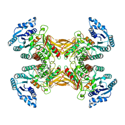 | | structure of G6PD-WT tetramer | | 分子名称: | Glucose-6-phosphate 1-dehydrogenase | | 著者 | Wei, X, Marmorstein, R. | | 登録日 | 2021-10-28 | | 公開日 | 2022-07-13 | | 最終更新日 | 2024-06-05 | | 実験手法 | ELECTRON MICROSCOPY (2.8 Å) | | 主引用文献 | Allosteric role of a structural NADP + molecule in glucose-6-phosphate dehydrogenase activity.
Proc.Natl.Acad.Sci.USA, 119, 2022
|
|
7TOE
 | |
7TOF
 | |
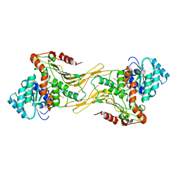
7SNH
 | | Structure of G6PD-D200N tetramer bound to NADP+ | | 分子名称: | Glucose-6-phosphate 1-dehydrogenase, NADP NICOTINAMIDE-ADENINE-DINUCLEOTIDE PHOSPHATE | | 著者 | Wei, X, Marmorstein, R. | | 登録日 | 2021-10-28 | | 公開日 | 2022-07-13 | | 最終更新日 | 2024-06-05 | | 実験手法 | ELECTRON MICROSCOPY (2.2 Å) | | 主引用文献 | Allosteric role of a structural NADP + molecule in glucose-6-phosphate dehydrogenase activity.
Proc.Natl.Acad.Sci.USA, 119, 2022
|
|
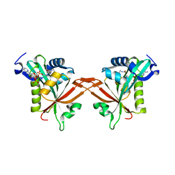
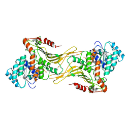
7SNF
 | | Structure of G6PD-WT dimer | | 分子名称: | Glucose-6-phosphate 1-dehydrogenase | | 著者 | Wei, X, Marmorstein, R. | | 登録日 | 2021-10-28 | | 公開日 | 2022-07-13 | | 最終更新日 | 2024-06-05 | | 実験手法 | ELECTRON MICROSCOPY (3.5 Å) | | 主引用文献 | Allosteric role of a structural NADP + molecule in glucose-6-phosphate dehydrogenase activity.
Proc.Natl.Acad.Sci.USA, 119, 2022
|
|
7UAL
 | |


