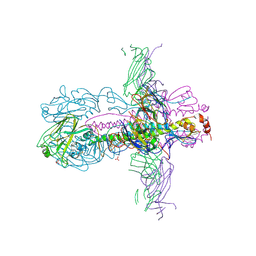
3J5Y
 
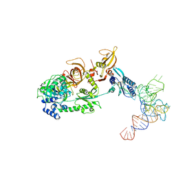 | | Structure of the mammalian ribosomal pre-termination complex associated with eRF1-eRF3-GDPNP | | 分子名称: | 5'-R(*AP*UP*UP*GP*UP*AP*AP*AP*AP*A)-3', Eukaryotic peptide chain release factor GTP-binding subunit ERF3A, Eukaryotic peptide chain release factor subunit 1, ... | | 著者 | des Georges, A, Hashem, Y, Unbehaun, A, Grassucci, R.A, Taylor, D, Hellen, C.U.T, Pestova, T.V, Frank, J. | | 登録日 | 2013-11-21 | | 公開日 | 2013-12-25 | | 最終更新日 | 2018-07-18 | | 実験手法 | ELECTRON MICROSCOPY (9.7 Å) | | 主引用文献 | Structure of the mammalian ribosomal pre-termination complex associated with eRF1*eRF3*GDPNP.
Nucleic Acids Res., 42, 2014
|
|
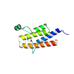
1FCX
 | | ISOTYPE SELECTIVITY OF THE HUMAN RETINOIC ACID NUCLEAR RECEPTOR HRAR: THE COMPLEX WITH THE RARGAMMA-SELECTIVE RETINOID BMS184394 | | 分子名称: | 6-[HYDROXY-(5,5,8,8-TETRAMETHYL-5,6,7,8-TETRAHYDRO-NAPHTALEN-2-YL)-METHYL]-NAPHTALENE-2-CARBOXYLIC ACID, DODECYL-ALPHA-D-MALTOSIDE, RETINOIC ACID RECEPTOR GAMMA-1 | | 著者 | Klaholz, B.P, Mitschler, A, Moras, D, Structural Proteomics in Europe (SPINE) | | 登録日 | 2000-07-19 | | 公開日 | 2000-09-11 | | 最終更新日 | 2023-08-09 | | 実験手法 | X-RAY DIFFRACTION (1.47 Å) | | 主引用文献 | Structural basis for isotype selectivity of the human retinoic acid nuclear receptor.
J.Mol.Biol., 302, 2000
|
|
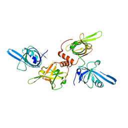
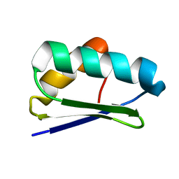
3K2J
 | | Crystal Structure of the 3rd Bromodomain of Human Poly-bromodomain containing protein 1 (PB1) | | 分子名称: | CHLORIDE ION, Protein polybromo-1, SULFATE ION | | 著者 | Filippakopoulos, P, Picaud, S, Keates, T, Chaikuad, A, Pike, A.C.W, Krojer, T, Sethi, R, von Delft, F, Arrowsmith, C.H, Edwards, A, Weigelt, J, Bountra, C, Knapp, S, Structural Genomics Consortium (SGC) | | 登録日 | 2009-09-30 | | 公開日 | 2009-10-13 | | 最終更新日 | 2023-11-01 | | 実験手法 | X-RAY DIFFRACTION (2.2 Å) | | 主引用文献 | Crystal Structure of the 3rd Bromodomain of Human Poly-bromodomain containing protein 1 (PB1)
To be Published
|
|
1FGU
 | |
5CFJ
 | | Structural and functional attributes of malaria parasite Ap4A hydrolase | | 分子名称: | BIS(5'-nucleosyl)-tetraphosphatase (Diadenosine tetraphosphatase), putative, DI(HYDROXYETHYL)ETHER, ... | | 著者 | Sharma, A, Yogavel, M, Sharma, A. | | 登録日 | 2015-07-08 | | 公開日 | 2016-02-10 | | 最終更新日 | 2024-03-20 | | 実験手法 | X-RAY DIFFRACTION (1.25 Å) | | 主引用文献 | Structural and functional attributes of malaria parasite diadenosine tetraphosphate hydrolase
Sci Rep, 6, 2016
|
|
3ZTN
 | | STRUCTURE OF INFLUENZA A NEUTRALIZING ANTIBODY SELECTED FROM CULTURES OF SINGLE HUMAN PLASMA CELLS IN COMPLEX WITH HUMAN H1 INFLUENZA HAEMAGGLUTININ. | | 分子名称: | 2-acetamido-2-deoxy-beta-D-glucopyranose, 2-acetamido-2-deoxy-beta-D-glucopyranose-(1-4)-2-acetamido-2-deoxy-beta-D-glucopyranose, FI6V3 ANTIBODY LIGHT CHAIN, ... | | 著者 | Hubbard, P.A, Ritchie, A.J, Corti, D, Voss, J.E, Gamblin, S.J, Codoni, G, Macagno, A, Jarrossay, D, Pinna, D, Minola, A, Vanzetta, F, Silacci, C, Fernandez-Rodriguez, B.M, Agatic, G, Giacchetto-Sasselli, I, Vachieri, S.G, Sallusto, F, Collins, P.J, Haire, L.F, Temperton, N, Langedijk, J.P.M, Skehel, J.J, Lanzavecchia, A. | | 登録日 | 2011-07-12 | | 公開日 | 2011-08-10 | | 最終更新日 | 2023-12-20 | | 実験手法 | X-RAY DIFFRACTION (3.001 Å) | | 主引用文献 | A Neutralizing Antibody Selected from Plasma Cells that Binds to Group 1 and Group 2 Influenza a Hemagglutinins.
Science, 333, 2011
|
|
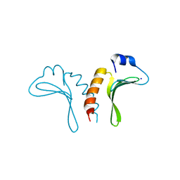
2K2P
 | | Solution NMR structure of protein Atu1203 from Agrobacterium tumefaciens. Northeast Structural Genomics Consortium (NESG) target AtT10, Ontario Center for Structural Proteomics target ATC1183 | | 分子名称: | Uncharacterized protein Atu1203 | | 著者 | Lemak, A, Gutmanas, A, Yee, A, Semesi, A, Arrowsmith, C.H, Northeast Structural Genomics Consortium (NESG) | | 登録日 | 2008-04-08 | | 公開日 | 2008-04-22 | | 最終更新日 | 2024-05-01 | | 実験手法 | SOLUTION NMR | | 主引用文献 | Solution structure of protein Atu1203 from Agrobacterium tumefaciens.
To be Published
|
|
3JTZ
 | | Structure of the arm-type binding domain of HPI integrase | | 分子名称: | Integrase, SODIUM ION | | 著者 | Szwagierczak, A, Antonenka, U, Popowicz, G.M, Sitar, T, Holak, T.A, Rakin, A. | | 登録日 | 2009-09-14 | | 公開日 | 2009-10-06 | | 最終更新日 | 2024-03-20 | | 実験手法 | X-RAY DIFFRACTION (1.3 Å) | | 主引用文献 | Structures of the arm-type binding domains of HPI and HAI7 integrases
J.Biol.Chem., 284, 2009
|
|
3R02
 | | The discovery of novel benzofuran-2-carboxylic acids as potent Pim-1 inhibitors | | 分子名称: | 7-[(cis-4-aminocyclohexyl)amino]-5-bromo-1-benzofuran-2-carboxylic acid, IMIDAZOLE, Proto-oncogene serine/threonine-protein kinase pim-1 | | 著者 | Xiang, Y, Hirth, B, Asmussen, G, Biemann, H.-P, Good, A, Fitzgerald, M, Gladysheva, T, Jancsics, K, Liu, J, Metz, M, Papoulis, A, Skerlj, R, Stepp, D.J, Wei, R.R. | | 登録日 | 2011-03-07 | | 公開日 | 2011-05-11 | | 最終更新日 | 2024-02-21 | | 実験手法 | X-RAY DIFFRACTION (1.95 Å) | | 主引用文献 | The discovery of novel benzofuran-2-carboxylic acids as potent Pim-1 inhibitors.
Bioorg.Med.Chem.Lett., 21, 2011
|
|
4YEH
 | | Crystal structure of Mg2+ ion containing hemopexin fold from Kabuli chana (chickpea white) at 2.45A resolution reveals a structural basis of metal ion transport | | 分子名称: | Lectin, MAGNESIUM ION | | 著者 | Kumar, S, Singh, A, Yamini, S, Bhushan, A, Dey, S, Sharma, S, Singh, T.P. | | 登録日 | 2015-02-24 | | 公開日 | 2015-03-25 | | 最終更新日 | 2023-11-08 | | 実験手法 | X-RAY DIFFRACTION (2.45 Å) | | 主引用文献 | Crystal Structure of Mg(2+) Containing Hemopexin-Fold Protein from Kabuli Chana (Chickpea-White, CW-25) at 2.45 angstrom Resolution Reveals Its Metal Ion Transport Property
Protein J., 34, 2015
|
|
4H8J
 | | Structure of GluA2-LBD in complex with MES | | 分子名称: | 1,2-ETHANEDIOL, 2-(N-MORPHOLINO)-ETHANESULFONIC ACID, Glutamate receptor 2, ... | | 著者 | Reiter, A, Skerra, A, Trauner, D, Schiefner, A. | | 登録日 | 2012-09-22 | | 公開日 | 2013-09-25 | | 最終更新日 | 2023-09-20 | | 実験手法 | X-RAY DIFFRACTION (1.8 Å) | | 主引用文献 | Structural basis of an artificial photoreceptor
To be Published
|
|
6S90
 | | BTK in complex with an inhibitor | | 分子名称: | 4-~{tert}-butyl-~{N}-[2-methyl-3-[6-[4-(4-methylpiperazin-1-yl)carbonylphenyl]-7~{H}-pyrrolo[2,3-d]pyrimidin-4-yl]phenyl]benzamide, Tyrosine-protein kinase BTK | | 著者 | Gutmann, S, Hinniger, A. | | 登録日 | 2019-07-11 | | 公開日 | 2019-09-18 | | 最終更新日 | 2024-05-15 | | 実験手法 | X-RAY DIFFRACTION (1.82 Å) | | 主引用文献 | Design of Potent and Selective Covalent Inhibitors of Bruton's Tyrosine Kinase Targeting an Inactive Conformation.
Acs Med.Chem.Lett., 10, 2019
|
|
3JZJ
 | | Crystal structures of the GacH receptor of Streptomyces glaucescens GLA.O in the unliganded form and in complex with acarbose and an acarbose homolog. Comparison with acarbose-loaded maltose binding protein of Salmonella typhimurium. | | 分子名称: | 4,6-dideoxy-4-{[(1S,4R,5S,6S)-4,5,6-trihydroxy-3-(hydroxymethyl)cyclohex-2-en-1-yl]amino}-alpha-D-glucopyranose-(1-4)-alpha-D-glucopyranose-(1-4)-alpha-D-glucopyranose, Acarbose/maltose binding protein GacH, SULFATE ION | | 著者 | Vahedi-Faridi, A, Licht, A, Bulut, H, Schneider, E. | | 登録日 | 2009-09-23 | | 公開日 | 2010-02-16 | | 最終更新日 | 2024-02-21 | | 実験手法 | X-RAY DIFFRACTION (1.4 Å) | | 主引用文献 | Crystal Structures of the Solute Receptor GacH of Streptomyces glaucescens in Complex with Acarbose and an Acarbose Homolog: Comparison with the Acarbose-Loaded Maltose-Binding Protein of Salmonella typhimurium.
J.Mol.Biol., 397, 2010
|
|
4YN8
 | | Crystal Structure of Response Regulator ChrA in Heme-Sensing Two Component System | | 分子名称: | MAGNESIUM ION, Response regulator ChrA, SULFATE ION | | 著者 | Doi, A, Nakamura, H, Shiro, Y, Sugimoto, H. | | 登録日 | 2015-03-09 | | 公開日 | 2015-08-19 | | 最終更新日 | 2023-11-08 | | 実験手法 | X-RAY DIFFRACTION (1.8 Å) | | 主引用文献 | Structure of the response regulator ChrA in the haem-sensing two-component system of Corynebacterium diphtheriae.
Acta Crystallogr.,Sect.F, 71, 2015
|
|
7AKR
 | |
7AKS
 | |
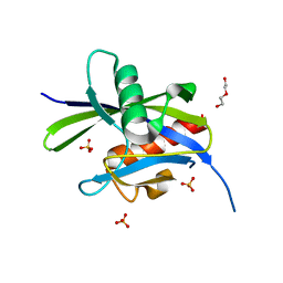
5CFI
 | |
5CHT
 | | Crystal structure of USP18 | | 分子名称: | Ubl carboxyl-terminal hydrolase 18, ZINC ION | | 著者 | Fritz, G, Basters, A. | | 登録日 | 2015-07-10 | | 公開日 | 2016-06-29 | | 最終更新日 | 2024-01-10 | | 実験手法 | X-RAY DIFFRACTION (2.8 Å) | | 主引用文献 | Structural basis of the specificity of USP18 toward ISG15.
Nat. Struct. Mol. Biol., 24, 2017
|
|
1K5D
 | | Crystal structure of Ran-GPPNHP-RanBP1-RanGAP complex | | 分子名称: | GTP-binding nuclear protein RAN, MAGNESIUM ION, PHOSPHOAMINOPHOSPHONIC ACID-GUANYLATE ESTER, ... | | 著者 | Seewald, M.J, Koerner, C, Wittinghofer, A, Vetter, I.R. | | 登録日 | 2001-10-10 | | 公開日 | 2002-02-13 | | 最終更新日 | 2023-08-16 | | 実験手法 | X-RAY DIFFRACTION (2.7 Å) | | 主引用文献 | RanGAP mediates GTP hydrolysis without an arginine finger.
Nature, 415, 2002
|
|
1FAI
 | | THREE-DIMENSIONAL STRUCTURE OF TWO CRYSTAL FORMS OF FAB R19.9, FROM A MONOCLONAL ANTI-ARSONATE ANTIBODY | | 分子名称: | IGG2B-KAPPA R19.9 FAB (HEAVY CHAIN), IGG2B-KAPPA R19.9 FAB (LIGHT CHAIN) | | 著者 | Lascombe, M.B, Alzari, P.M, Poljak, R.J, Nisonoff, A. | | 登録日 | 1992-05-27 | | 公開日 | 1992-10-15 | | 最終更新日 | 2024-06-05 | | 実験手法 | X-RAY DIFFRACTION (2.7 Å) | | 主引用文献 | Three-dimensional structure of two crystal forms of FabR19.9 from a monoclonal anti-arsonate antibody.
Proc.Natl.Acad.Sci.USA, 89, 1992
|
|
3JU0
 | | Structure of the arm-type binding domain of HAI7 integrase | | 分子名称: | Phage integrase | | 著者 | Szwagierczak, A, Antonenka, U, Popowicz, G.M, Sitar, T, Holak, T.A, Rakin, A. | | 登録日 | 2009-09-14 | | 公開日 | 2009-10-06 | | 最終更新日 | 2023-11-01 | | 実験手法 | X-RAY DIFFRACTION (1.6 Å) | | 主引用文献 | Structures of the arm-type binding domains of HPI and HAI7 integrases
J.Biol.Chem., 284, 2009
|
|
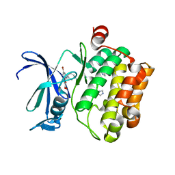
2WEJ
 | | Thermodynamic Optimisation of Carbonic Anhydrase Fragment Inhibitors | | 分子名称: | CARBONIC ANHYDRASE 2, GLYCEROL, ZINC ION, ... | | 著者 | Scott, A.D, Phillips, C, Alex, A, Bent, A, O'Brien, R, Damian, L, Jones, L.H. | | 登録日 | 2009-03-31 | | 公開日 | 2009-11-10 | | 最終更新日 | 2024-05-08 | | 実験手法 | X-RAY DIFFRACTION (1.45 Å) | | 主引用文献 | Thermodynamic Optimisation in Drug Discovery: A Case Study Using Carbonic Anhydrase Inhibitors.
Chemmedchem, 4, 2009
|
|
3J7H
 | | Structure of beta-galactosidase at 3.2-A resolution obtained by cryo-electron microscopy | | 分子名称: | Beta-galactosidase, MAGNESIUM ION | | 著者 | Bartesaghi, A, Matthies, D, Banerjee, S, Merk, A, Subramaniam, S. | | 登録日 | 2014-06-30 | | 公開日 | 2014-07-30 | | 最終更新日 | 2024-02-21 | | 実験手法 | ELECTRON MICROSCOPY (3.2 Å) | | 主引用文献 | Structure of beta-galactosidase at 3.2- angstrom resolution obtained by cryo-electron microscopy.
Proc.Natl.Acad.Sci.USA, 111, 2014
|
|
5CNN
 | | Crystal structure of the EGFR kinase domain mutant I682Q | | 分子名称: | Epidermal growth factor receptor, MAGNESIUM ION, PHOSPHOAMINOPHOSPHONIC ACID-ADENYLATE ESTER | | 著者 | Kovacs, E, Das, R, Mirza, A, Jura, N, Barros, T, Kuriyan, J. | | 登録日 | 2015-07-17 | | 公開日 | 2015-07-29 | | 最終更新日 | 2023-09-27 | | 実験手法 | X-RAY DIFFRACTION (1.9 Å) | | 主引用文献 | Analysis of the Role of the C-Terminal Tail in the Regulation of the Epidermal Growth Factor Receptor.
Mol.Cell.Biol., 35, 2015
|
|
3JZA
 | | Crystal structure of human Rab1b in complex with the GEF domain of DrrA/SidM from Legionella pneumophila | | 分子名称: | PHOSPHATE ION, Ras-related protein Rab-1B, Uncharacterized protein DrrA | | 著者 | Schoebel, S, Oesterlin, L.K, Blankenfeldt, W, Goody, R.S, Itzen, A. | | 登録日 | 2009-09-23 | | 公開日 | 2010-01-19 | | 最終更新日 | 2024-02-21 | | 実験手法 | X-RAY DIFFRACTION (1.8 Å) | | 主引用文献 | RabGDI displacement by DrrA from Legionella is a consequence of its guanine nucleotide exchange activity.
Mol.Cell, 36, 2009
|
|


