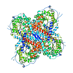
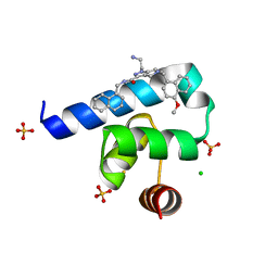
6MBW
 
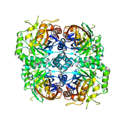
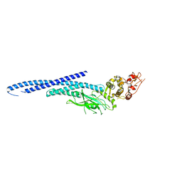 | | Structure of Transcription Factor | | 分子名称: | Signal transducer and activator of transcription 5B | | 著者 | Seo, H.-S, Dhe-Paganon, S. | | 登録日 | 2018-08-30 | | 公開日 | 2019-06-19 | | 最終更新日 | 2023-10-11 | | 実験手法 | X-RAY DIFFRACTION (3.29 Å) | | 主引用文献 | Structural and functional consequences of the STAT5BN642H driver mutation.
Nat Commun, 10, 2019
|
|
4BNA
 | |
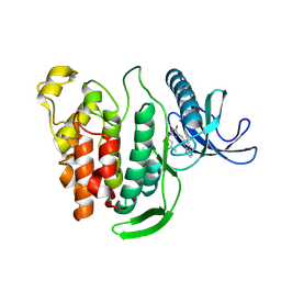
8HUB
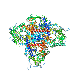 | | AMP deaminase 2 in complex with an inhibitor | | 分子名称: | 3,3-dimethyl-4-(phenylmethyl)-2~{H}-quinoxaline-1-carboxamide, AMP deaminase 2, ZINC ION | | 著者 | Adachi, T, Doi, S. | | 登録日 | 2022-12-23 | | 公開日 | 2023-01-18 | | 最終更新日 | 2024-05-29 | | 実験手法 | X-RAY DIFFRACTION (3.25 Å) | | 主引用文献 | The discovery of 3,3-dimethyl-1,2,3,4-tetrahydroquinoxaline-1-carboxamides as AMPD2 inhibitors with a novel mechanism of action.
Bioorg.Med.Chem.Lett., 80, 2023
|
|
6R76
 | |
7DRE
 | | Cryo-EM structure of DfgA-B at 2.54 angstrom resolution | | 分子名称: | DfgB, Sugar phosphate isomerase/epimerase | | 著者 | Mori, T, Moriya, T, Adachi, N, Senda, T, Abe, I. | | 登録日 | 2020-12-28 | | 公開日 | 2021-12-08 | | 最終更新日 | 2024-06-05 | | 実験手法 | ELECTRON MICROSCOPY (2.54 Å) | | 主引用文献 | C-Glycoside metabolism in the gut and in nature: Identification, characterization, structural analyses and distribution of C-C bond-cleaving enzymes.
Nat Commun, 12, 2021
|
|
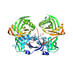
8HU6
 | | AMP deaminase 2 in complex with AMP | | 分子名称: | ADENOSINE MONOPHOSPHATE, AMP deaminase 2, SULFATE ION, ... | | 著者 | Adachi, T, Doi, S. | | 登録日 | 2022-12-22 | | 公開日 | 2023-01-18 | | 最終更新日 | 2024-05-29 | | 実験手法 | X-RAY DIFFRACTION (2.33 Å) | | 主引用文献 | The discovery of 3,3-dimethyl-1,2,3,4-tetrahydroquinoxaline-1-carboxamides as AMPD2 inhibitors with a novel mechanism of action.
Bioorg.Med.Chem.Lett., 80, 2023
|
|
7DRD
 | | Cryo-EM structure of DgpB-C at 2.85 angstrom resolution | | 分子名称: | AP_endonuc_2 domain-containing protein, DgpB | | 著者 | Mori, T, Moriya, T, Adachi, N, Senda, T, Abe, I. | | 登録日 | 2020-12-28 | | 公開日 | 2021-12-08 | | 最終更新日 | 2024-06-05 | | 実験手法 | ELECTRON MICROSCOPY (2.85 Å) | | 主引用文献 | C-Glycoside metabolism in the gut and in nature: Identification, characterization, structural analyses and distribution of C-C bond-cleaving enzymes.
Nat Commun, 12, 2021
|
|
5T8L
 | |
7BVS
 | | DfgA-DfgB complex apo | | 分子名称: | DfgB, GLYCEROL, MANGANESE (II) ION, ... | | 著者 | Mori, T, He, H, Abe, I. | | 登録日 | 2020-04-11 | | 公開日 | 2021-04-14 | | 最終更新日 | 2023-11-29 | | 実験手法 | X-RAY DIFFRACTION (2.85 Å) | | 主引用文献 | C-Glycoside metabolism in the gut and in nature: Identification, characterization, structural analyses and distribution of C-C bond-cleaving enzymes.
Nat Commun, 12, 2021
|
|
3BNA
 | |
5N8V
 | | Targeting the PEX14-PEX5 interaction by small molecules provides novel therapeutic routes to treat trypanosomiases. | | 分子名称: | 1-(2-azanylethyl)-5-[(4-methoxynaphthalen-1-yl)methyl]-~{N}-(naphthalen-1-ylmethyl)-6,7-dihydro-4~{H}-pyrazolo[4,3-c]pyridine-3-carboxamide, BETA-MERCAPTOETHANOL, CHLORIDE ION, ... | | 著者 | Dawidowski, M, Emmanouilidis, L, Sattler, M, Popowicz, G.M. | | 登録日 | 2017-02-24 | | 公開日 | 2017-03-15 | | 最終更新日 | 2024-05-08 | | 実験手法 | X-RAY DIFFRACTION (1.55 Å) | | 主引用文献 | Inhibitors of PEX14 disrupt protein import into glycosomes and kill Trypanosoma parasites.
Science, 355, 2017
|
|
5T8N
 | |
8P08
 | | Crystal structure of human CLK1 in complex with Leucettinib-21 | | 分子名称: | (4~{Z})-4-(1,3-benzothiazol-6-ylmethylidene)-2-[[(2~{R})-1-methoxy-4-methyl-pentan-2-yl]amino]-1~{H}-imidazol-5-one, Dual specificity protein kinase CLK1 | | 著者 | Kraemer, A, Schroeder, M, Meijer, L, Knapp, S, Structural Genomics Consortium (SGC) | | 登録日 | 2023-05-09 | | 公開日 | 2023-05-17 | | 最終更新日 | 2024-07-31 | | 実験手法 | X-RAY DIFFRACTION (2.4 Å) | | 主引用文献 | Chemical, Biochemical, Cellular, and Physiological Characterization of Leucettinib-21, a Down Syndrome and Alzheimer's Disease Drug Candidate.
J.Med.Chem., 66, 2023
|
|
7N5H
 | | Cryo-EM structure of broadly neutralizing antibody 2-36 in complex with prefusion SARS-CoV-2 spike glycoprotein | | 分子名称: | 2-36 Fab heavy chain, 2-36 Fab light chain, 2-acetamido-2-deoxy-beta-D-glucopyranose, ... | | 著者 | Casner, R.G, Cerutti, G, Shapiro, L. | | 登録日 | 2021-06-05 | | 公開日 | 2021-11-03 | | 最終更新日 | 2024-10-16 | | 実験手法 | ELECTRON MICROSCOPY (3.24 Å) | | 主引用文献 | A monoclonal antibody that neutralizes SARS-CoV-2 variants, SARS-CoV, and other sarbecoviruses.
Emerg Microbes Infect, 11, 2022
|
|
2V1X
 | | Crystal structure of human RECQ-like DNA helicase | | 分子名称: | 1,2-ETHANEDIOL, ADENOSINE-5'-DIPHOSPHATE, ATP-DEPENDENT DNA HELICASE Q1, ... | | 著者 | Pike, A.C.W, Shrestha, B, Burgess-Brown, N, King, O, Ugochukwu, E, Watt, S, Edwards, A, Arrowsmith, C.H, Weigelt, J, Sundstrom, M, Gileadi, O. | | 登録日 | 2007-05-30 | | 公開日 | 2007-07-03 | | 最終更新日 | 2023-12-13 | | 実験手法 | X-RAY DIFFRACTION (2 Å) | | 主引用文献 | Structure of the Human Recq1 Helicase Reveals a Putative Strand-Separation Pin.
Proc.Natl.Acad.Sci.USA, 106, 2009
|
|
2DVJ
 | | phosphorylated Crk-II | | 分子名称: | V-crk sarcoma virus CT10 oncogene homolog, isoform a | | 著者 | Kobashigawa, Y, Inagaki, F. | | 登録日 | 2006-07-31 | | 公開日 | 2007-05-08 | | 最終更新日 | 2022-03-09 | | 実験手法 | SOLUTION NMR | | 主引用文献 | Structural basis for the transforming activity of human cancer-related signaling adaptor protein CRK.
Nat.Struct.Mol.Biol., 14, 2007
|
|
2RV0
 | | Solution structures of the DNA-binding domain (ZF12) of immune-related zinc-finger protein ZFAT | | 分子名称: | ZINC ION, Zinc finger protein ZFAT | | 著者 | Tochio, N, Umehara, T, Kigawa, T, Yokoyama, S. | | 登録日 | 2015-01-26 | | 公開日 | 2015-04-08 | | 最終更新日 | 2024-05-01 | | 実験手法 | SOLUTION NMR | | 主引用文献 | Solution structures of the DNA-binding domains of immune-related zinc-finger protein ZFAT
J.Struct.Funct.Genom., 16, 2015
|
|
2RV6
 | | Solution structures of the DNA-binding domains (ZF2-ZF3-ZF4) of immune-related zinc-finger protein ZFAT | | 分子名称: | ZINC ION, Zinc finger protein ZFAT | | 著者 | Tochio, N, Umehara, T, Kigawa, T, Yokoyama, S. | | 登録日 | 2015-01-26 | | 公開日 | 2015-04-08 | | 最終更新日 | 2024-05-01 | | 実験手法 | SOLUTION NMR | | 主引用文献 | Solution structures of the DNA-binding domains of immune-related zinc-finger protein ZFAT
J.Struct.Funct.Genom., 16, 2015
|
|
8CJ7
 | | HDAC6 selective degraded (difluoromethyl)-1,3,4-oxadiazole substrate inhibitor | | 分子名称: | 6-[(5-pyridin-2-yl-1,2$l^{4},3,4-tetrazacyclopenta-1,3-dien-2-yl)methyl]pyridine-3-carbohydrazide, Histone deacetylase 6, IODIDE ION, ... | | 著者 | Sandmark, J, Ek, M, Ripa, L. | | 登録日 | 2023-02-12 | | 公開日 | 2023-10-18 | | 最終更新日 | 2023-11-01 | | 実験手法 | X-RAY DIFFRACTION (1.51 Å) | | 主引用文献 | Selective and Bioavailable HDAC6 2-(Difluoromethyl)-1,3,4-oxadiazole Substrate Inhibitors and Modeling of Their Bioactivation Mechanism.
J.Med.Chem., 66, 2023
|
|
2ZPD
 | | Crystal structure of the R43L mutant of LolA in the open form | | 分子名称: | Outer-membrane lipoprotein carrier protein | | 著者 | Takeda, K, Yokota, N, Oguchi, Y, Tokuda, H, Miki, K. | | 登録日 | 2008-07-10 | | 公開日 | 2008-08-05 | | 最終更新日 | 2023-11-01 | | 実験手法 | X-RAY DIFFRACTION (1.85 Å) | | 主引用文献 | Opening and closing of the hydrophobic cavity of LolA coupled to lipoprotein binding and release.
J.Biol.Chem., 283, 2008
|
|
2ZPC
 | | Crystal structure of the R43L mutant of LolA in the closed form | | 分子名称: | Outer-membrane lipoprotein carrier protein | | 著者 | Takeda, K, Yokota, N, Oguchi, Y, Tokuda, H, Miki, K. | | 登録日 | 2008-07-10 | | 公開日 | 2008-08-05 | | 最終更新日 | 2023-11-01 | | 実験手法 | X-RAY DIFFRACTION (2.35 Å) | | 主引用文献 | Opening and closing of the hydrophobic cavity of LolA coupled to lipoprotein binding and release.
J.Biol.Chem., 283, 2008
|
|
3VHQ
 | | Crystal structure of the Ca6 site mutant of Pro-SA-subtilisin | | 分子名称: | CALCIUM ION, Tk-subtilisin | | 著者 | Uehara, R, Takeuchi, Y, Tanaka, S, Matsumura, H, Koga, Y, Takano, K, Kanaya, S. | | 登録日 | 2011-09-01 | | 公開日 | 2012-07-11 | | 最終更新日 | 2024-10-16 | | 実験手法 | X-RAY DIFFRACTION (2.15 Å) | | 主引用文献 | Requirement of Ca(2+) Ions for the Hyperthermostability of Tk-Subtilisin from Thermococcus kodakarensis
Biochemistry, 51, 2012
|
|
1ZCA
 | |
3FKR
 | |
3FKK
 | | Structure of L-2-keto-3-deoxyarabonate dehydratase | | 分子名称: | L-2-keto-3-deoxyarabonate dehydratase, PHOSPHATE ION | | 著者 | Shimada, N, Mikami, B. | | 登録日 | 2008-12-17 | | 公開日 | 2010-01-12 | | 最終更新日 | 2024-03-20 | | 実験手法 | X-RAY DIFFRACTION (2.1 Å) | | 主引用文献 | Structural analysis of L -2-keto-3-deoxyarabonate dehydratase an enzyme involved in an alternative bacterial pathway of L-arabinose metabolism in complex with pyruvate
To be Published
|
|


