7BCY
 
 | |
7WIN
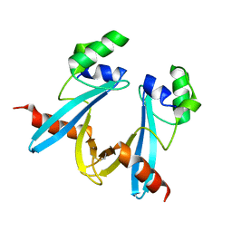 | |
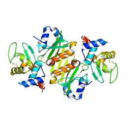
3O98
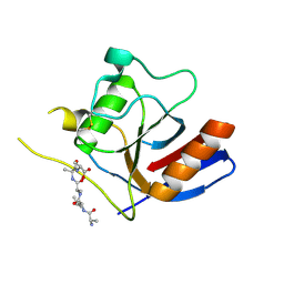
 | | Glutathionylspermidine synthetase/amidase C59A complex with ADP and Gsp | | 分子名称: | ADENOSINE-5'-DIPHOSPHATE, Bifunctional glutathionylspermidine synthetase/amidase, GLUTATHIONYLSPERMIDINE, ... | | 著者 | Pai, C.H, Lin, C.H, Wang, A.H.-J. | | 登録日 | 2010-08-04 | | 公開日 | 2011-03-02 | | 最終更新日 | 2023-11-01 | | 実験手法 | X-RAY DIFFRACTION (2.8 Å) | | 主引用文献 | Structure and mechanism of Escherichia coli glutathionylspermidine amidase belonging to the family of cysteine; histidine-dependent amidohydrolases/peptidases
Protein Sci., 20, 2011
|
|
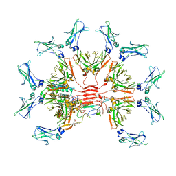
7Y09
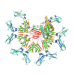 | | Cryo-EM structure of human IgM-Fc in complex with the J chain and the DBL domain of DBLMSP | | 分子名称: | 2-acetamido-2-deoxy-beta-D-glucopyranose, 2-acetamido-2-deoxy-beta-D-glucopyranose-(1-4)-2-acetamido-2-deoxy-beta-D-glucopyranose, Immunoglobulin J chain, ... | | 著者 | Shen, H, Ji, C, Xiao, J. | | 登録日 | 2022-06-03 | | 公開日 | 2023-03-29 | | 最終更新日 | 2024-05-08 | | 実験手法 | ELECTRON MICROSCOPY (3.71 Å) | | 主引用文献 | Plasmodium falciparum has evolved multiple mechanisms to hijack human immunoglobulin M.
Nat Commun, 14, 2023
|
|
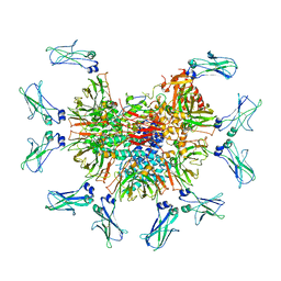
7YG2
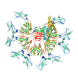 | | Cryo-EM structure of human IgM-Fc in complex with the J chain and the DBL domain of DBLMSP2 | | 分子名称: | 2-acetamido-2-deoxy-beta-D-glucopyranose, 2-acetamido-2-deoxy-beta-D-glucopyranose-(1-4)-2-acetamido-2-deoxy-beta-D-glucopyranose, DBLMSP2, ... | | 著者 | Shen, H, Ji, C, Xiao, J. | | 登録日 | 2022-07-09 | | 公開日 | 2023-03-29 | | 最終更新日 | 2024-05-08 | | 実験手法 | ELECTRON MICROSCOPY (3.32 Å) | | 主引用文献 | Plasmodium falciparum has evolved multiple mechanisms to hijack human immunoglobulin M.
Nat Commun, 14, 2023
|
|
7Y0J
 | |
7Y0H
 | |
7Y4H
 | |
3EQX
 | |
2IIZ
 | |
1AHC
 | |
7FEF
 | | Crystal structure of AtMBD6 with DNA | | 分子名称: | DNA (5'-D(*GP*CP*CP*AP*AP*(5CM)P*GP*TP*TP*GP*GP*C)-3'), Methyl-CpG-binding domain-containing protein 6 | | 著者 | Wu, Z.B, Liu, K, Min, J.R. | | 登録日 | 2021-07-19 | | 公開日 | 2021-12-29 | | 最終更新日 | 2023-11-29 | | 実験手法 | X-RAY DIFFRACTION (2.39 Å) | | 主引用文献 | Family-wide Characterization of Methylated DNA Binding Ability of Arabidopsis MBDs.
J.Mol.Biol., 434, 2022
|
|
7FEO
 | | Crystal structure of AtMBD5 MBD domain | | 分子名称: | Methyl-CpG-binding domain-containing protein 5, SULFATE ION | | 著者 | Zhou, M.Q, Wu, Z.B, Liu, K, Min, J.R, Structural Genomics Consortium (SGC) | | 登録日 | 2021-07-21 | | 公開日 | 2021-12-29 | | 最終更新日 | 2023-11-29 | | 実験手法 | X-RAY DIFFRACTION (2.2 Å) | | 主引用文献 | Family-wide Characterization of Methylated DNA Binding Ability of Arabidopsis MBDs.
J.Mol.Biol., 434, 2022
|
|
1AHA
 | | THE N-GLYCOSIDASE MECHANISM OF RIBOSOME-INACTIVATING PROTEINS IMPLIED BY CRYSTAL STRUCTURES OF ALPHA-MOMORCHARIN | | 分子名称: | ADENINE, ALPHA-MOMORCHARIN | | 著者 | Ren, J, Wang, Y, Dong, Y, Stuart, D.I. | | 登録日 | 1994-01-07 | | 公開日 | 1994-06-22 | | 最終更新日 | 2024-02-07 | | 実験手法 | X-RAY DIFFRACTION (2.2 Å) | | 主引用文献 | The N-glycosidase mechanism of ribosome-inactivating proteins implied by crystal structures of alpha-momorcharin.
Structure, 2, 1994
|
|
1AHB
 | | THE N-GLYCOSIDASE MECHANISM OF RIBOSOME-INACTIVATING PROTEINS IMPLIED BY CRYSTAL STRUCTURES OF ALPHA-MOMORCHARIN | | 分子名称: | ALPHA-MOMORCHARIN, FORMYCIN-5'-MONOPHOSPHATE | | 著者 | Ren, J, Wang, Y, Dong, Y, Stuart, D.I. | | 登録日 | 1994-01-07 | | 公開日 | 1994-06-22 | | 最終更新日 | 2024-02-07 | | 実験手法 | X-RAY DIFFRACTION (2.2 Å) | | 主引用文献 | The N-glycosidase mechanism of ribosome-inactivating proteins implied by crystal structures of alpha-momorcharin.
Structure, 2, 1994
|
|
7F2D
 | |
7F2I
 | |
5MKI
 | |
5MKL
 | |
7X2P
 | |
5MKM
 | |
3NG7
 | |
1I2A
 | |
1GPQ
 | | Structure of ivy complexed with its target, HEWL | | 分子名称: | INHIBITOR OF VERTEBRATE LYSOZYME, LYSOZYME C | | 著者 | Abergel, C, Monchois, V, Claverie, J.-M. | | 登録日 | 2001-11-08 | | 公開日 | 2003-03-11 | | 最終更新日 | 2023-12-13 | | 実験手法 | X-RAY DIFFRACTION (1.6 Å) | | 主引用文献 | Structure and Evolution of the Ivy Protein Family, Unexpected Lysozyme Inhibitors in Gram-Negative Bacteria.
Proc.Natl.Acad.Sci.USA, 104, 2007
|
|
8AF1
 | | Beta-Lytic Protease from Lysobacter capsici | | 分子名称: | CHLORIDE ION, FORMIC ACID, GLYCEROL, ... | | 著者 | Gabdulkhakov, A.G, Tishchenko, T.V, Kudryakova, I.V, Afoshin, A.S, Vasilyeva, N.V. | | 登録日 | 2022-07-15 | | 公開日 | 2023-08-16 | | 最終更新日 | 2024-02-28 | | 実験手法 | X-RAY DIFFRACTION (1.57 Å) | | 主引用文献 | Structural and Functional Characterization of beta-lytic Protease from Lysobacter capsici VKM B-2533 T.
Int J Mol Sci, 23, 2022
|
|


