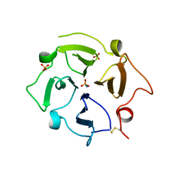
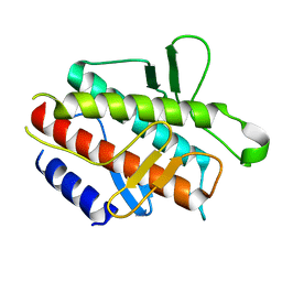
2MCU
 
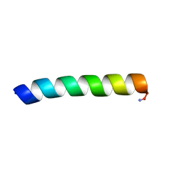 | | Solid-state NMR structure of piscidin 1 in aligned 3:1 phosphatidylcholine/phosphoglycerol lipid bilayers | | 分子名称: | Moronecidin | | 著者 | Fu, R, Tian, Y, Perrin Jr, B.S, Grant, C.V, Pastor, R.W, Cotten, M.L. | | 登録日 | 2013-08-27 | | 公開日 | 2014-01-22 | | 最終更新日 | 2014-03-19 | | 実験手法 | SOLID-STATE NMR | | 主引用文献 | High-resolution structures and orientations of antimicrobial peptides piscidin 1 and piscidin 3 in fluid bilayers reveal tilting, kinking, and bilayer immersion.
J.Am.Chem.Soc., 136, 2014
|
|
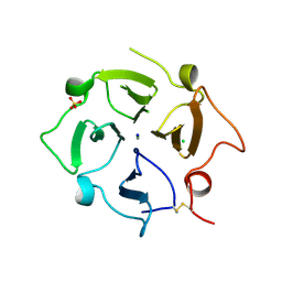
2M67
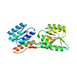
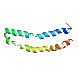 | | Full-length mercury transporter protein MerF in lipid bilayer membranes | | 分子名称: | MerF | | 著者 | Lu, G.J, Tian, Y, Vora, N, Marassi, F.M, Opella, S.J. | | 登録日 | 2013-03-27 | | 公開日 | 2013-07-03 | | 最終更新日 | 2024-05-15 | | 実験手法 | SOLID-STATE NMR | | 主引用文献 | The Structure of the Mercury Transporter MerF in Phospholipid Bilayers: A Large Conformational Rearrangement Results from N-Terminal Truncation.
J.Am.Chem.Soc., 135, 2013
|
|
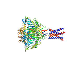
5ZCS
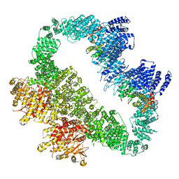
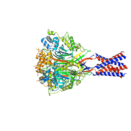
 | | 4.9 Angstrom Cryo-EM structure of human mTOR complex 2 | | 分子名称: | Rapamycin-insensitive companion of mTOR, Serine/threonine-protein kinase mTOR, Target of rapamycin complex 2 subunit MAPKAP1, ... | | 著者 | Chen, X, Liu, M, Tian, Y, Wang, H, Wang, J, Xu, Y. | | 登録日 | 2018-02-20 | | 公開日 | 2018-03-21 | | 最終更新日 | 2024-03-27 | | 実験手法 | ELECTRON MICROSCOPY (4.9 Å) | | 主引用文献 | Cryo-EM structure of human mTOR complex 2.
Cell Res., 28, 2018
|
|
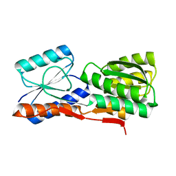
5YZ0
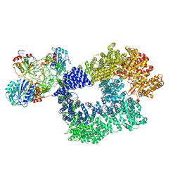
 | | Cryo-EM Structure of human ATR-ATRIP complex | | 分子名称: | ATR-interacting protein, Serine/threonine-protein kinase ATR | | 著者 | Rao, Q, Liu, M, Tian, Y, Wu, Z, Wang, H, Wang, J, Xu, Y. | | 登録日 | 2017-12-11 | | 公開日 | 2018-01-31 | | 最終更新日 | 2019-11-06 | | 実験手法 | ELECTRON MICROSCOPY (4.7 Å) | | 主引用文献 | Cryo-EM structure of human ATR-ATRIP complex.
Cell Res., 28, 2018
|
|
5Y3R
 | | Cryo-EM structure of Human DNA-PK Holoenzyme | | 分子名称: | DNA (34-MER), DNA (36-MER), DNA-dependent protein kinase catalytic subunit, ... | | 著者 | Yin, X, Liu, M, Tian, Y, Wang, J, Xu, Y. | | 登録日 | 2017-07-29 | | 公開日 | 2017-09-06 | | 最終更新日 | 2024-03-27 | | 実験手法 | ELECTRON MICROSCOPY (6.6 Å) | | 主引用文献 | Cryo-EM structure of human DNA-PK holoenzyme
Cell Res., 27, 2017
|
|
7FI6
 | | Crystal structure of human MICA mutants in complex with natural killer cell receptor NKG2D | | 分子名称: | MHC class I polypeptide-related sequence A, NKG2-D type II integral membrane protein | | 著者 | Cai, W, Peng, S, Xu, T, Tian, Y, Li, Y, Liu, J. | | 登録日 | 2021-07-30 | | 公開日 | 2022-08-31 | | 最終更新日 | 2023-11-29 | | 実験手法 | X-RAY DIFFRACTION (2.9 Å) | | 主引用文献 | Crystal structure of human MICA mutants in complex with natural killer cell receptor NKG2D
to be published
|
|
7FI7
 | | Crystal structure of human MICA mutants in complex with natural killer cell receptor NKG2D | | 分子名称: | MHC class I polypeptide-related sequence A, NKG2-D type II integral membrane protein | | 著者 | Cai, W, Peng, S, Xu, T, Tian, Y, Li, Y, Liu, J. | | 登録日 | 2021-07-30 | | 公開日 | 2022-08-31 | | 最終更新日 | 2023-11-29 | | 実験手法 | X-RAY DIFFRACTION (2.78 Å) | | 主引用文献 | Crystal structure of human MICA mutants in complex with natural killer cell receptor NKG2D
to be published
|
|
7FI8
 | | Crystal structure of human MICA mutants in complex with natural killer cell receptor NKG2D | | 分子名称: | MHC class I polypeptide-related sequence A, NKG2-D type II integral membrane protein | | 著者 | Cai, W, Peng, S, Xu, T, Tian, Y, Li, Y, Liu, J. | | 登録日 | 2021-07-30 | | 公開日 | 2022-08-31 | | 最終更新日 | 2023-11-29 | | 実験手法 | X-RAY DIFFRACTION (2.8 Å) | | 主引用文献 | Crystal structure of human MICA mutants in complex with natural killer cell receptor NKG2D
to be published
|
|
7FI9
 | | Crystal structure of human MICA mutants in complex with natural killer cell receptor NKG2D | | 分子名称: | GLYCEROL, MHC class I polypeptide-related sequence A, NKG2-D type II integral membrane protein | | 著者 | Cai, W, Peng, S, Xu, T, Tian, Y, Li, Y, Liu, J. | | 登録日 | 2021-07-30 | | 公開日 | 2022-08-31 | | 最終更新日 | 2023-11-29 | | 実験手法 | X-RAY DIFFRACTION (2.16 Å) | | 主引用文献 | Crystal structure of human MICA mutants in complex with natural killer cell receptor NKG2D
to be published
|
|
7FI5
 | | Crystal structure of human MICA mutants in complex with natural killer cell receptor NKG2D | | 分子名称: | GLYCEROL, MHC class I polypeptide-related sequence A, NKG2-D type II integral membrane protein | | 著者 | Cai, W, Peng, S, Xu, T, Tian, Y, Li, Y, Liu, J. | | 登録日 | 2021-07-30 | | 公開日 | 2022-08-31 | | 最終更新日 | 2023-11-29 | | 実験手法 | X-RAY DIFFRACTION (2.39 Å) | | 主引用文献 | Crystal structure of human MICA mutants in complex with natural killer cell receptor NKG2D
to be published
|
|
7U68
 | |
7TXR
 | |
2GX6
 | |
7YVC
 | | Aplysia californica FaNaC in apo state | | 分子名称: | 2-acetamido-2-deoxy-beta-D-glucopyranose, 2-acetamido-2-deoxy-beta-D-glucopyranose-(1-4)-2-acetamido-2-deoxy-beta-D-glucopyranose, CALCIUM ION, ... | | 著者 | Chen, Q.F, Liu, F.L, Dang, Y, Feng, H, Zhang, Z, Ye, S. | | 登録日 | 2022-08-19 | | 公開日 | 2023-08-09 | | 最終更新日 | 2023-10-11 | | 実験手法 | ELECTRON MICROSCOPY (3 Å) | | 主引用文献 | Structure and mechanism of a neuropeptide-activated channel in the ENaC/DEG superfamily.
Nat.Chem.Biol., 19, 2023
|
|
7YVB
 | | Aplysia californica FaNaC in ligand bound state | | 分子名称: | 2-acetamido-2-deoxy-beta-D-glucopyranose, 2-acetamido-2-deoxy-beta-D-glucopyranose-(1-4)-2-acetamido-2-deoxy-beta-D-glucopyranose, FMRFamide-gated Na+ channel, ... | | 著者 | Chen, Q.F, Liu, F.L, Dang, Y, Feng, H, Zhang, Z, Ye, S. | | 登録日 | 2022-08-19 | | 公開日 | 2023-08-09 | | 最終更新日 | 2023-10-11 | | 実験手法 | ELECTRON MICROSCOPY (2.9 Å) | | 主引用文献 | Structure and mechanism of a neuropeptide-activated channel in the ENaC/DEG superfamily.
Nat.Chem.Biol., 19, 2023
|
|
4F9G
 | | Crystal structure of STING complex with Cyclic di-GMP. | | 分子名称: | 9,9'-[(2R,3R,3aS,5S,7aR,9R,10R,10aS,12S,14aR)-3,5,10,12-tetrahydroxy-5,12-dioxidooctahydro-2H,7H-difuro[3,2-d:3',2'-j][1,3,7,9,2,8]tetraoxadiphosphacyclododecine-2,9-diyl]bis(2-amino-1,9-dihydro-6H-purin-6-one), Transmembrane protein 173 | | 著者 | Kabaleeswaran, V, Wu, H. | | 登録日 | 2012-05-18 | | 公開日 | 2012-07-25 | | 最終更新日 | 2023-09-13 | | 実験手法 | X-RAY DIFFRACTION (2.95 Å) | | 主引用文献 | Cyclic di-GMP Sensing via the Innate Immune Signaling Protein STING.
Mol.Cell, 46, 2012
|
|
4F9E
 | |
5JZV
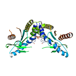
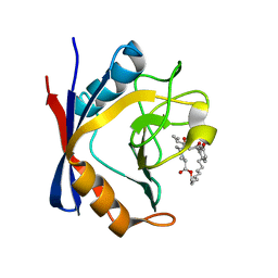
 | | The structure of D77G hCINAP-ADP | | 分子名称: | ADENOSINE-5'-DIPHOSPHATE, Adenylate kinase isoenzyme 6 | | 著者 | Liu, Y, Yang, Z, Yang, Y, Cai, X, Zheng, X. | | 登録日 | 2016-05-17 | | 公開日 | 2016-08-10 | | 最終更新日 | 2023-11-08 | | 実験手法 | X-RAY DIFFRACTION (2.07 Å) | | 主引用文献 | The ATPase hCINAP regulates 18S rRNA processing and is essential for embryogenesis and tumour growth.
Nat Commun, 7, 2016
|
|
2FN9
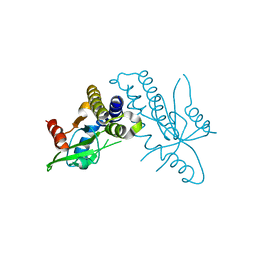
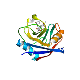
 | | Thermotoga maritima Ribose Binding Protein Unliganded Form | | 分子名称: | ribose ABC transporter, periplasmic ribose-binding protein | | 著者 | Cuneo, M.J, Changela, A, Tian, Y, Beese, L.S, Hellinga, H.W. | | 登録日 | 2006-01-10 | | 公開日 | 2007-01-16 | | 最終更新日 | 2023-08-30 | | 実験手法 | X-RAY DIFFRACTION (1.4 Å) | | 主引用文献 | Ligand-induced conformational changes in a thermophilic ribose-binding protein.
Bmc Struct.Biol., 8, 2008
|
|
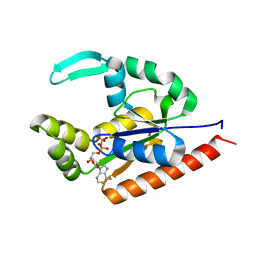
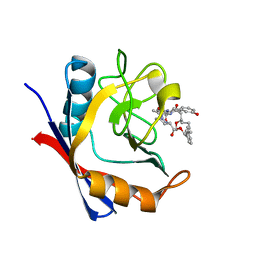
5T9W
 | | Discovery of a Potent Cyclophilin Inhibitor (Compound 5) based on Structural Simplification of Sanglifehrin A | | 分子名称: | 3-[(3-hydroxyphenyl)methyl]-6-(propan-2-yl)-19-oxa-1,4,7,25-tetraazabicyclo[19.3.1]pentacosa-13,15-diene-2,5,8,20-tetrone, Peptidyl-prolyl cis-trans isomerase A | | 著者 | Appleby, T.C, Steadman, V, Pettit, P, Schmitz, U, Mackman, R.L, Schultz, B. | | 登録日 | 2016-09-09 | | 公開日 | 2017-01-25 | | 最終更新日 | 2024-03-06 | | 実験手法 | X-RAY DIFFRACTION (2 Å) | | 主引用文献 | Discovery of Potent Cyclophilin Inhibitors Based on the Structural Simplification of Sanglifehrin A.
J. Med. Chem., 60, 2017
|
|
5T9U
 | | Discovery of a Potent Cyclophilin Inhibitor (Compound 3) based on Structural Simplification of Sanglifehrin A | | 分子名称: | 3-[(3-hydroxyphenyl)methyl]-10,12-dimethoxy-9,11-dimethyl-6-(propan-2-yl)-19-oxa-1,4,7,25-tetraazabicyclo[19.3.1]pentacosa-13,15-diene-2,5,8,20-tetrone, Peptidyl-prolyl cis-trans isomerase A | | 著者 | Appleby, T.C, Steadman, V, Pettit, S, Schmitz, U, Mackman, R.L, Schultz, B. | | 登録日 | 2016-09-09 | | 公開日 | 2017-01-25 | | 最終更新日 | 2024-03-06 | | 実験手法 | X-RAY DIFFRACTION (2.301 Å) | | 主引用文献 | Discovery of Potent Cyclophilin Inhibitors Based on the Structural Simplification of Sanglifehrin A.
J. Med. Chem., 60, 2017
|
|
5T9Z
 | | Discovery of a Potent Cyclophilin Inhibitor (Compound 6) based on Structural Simplification of Sanglifehrin A | | 分子名称: | 11-[(3-hydroxyphenyl)methyl]-18-methoxy-17-methyl-14-(propan-2-yl)-3-oxa-9,12,15,28-tetraazatricyclo[21.3.1.1~5,9~]octacosa-1(27),21,23,25-tetraene-4,10,13,16-tetrone, Peptidyl-prolyl cis-trans isomerase A | | 著者 | Appleby, T.C, Steadman, V, Pettit, S, Schmitz, U, Mackman, R.L, Schultz, B. | | 登録日 | 2016-09-09 | | 公開日 | 2017-01-25 | | 最終更新日 | 2024-03-06 | | 実験手法 | X-RAY DIFFRACTION (1.4 Å) | | 主引用文献 | Discovery of Potent Cyclophilin Inhibitors Based on the Structural Simplification of Sanglifehrin A.
J. Med. Chem., 60, 2017
|
|
4DDP
 | |
5TA2
 | | Discovery of a Potent Cyclophilin Inhibitor (Compound 7) based on Structural Simplification of Sanglifehrin A | | 分子名称: | 11-[(3-hydroxyphenyl)methyl]-18-methoxy-2,17-dimethyl-14-(propan-2-yl)-3-oxa-9,12,15,28-tetraazatricyclo[21.3.1.1~5,9~]octacosa-1(27),21,23,25-tetraene-4,10,13,16-tetrone, Peptidyl-prolyl cis-trans isomerase A | | 著者 | Appleby, T.C, Steadman, V, Pettit, S, Schmitz, U, Mackman, R.L, Schultz, B. | | 登録日 | 2016-09-09 | | 公開日 | 2017-01-25 | | 最終更新日 | 2024-03-06 | | 実験手法 | X-RAY DIFFRACTION (1.48 Å) | | 主引用文献 | Discovery of Potent Cyclophilin Inhibitors Based on the Structural Simplification of Sanglifehrin A.
J. Med. Chem., 60, 2017
|
|
5TA4
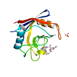 | | Discovery of a Potent Cyclophilin Inhibitor (Compound 8) based on Structural Simplification of Sanglifehrin A | | 分子名称: | 18-methoxy-2,11,17-trimethyl-14-(propan-2-yl)-3-oxa-9,12,15,28-tetraazatricyclo[21.3.1.1~5,9~]octacosa-1(27),21,23,25-tetraene-4,10,13,16-tetrone, Peptidyl-prolyl cis-trans isomerase A, SULFATE ION | | 著者 | Appleby, T.C, Steadman, V, Pettit, S, Schmitz, U, Mackman, R.L, Schultz, B. | | 登録日 | 2016-09-09 | | 公開日 | 2017-01-25 | | 最終更新日 | 2024-03-06 | | 実験手法 | X-RAY DIFFRACTION (1.5 Å) | | 主引用文献 | Discovery of Potent Cyclophilin Inhibitors Based on the Structural Simplification of Sanglifehrin A.
J. Med. Chem., 60, 2017
|
|


