3J5S
 
 | |
3I6D
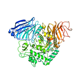
 | | Crystal structure of PPO from bacillus subtilis with AF | | 分子名称: | 5-[2-CHLORO-4-(TRIFLUOROMETHYL)PHENOXY]-2-NITROBENZOIC ACID, FLAVIN-ADENINE DINUCLEOTIDE, PHOSPHATE ION, ... | | 著者 | Shen, Y. | | 登録日 | 2009-07-06 | | 公開日 | 2009-12-08 | | 最終更新日 | 2024-03-20 | | 実験手法 | X-RAY DIFFRACTION (2.9 Å) | | 主引用文献 | Structural insight into unique properties of protoporphyrinogen oxidase from Bacillus subtilis
J.Struct.Biol., 170, 2010
|
|
3KKR
 | |
3KKS
 | |
6HX0
 | | Cell wall binding domain of endolysin from Listeria phage A500. | | 分子名称: | L-alanyl-D-glutamate peptidase | | 著者 | Dunne, M, Taylor, N.M.I, Shen, Y, Loessner, M.J, Leiman, P.G. | | 登録日 | 2018-10-15 | | 公開日 | 2019-10-30 | | 最終更新日 | 2024-01-24 | | 実験手法 | X-RAY DIFFRACTION (1.59 Å) | | 主引用文献 | Structural basis for recognition of bacterial cell wall teichoic acid by pseudo-symmetric SH3b-like repeats of a viral peptidoglycan hydrolase
Chem Sci, 2020
|
|
4ZRZ
 | |
3IZ4
 | |
4HWF
 | | Crystal structure of ATBAG3 | | 分子名称: | BAG family molecular chaperone regulator 3, SULFATE ION | | 著者 | Shen, Y, Fang, S. | | 登録日 | 2012-11-07 | | 公開日 | 2013-05-29 | | 最終更新日 | 2024-02-28 | | 実験手法 | X-RAY DIFFRACTION (2 Å) | | 主引用文献 | Structural insight into plant programmed cell death mediated by BAG proteins in Arabidopsis thaliana.
Acta Crystallogr.,Sect.D, 69, 2013
|
|
4M6T
 | | Structure of human Paf1 and Leo1 complex | | 分子名称: | RNA polymerase II-associated factor 1 homolog, Linker, RNA polymerase-associated protein LEO1, ... | | 著者 | Shen, Y, Qin, X. | | 登録日 | 2013-08-11 | | 公開日 | 2013-10-02 | | 最終更新日 | 2024-03-20 | | 実験手法 | X-RAY DIFFRACTION (2.498 Å) | | 主引用文献 | Structural insights into Paf1 complex assembly and histone binding
Nucleic Acids Res., 41, 2013
|
|
4HWC
 | | Structure of ATBAG1 | | 分子名称: | BAG family molecular chaperone regulator 1, CHLORIDE ION | | 著者 | Shen, Y, Fang, S. | | 登録日 | 2012-11-07 | | 公開日 | 2013-05-29 | | 最終更新日 | 2024-02-28 | | 実験手法 | X-RAY DIFFRACTION (1.798 Å) | | 主引用文献 | Structural insight into plant programmed cell death mediated by BAG proteins in Arabidopsis thaliana.
Acta Crystallogr.,Sect.D, 69, 2013
|
|
4HWH
 | | Crystal structure of ATBAG4 | | 分子名称: | ACETATE ION, BAG family molecular chaperone regulator 4, GLYCEROL, ... | | 著者 | Shen, Y, Fang, S. | | 登録日 | 2012-11-07 | | 公開日 | 2013-05-29 | | 最終更新日 | 2024-02-28 | | 実験手法 | X-RAY DIFFRACTION (1.895 Å) | | 主引用文献 | Structural insight into plant programmed cell death mediated by BAG proteins in Arabidopsis thaliana.
Acta Crystallogr.,Sect.D, 69, 2013
|
|
4HWI
 | | Crystal structure of ATBAG1 in complex with HSP70 | | 分子名称: | BAG family molecular chaperone regulator 1, Heat shock cognate 71 kDa protein | | 著者 | Shen, Y, Fang, S. | | 登録日 | 2012-11-07 | | 公開日 | 2013-05-29 | | 最終更新日 | 2023-09-20 | | 実験手法 | X-RAY DIFFRACTION (2.273 Å) | | 主引用文献 | Structural insight into plant programmed cell death mediated by BAG proteins in Arabidopsis thaliana.
Acta Crystallogr.,Sect.D, 69, 2013
|
|
4HWD
 | | Crystal structure of ATBAG2 | | 分子名称: | BAG family molecular chaperone regulator 2 | | 著者 | Shen, Y, Fang, S. | | 登録日 | 2012-11-07 | | 公開日 | 2013-05-29 | | 最終更新日 | 2024-02-28 | | 実験手法 | X-RAY DIFFRACTION (2.312 Å) | | 主引用文献 | Structural insight into plant programmed cell death mediated by BAG proteins in Arabidopsis thaliana.
Acta Crystallogr.,Sect.D, 69, 2013
|
|
3SLU
 | | Crystal structure of NMB0315 | | 分子名称: | M23 peptidase domain protein, NICKEL (II) ION | | 著者 | Shen, Y, Wang, X, Yang, X, Xu, H. | | 登録日 | 2011-06-26 | | 公開日 | 2012-02-01 | | 最終更新日 | 2024-03-20 | | 実験手法 | X-RAY DIFFRACTION (2.41 Å) | | 主引用文献 | Crystal structure of outer membrane protein NMB0315 from Neisseria meningitidis.
Plos One, 6, 2011
|
|
3SV1
 | | Crystal structure of APP peptide bound rat Mint2 PARM | | 分子名称: | Amyloid beta A4 precursor protein-binding family A member 2, Amyloid beta A4 protein | | 著者 | Shen, Y, Long, J, Yan, X, Xie, X. | | 登録日 | 2011-07-12 | | 公開日 | 2012-07-11 | | 最終更新日 | 2023-11-01 | | 実験手法 | X-RAY DIFFRACTION (3.3 Å) | | 主引用文献 | Open-closed motion of Mint2 regulates APP metabolism
J Mol Cell Biol, 5, 2013
|
|
3SUZ
 | |
3UB2
 | | TIR domain of Mal/TIRAP | | 分子名称: | 2,3-DIHYDROXY-1,4-DITHIOBUTANE, Toll/interleukin-1 receptor domain-containing adapter protein | | 著者 | Shen, Y, Lin, Z. | | 登録日 | 2011-10-23 | | 公開日 | 2012-05-16 | | 実験手法 | X-RAY DIFFRACTION (2.4 Å) | | 主引用文献 | Structural Insights into TIR Domain Specificity of the Bridging Adaptor Mal in TLR4 Signaling
Plos One, 7, 2012
|
|
3TON
 | |
3UB4
 | | S180L variant of TIR domain of Mal/TIRAP | | 分子名称: | (2S,3S)-1,4-DIMERCAPTOBUTANE-2,3-DIOL, Toll/interleukin-1 receptor domain-containing adapter protein | | 著者 | Shen, Y, Lin, Z. | | 登録日 | 2011-10-23 | | 公開日 | 2012-05-16 | | 最終更新日 | 2023-11-01 | | 実験手法 | X-RAY DIFFRACTION (3.1 Å) | | 主引用文献 | Structural Insights into TIR Domain Specificity of the Bridging Adaptor Mal in TLR4 Signaling
Plos One, 7, 2012
|
|
3UB3
 | | D96N variant of TIR domain of Mal/TIRAP | | 分子名称: | 2,3-DIHYDROXY-1,4-DITHIOBUTANE, Toll/interleukin-1 receptor domain-containing adapter protein | | 著者 | Shen, Y, Lin, Z. | | 登録日 | 2011-10-23 | | 公開日 | 2012-05-16 | | 最終更新日 | 2023-11-01 | | 実験手法 | X-RAY DIFFRACTION (2.75 Å) | | 主引用文献 | Structural Insights into TIR Domain Specificity of the Bridging Adaptor Mal in TLR4 Signaling
Plos One, 7, 2012
|
|
3TOP
 | | Crystral Structure of the C-terminal Subunit of Human Maltase-Glucoamylase in Complex with Acarbose | | 分子名称: | 4,6-dideoxy-4-{[(1S,4R,5S,6S)-4,5,6-trihydroxy-3-(hydroxymethyl)cyclohex-2-en-1-yl]amino}-alpha-D-glucopyranose-(1-4)-alpha-D-glucopyranose-(1-4)-alpha-D-glucopyranose, Maltase-glucoamylase, intestinal | | 著者 | Shen, Y, Qin, X.H, Ren, L.M. | | 登録日 | 2011-09-06 | | 公開日 | 2011-11-23 | | 最終更新日 | 2023-11-01 | | 実験手法 | X-RAY DIFFRACTION (2.881 Å) | | 主引用文献 | Structural insight into substrate specificity of human intestinal maltase-glucoamylase
Protein Cell, 2, 2011
|
|
2K2D
 | | Solution NMR structure of C-terminal domain of human pirh2. Northeast Structural Genomics Consortium (NESG) target HT2C | | 分子名称: | RING finger and CHY zinc finger domain-containing protein 1, ZINC ION | | 著者 | Lemak, A, Sheng, Y, Karra, M, Srisailam, S, Laister, R.C, Duan, S, Arrowsmith, C.H, Northeast Structural Genomics Consortium (NESG) | | 登録日 | 2008-03-31 | | 公開日 | 2008-04-15 | | 最終更新日 | 2024-05-01 | | 実験手法 | SOLUTION NMR | | 主引用文献 | Molecular basis of Pirh2-mediated p53 ubiquitylation.
Nat.Struct.Mol.Biol., 15, 2008
|
|
2K2C
 | | Solution NMR structure of N-terminal domain of human pirh2. Northeast Structural Genomics Consortium (NESG) target HT2A | | 分子名称: | RING finger and CHY zinc finger domain-containing protein 1, ZINC ION | | 著者 | Wu, B, Lemak, A, Sheng, Y, Karra, M, Srisailam, S, Sunnerhagen, M, Arrowsmith, C.H, Northeast Structural Genomics Consortium (NESG) | | 登録日 | 2008-03-31 | | 公開日 | 2008-04-15 | | 最終更新日 | 2024-05-08 | | 実験手法 | SOLUTION NMR | | 主引用文献 | Molecular basis of Pirh2-mediated p53 ubiquitylation.
Nat.Struct.Mol.Biol., 15, 2008
|
|
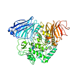
3NKS
 | | Structure of human protoporphyrinogen IX oxidase | | 分子名称: | 5-[2-CHLORO-4-(TRIFLUOROMETHYL)PHENOXY]-2-NITROBENZOIC ACID, FLAVIN-ADENINE DINUCLEOTIDE, GLYCEROL, ... | | 著者 | Shen, Y. | | 登録日 | 2010-06-21 | | 公開日 | 2011-04-20 | | 最終更新日 | 2023-11-01 | | 実験手法 | X-RAY DIFFRACTION (1.9 Å) | | 主引用文献 | Structural insight into human variegate porphyria disease
Faseb J., 25, 2011
|
|
2FOJ
 | | The Crystal Structure of the N-terminal domain of HAUSP/USP7 complexed with p53 peptide 364-367 | | 分子名称: | Ubiquitin carboxyl-terminal hydrolase 7, p53 peptide | | 著者 | Saridakis, V, Sheng, Y, Sarkari, F, Duan, S, Wu, T, Arrowsmith, C.H, Frappier, L. | | 登録日 | 2006-01-13 | | 公開日 | 2006-02-14 | | 最終更新日 | 2023-08-30 | | 実験手法 | X-RAY DIFFRACTION (1.6 Å) | | 主引用文献 | Molecular recognition of p53 and MDM2 by USP7/HAUSP
Nat.Struct.Mol.Biol., 13, 2006
|
|


