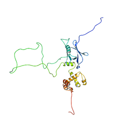
375D
 
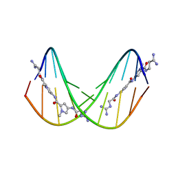 | | A NOVEL END-TO-END BINDING OF TWO NETROPSINS TO THE DNA DECAMER D(CCCCCIIIII)2 | | 分子名称: | DNA (5'-D(*CP*CP*CP*CP*CP*IP*IP*IP*IP*I)-3'), NETROPSIN | | 著者 | Chen, X, Rao, S.T, Sekar, K, Sundaralingam, M. | | 登録日 | 1998-01-14 | | 公開日 | 1998-12-02 | | 最終更新日 | 2024-02-21 | | 実験手法 | X-RAY DIFFRACTION (2.4 Å) | | 主引用文献 | A Novel End-to-End Binding of Two Netropsins to the DNA Decamers d(CCCCCIIIII) 2, d(CCCBr5CCIIIII)2, d(CBr5CCCCIIIII)2
Nucleic Acids Res., 26, 1998
|
|
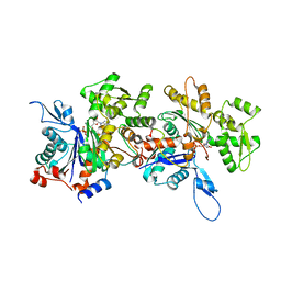
8YZ5
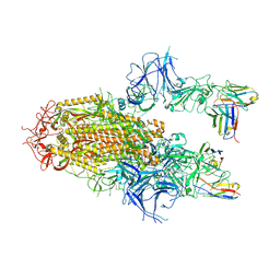 | | SARS-CoV-2 Delta Spike in complex with Fab of JE-5C | | 分子名称: | Fab heavy chain of JE-5C, Fab light chain of JE-5C, Spike glycoprotein | | 著者 | Chen, X, Wu, Y.-M. | | 登録日 | 2024-04-06 | | 公開日 | 2024-06-05 | | 最終更新日 | 2024-11-06 | | 実験手法 | ELECTRON MICROSCOPY (3.93 Å) | | 主引用文献 | The presence of broadly neutralizing anti-SARS-CoV-2 RBD antibodies elicited by primary series and booster dose of COVID-19 vaccine.
Plos Pathog., 20, 2024
|
|
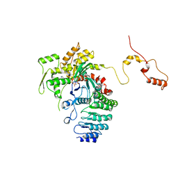
8YZ6
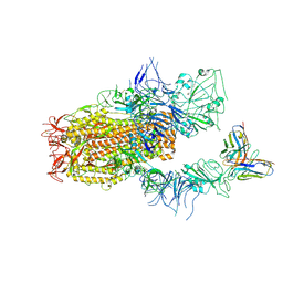 | | SARS-CoV-2 Spike (BA.1) in complex with Fab of JH-8B | | 分子名称: | 2-acetamido-2-deoxy-beta-D-glucopyranose, 2-acetamido-2-deoxy-beta-D-glucopyranose-(1-4)-2-acetamido-2-deoxy-beta-D-glucopyranose, Fab heavy chain of JH-8B, ... | | 著者 | Chen, X, Wu, Y.-M. | | 登録日 | 2024-04-06 | | 公開日 | 2024-06-05 | | 最終更新日 | 2024-11-06 | | 実験手法 | ELECTRON MICROSCOPY (4.55 Å) | | 主引用文献 | The presence of broadly neutralizing anti-SARS-CoV-2 RBD antibodies elicited by primary series and booster dose of COVID-19 vaccine.
Plos Pathog., 20, 2024
|
|
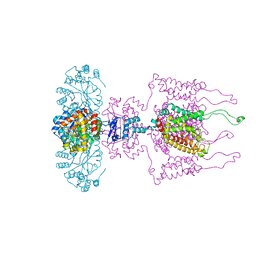
6V0V
 | | Cryo-EM structure of mouse WT RAG1/2 NFC complex (DNA0) | | 分子名称: | CALCIUM ION, DNA (30-MER), V(D)J recombination-activating protein 1, ... | | 著者 | Chen, X, Yang, W, Gellert, M. | | 登録日 | 2019-11-19 | | 公開日 | 2020-01-29 | | 最終更新日 | 2025-05-28 | | 実験手法 | ELECTRON MICROSCOPY (3.61 Å) | | 主引用文献 | Cutting antiparallel DNA strands in a single active site.
Nat.Struct.Mol.Biol., 27, 2020
|
|
4ZD3
 | | Structure of a transglutaminase 2-specific autoantibody Fab fragment | | 分子名称: | 679-14-14E06 Fab fragment heavy chain, 679-14-14E06 Fab fragment light chain | | 著者 | Chen, X, Dalhus, B, Hnida, K, Iversen, R, Sollid, L.M. | | 登録日 | 2015-04-16 | | 公開日 | 2015-07-22 | | 最終更新日 | 2024-11-06 | | 実験手法 | X-RAY DIFFRACTION (2.4 Å) | | 主引用文献 | Structural Basis for Antigen Recognition by Transglutaminase 2-specific Autoantibodies in Celiac Disease.
J.Biol.Chem., 290, 2015
|
|
2KR0
 | |
2KQZ
 | |
2NBU
 | |
2NBW
 | |
2NBV
 | |
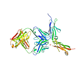
4JHD
 | | Crystal Structure of an Actin Dimer in Complex with the Actin Nucleator Cordon-Bleu | | 分子名称: | Actin-5C, MAGNESIUM ION, PHOSPHOAMINOPHOSPHONIC ACID-ADENYLATE ESTER, ... | | 著者 | Chen, X, Ni, F, Wang, Q. | | 登録日 | 2013-03-04 | | 公開日 | 2013-06-19 | | 最終更新日 | 2024-02-28 | | 実験手法 | X-RAY DIFFRACTION (2.91 Å) | | 主引用文献 | Structural basis of actin filament nucleation by tandem w domains.
Cell Rep, 3, 2013
|
|
4RWT
 | | Structure of actin-Lmod complex | | 分子名称: | Actin-5C, Leiomodin-2, MAGNESIUM ION, ... | | 著者 | Chen, X, Ni, F, Wang, Q. | | 登録日 | 2014-12-05 | | 公開日 | 2015-10-14 | | 最終更新日 | 2024-02-28 | | 実験手法 | X-RAY DIFFRACTION (2.98 Å) | | 主引用文献 | Mechanisms of leiomodin 2-mediated regulation of actin filament in muscle cells.
Proc.Natl.Acad.Sci.USA, 112, 2015
|
|
6AKO
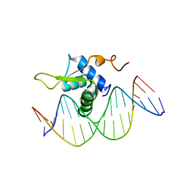 | | Crystal Structure of FOXC2 DBD Bound to DBE2 DNA | | 分子名称: | DNA (5'-D(CP*AP*AP*AP*AP*TP*GP*TP*AP*AP*AP*CP*AP*AP*GP*A)-3'), DNA (5'-D(TP*CP*TP*TP*GP*TP*TP*TP*AP*CP*AP*TP*TP*TP*TP*G)-3'), Forkhead box protein C2, ... | | 著者 | Chen, X, Wei, H, Li, J, Liang, X, Dai, S, Jiang, L, Guo, M, Chen, Y. | | 登録日 | 2018-09-03 | | 公開日 | 2019-02-06 | | 最終更新日 | 2023-11-22 | | 実験手法 | X-RAY DIFFRACTION (2.396 Å) | | 主引用文献 | Structural basis for DNA recognition by FOXC2.
Nucleic Acids Res., 47, 2019
|
|
6AKP
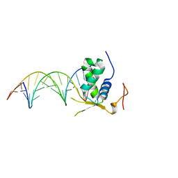 | | Crystal Structural of FOXC2 DNA binding domain bound to PC promoter | | 分子名称: | DNA (5'-D(AP*CP*AP*CP*AP*AP*AP*TP*AP*TP*TP*TP*GP*TP*GP*T)-3'), Forkhead box protein C2, MAGNESIUM ION | | 著者 | Chen, X, Wei, H, Li, J, Liang, X, Dai, S, Jiang, L, Guo, M, Chen, Y. | | 登録日 | 2018-09-03 | | 公開日 | 2019-02-06 | | 最終更新日 | 2023-11-22 | | 実験手法 | X-RAY DIFFRACTION (2.323 Å) | | 主引用文献 | Structural basis for DNA recognition by FOXC2.
Nucleic Acids Res., 47, 2019
|
|
6LRB
 | |
3LUT
 | | A Structural Model for the Full-length Shaker Potassium Channel Kv1.2 | | 分子名称: | NADP NICOTINAMIDE-ADENINE-DINUCLEOTIDE PHOSPHATE, POTASSIUM ION, Potassium voltage-gated channel subfamily A member 2, ... | | 著者 | Chen, X, Ni, F, Wang, Q, Ma, J. | | 登録日 | 2010-02-18 | | 公開日 | 2010-06-23 | | 最終更新日 | 2024-02-21 | | 実験手法 | X-RAY DIFFRACTION (2.9 Å) | | 主引用文献 | Structure of the full-length Shaker potassium channel Kv1.2 by normal-mode-based X-ray crystallographic refinement.
Proc.Natl.Acad.Sci.USA, 107, 2010
|
|
6LYN
 | | CD146 D4-D5/AA98 Fab | | 分子名称: | 2-acetamido-2-deoxy-beta-D-glucopyranose, 4-(2-HYDROXYETHYL)-1-PIPERAZINE ETHANESULFONIC ACID, AA98 Fab heavy chain, ... | | 著者 | Chen, X, Yan, X. | | 登録日 | 2020-02-14 | | 公開日 | 2021-02-24 | | 最終更新日 | 2024-10-23 | | 実験手法 | X-RAY DIFFRACTION (2.776 Å) | | 主引用文献 | Structure basis for AA98 inhibition on the activation of endothelial cells mediated by CD146.
Iscience, 24, 2021
|
|
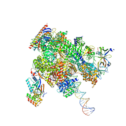
8WAN
 | | Structure of transcribing complex 4 (TC4), the initially transcribing complex with Pol II positioned 4nt downstream of TSS. | | 分子名称: | Alpha-amanitin, CDK-activating kinase assembly factor MAT1, DNA-directed RNA polymerase II subunit E, ... | | 著者 | Chen, X, Liu, W, Wang, Q, Wang, X, Ren, Y, Qu, X, Li, W, Xu, Y. | | 登録日 | 2023-09-07 | | 公開日 | 2023-12-06 | | 最終更新日 | 2024-01-03 | | 実験手法 | ELECTRON MICROSCOPY (6.07 Å) | | 主引用文献 | Structural visualization of transcription initiation in action.
Science, 382, 2023
|
|
8WAV
 | | De novo transcribing complex 12 (TC12), the early elongation complex with Pol II positioned 12nt downstream of TSS | | 分子名称: | Alpha-amanitin, DNA-directed RNA polymerase II subunit E, DNA-directed RNA polymerase II subunit F, ... | | 著者 | Chen, X, Liu, W, Wang, Q, Wang, X, Ren, Y, Qu, X, Li, W, Xu, Y. | | 登録日 | 2023-09-08 | | 公開日 | 2023-12-06 | | 最終更新日 | 2024-01-03 | | 実験手法 | ELECTRON MICROSCOPY (2.72 Å) | | 主引用文献 | Structural visualization of transcription initiation in action.
Science, 382, 2023
|
|
8WAS
 | | Structure of transcribing complex 9 (TC9), the initially transcribing complex with Pol II positioned 9nt downstream of TSS. | | 分子名称: | Alpha-amanitin, CDK-activating kinase assembly factor MAT1, DNA-directed RNA polymerase II subunit E, ... | | 著者 | Chen, X, Liu, W, Wang, Q, Wang, X, Ren, Y, Qu, X, Li, W, Xu, Y. | | 登録日 | 2023-09-08 | | 公開日 | 2023-12-06 | | 最終更新日 | 2024-01-03 | | 実験手法 | ELECTRON MICROSCOPY (6.13 Å) | | 主引用文献 | Structural visualization of transcription initiation in action.
Science, 382, 2023
|
|
8WAP
 | | Structure of transcribing complex 6 (TC6), the initially transcribing complex with Pol II positioned 6nt downstream of TSS. | | 分子名称: | Alpha-amanitin, CDK-activating kinase assembly factor MAT1, DNA-directed RNA polymerase II subunit E, ... | | 著者 | Chen, X, Liu, W, Wang, Q, Wang, X, Ren, Y, Qu, X, Li, W, Xu, Y. | | 登録日 | 2023-09-08 | | 公開日 | 2023-12-06 | | 最終更新日 | 2024-01-03 | | 実験手法 | ELECTRON MICROSCOPY (5.85 Å) | | 主引用文献 | Structural visualization of transcription initiation in action.
Science, 382, 2023
|
|
8WAL
 | | Structure of transcribing complex 3 (TC3), the initially transcribing complex with Pol II positioned 3nt downstream of TSS. | | 分子名称: | Alpha-amanitin, CDK-activating kinase assembly factor MAT1, DNA-directed RNA polymerase II subunit E, ... | | 著者 | Chen, X, Liu, W, Wang, Q, Wang, X, Ren, Y, Qu, X, Li, W, Xu, Y. | | 登録日 | 2023-09-07 | | 公開日 | 2023-12-06 | | 最終更新日 | 2024-01-03 | | 実験手法 | ELECTRON MICROSCOPY (8.52 Å) | | 主引用文献 | Structural visualization of transcription initiation in action.
Science, 382, 2023
|
|
8WAO
 | | Structure of transcribing complex 5 (TC5), the initially transcribing complex with Pol II positioned 5nt downstream of TSS. | | 分子名称: | Alpha-amanitin, CDK-activating kinase assembly factor MAT1, DNA-directed RNA polymerase II subunit E, ... | | 著者 | Chen, X, Liu, W, Wang, Q, Wang, X, Ren, Y, Qu, X, Li, W, Xu, Y. | | 登録日 | 2023-09-07 | | 公開日 | 2023-12-06 | | 最終更新日 | 2024-01-03 | | 実験手法 | ELECTRON MICROSCOPY (6.4 Å) | | 主引用文献 | Structural visualization of transcription initiation in action.
Science, 382, 2023
|
|
8WAQ
 | | Structure of transcribing complex 7 (TC7), the initially transcribing complex with Pol II positioned 7nt downstream of TSS. | | 分子名称: | Alpha-amanitin, CDK-activating kinase assembly factor MAT1, DNA-directed RNA polymerase II subunit E, ... | | 著者 | Chen, X, Liu, W, Wang, Q, Wang, X, Ren, Y, Qu, X, Li, W, Xu, Y. | | 登録日 | 2023-09-08 | | 公開日 | 2023-12-06 | | 最終更新日 | 2024-01-03 | | 実験手法 | ELECTRON MICROSCOPY (6.29 Å) | | 主引用文献 | Structural visualization of transcription initiation in action.
Science, 382, 2023
|
|
8WAX
 | | De novo transcribing complex 14 (TC14), the early elongation complex with Pol II positioned 14nt downstream of TSS | | 分子名称: | Alpha-amanitin, DNA-directed RNA polymerase II subunit E, DNA-directed RNA polymerase II subunit F, ... | | 著者 | Chen, X, Liu, W, Wang, Q, Wang, X, Ren, Y, Qu, X, Li, W, Xu, Y. | | 登録日 | 2023-09-08 | | 公開日 | 2023-12-06 | | 最終更新日 | 2024-01-03 | | 実験手法 | ELECTRON MICROSCOPY (2.75 Å) | | 主引用文献 | Structural visualization of transcription initiation in action.
Science, 382, 2023
|
|


