5KJI
 
 | |
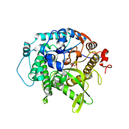
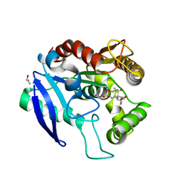
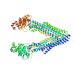
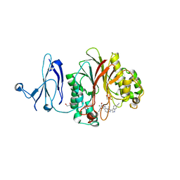
4TXM
 | | Crystal structure of uridine phosphorylase from Schistosoma mansoni in complex with thymine | | 分子名称: | SULFATE ION, THYMINE, Uridine phosphorylase | | 著者 | Marinho, A, Torini, J, Romanello, L, Cassago, A, DeMarco, R, Brandao-Neto, J, Pereira, H.M. | | 登録日 | 2014-07-03 | | 公開日 | 2015-10-14 | | 最終更新日 | 2023-09-27 | | 実験手法 | X-RAY DIFFRACTION (1.93 Å) | | 主引用文献 | Analysis of two Schistosoma mansoni uridine phosphorylases isoforms suggests the emergence of a protein with a non-canonical function.
Biochimie, 125, 2016
|
|
2AMS
 | | Structure of the oxidized Hipip from thermochromatium tepidum at 1.4 angstrom resolution | | 分子名称: | GLYCEROL, High potential iron-sulfur protein, IRON/SULFUR CLUSTER, ... | | 著者 | Hunsicker-Wang, L.M, Han, W, Stout, C.D, Noodleman, L, Fee, J.A. | | 登録日 | 2005-08-10 | | 公開日 | 2006-08-15 | | 最終更新日 | 2023-08-23 | | 実験手法 | X-RAY DIFFRACTION (1.4 Å) | | 主引用文献 | Geometric factors determine, in part, the electronic state of the 4Fe-4S cluster of Hipip from thermochromtium tepidum: a geomteric, crystallographic, and theoretical study.
To be Published
|
|
2AN9
 | | Crystal Structure Of Oligomeric E.coli Guanylate Kinase In Complex With GDP | | 分子名称: | GUANOSINE, GUANOSINE-5'-DIPHOSPHATE, Guanylate kinase, ... | | 著者 | Hible, G, Renault, L, Schaeffer, F, Christova, P, Radulescu, A.Z, Evrin, C, Gilles, A.M, Cherfils, J. | | 登録日 | 2005-08-11 | | 公開日 | 2005-08-30 | | 最終更新日 | 2023-10-25 | | 実験手法 | X-RAY DIFFRACTION (2.35 Å) | | 主引用文献 | Calorimetric and crystallographic analysis of the oligomeric structure of Escherichia coli GMP kinase
J.Mol.Biol., 352, 2005
|
|
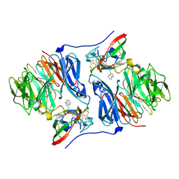
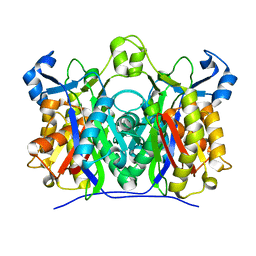
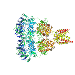
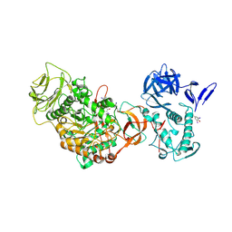
4YXX
 | |
7UZ1
 | | Structure of beta-glycosidase from Sulfolobus solfataricus in complex with C5a-bromo-valienide. | | 分子名称: | (1R,2S,3R,4R)-5-bromo-6-(hydroxymethyl)cyclohex-5-ene-1,2,3,4-tetrol, 1,2-ETHANEDIOL, Beta-galactosidase | | 著者 | Danby, P.M, Jeong, A, Sim, L, Sweeney, R.P, Wardman, J.F, Karimi, R, Geissner, A, Worrall, L.J, Strynadka, N.C.J, Withers, S.G. | | 登録日 | 2022-05-08 | | 公開日 | 2023-04-05 | | 最終更新日 | 2023-10-25 | | 実験手法 | X-RAY DIFFRACTION (1.58 Å) | | 主引用文献 | Vinyl Halide-Modified Unsaturated Cyclitols are Mechanism-Based Glycosidase Inhibitors.
Angew.Chem.Int.Ed.Engl., 62, 2023
|
|
2AH0
 | | Crystal structure of the carbinolamine intermediate in the reductive half-reaction of aromatic amine dehydrogenase (AADH) with tryptamine. Monoclinic form | | 分子名称: | (1S)-1-AMINO-2-(1H-INDOL-3-YL)ETHANOL, 2-(1H-INDOL-3-YL)ETHANIMINE, Aromatic amine dehydrogenase | | 著者 | Masgrau, L, Roujeinikova, A, Johannissen, L.O, Hothi, P, Basran, J, Ranaghan, K.E, Mulholland, A.J, Sutcliffe, M.J, Scrutton, N.S, Leys, D. | | 登録日 | 2005-07-27 | | 公開日 | 2006-04-25 | | 最終更新日 | 2011-07-13 | | 実験手法 | X-RAY DIFFRACTION (1.45 Å) | | 主引用文献 | Atomic description of an enzyme reaction dominated by proton tunneling
Science, 312, 2006
|
|
2ARJ
 | | CD8alpha-alpha in complex with YTS 105.18 Fab | | 分子名称: | T-cell surface glycoprotein CD8 alpha chain, YTS 105.18 antigen binding region Heavy chain, YTS 105.18 antigen binding region Light chain | | 著者 | Shore, D.A, Teyton, L, Dwek, R.A, Rudd, P.M, Wilson, I.A. | | 登録日 | 2005-08-19 | | 公開日 | 2006-05-30 | | 最終更新日 | 2023-08-23 | | 実験手法 | X-RAY DIFFRACTION (2.88 Å) | | 主引用文献 | Crystal structure of the TCR co-receptor CD8alphaalpha in complex with monoclonal antibody YTS 105.18 Fab fragment at 2.88 A resolution.
J.Mol.Biol., 358, 2006
|
|
2AJ2
 | | X-Ray Crystal Structure of Protein VC0467 from Vibrio cholerae. Northeast Structural Genomics Consortium Target VcR8. | | 分子名称: | Hypothetical UPF0301 protein VC0467 | | 著者 | Kuzin, A.P, Abashidze, M, Vorobiev, S.M, Acton, T, Xiao, R, Conover, K, Ma, L.-C, Kellie, R, Montelione, G.T, Hunt, J.F, Tong, L, Northeast Structural Genomics Consortium (NESG) | | 登録日 | 2005-08-01 | | 公開日 | 2005-09-20 | | 最終更新日 | 2011-07-13 | | 実験手法 | X-RAY DIFFRACTION (3.21 Å) | | 主引用文献 | X-Ray structure of hypothetical protein VC0467 from Vibrio cholerae: new fold. Northeast Structural Genomics Consortium target VcR8.
To be Published
|
|
5OCJ
 | | Crystal structure of Ag85C bound to cyclophostin 8beta inhibitor | | 分子名称: | DIMETHYL SULFOXIDE, Diacylglycerol acyltransferase/mycolyltransferase Ag85C, methoxy-[(3~{R})-3-[(2~{R})-1-methoxy-1,3-bis(oxidanylidene)butan-2-yl]pentadecyl]phosphinic acid | | 著者 | Viljoen, A, Richard, M, Nguyen, P.C, Spilling, C.D, Canaan, S, Cavalier, J.F, Blaise, M, Kremer, L. | | 登録日 | 2017-07-03 | | 公開日 | 2018-01-24 | | 最終更新日 | 2024-01-17 | | 実験手法 | X-RAY DIFFRACTION (1.8 Å) | | 主引用文献 | Cyclipostins and cyclophostin analogs inhibit the antigen 85C from
J. Biol. Chem., 293, 2018
|
|
2AJ9
 | | X-ray crystal structure of W42A,R161A double mutant of Mycobacterium tuberculosis beta-ketoacyl-ACP synthase III | | 分子名称: | 3-oxoacyl-[acyl-carrier-protein] synthase III | | 著者 | Brown, A.K, Sridharan, S, Kremer, L, Lindenberg, S, Dover, L.G, Sacchettini, J.C, Besra, G.S. | | 登録日 | 2005-08-01 | | 公開日 | 2005-08-30 | | 最終更新日 | 2023-08-23 | | 実験手法 | X-RAY DIFFRACTION (2.5 Å) | | 主引用文献 | Probing the Mechanism of the Mycobacterium tuberculosis beta-Ketoacyl-Acyl Carrier Protein Synthase III mtFabH: Factors Influencing Catalysis And Substrate Specificity.
J.Biol.Chem., 280, 2005
|
|
1CES
 | | CRYSTALS OF DEMETALLIZED CONCANAVALIN A SOAKED WITH ZINC HAVE A ZINC ION BOUND IN THE S1 SITE | | 分子名称: | CONCANAVALIN A, ZINC ION | | 著者 | Bouckaert, J, Loris, R, Poortmans, F, Wyns, L. | | 登録日 | 1996-02-15 | | 公開日 | 1997-02-15 | | 最終更新日 | 2024-05-22 | | 実験手法 | X-RAY DIFFRACTION (2.7 Å) | | 主引用文献 | Sequential structural changes upon zinc and calcium binding to metal-free concanavalin A.
J.Biol.Chem., 271, 1996
|
|
5KPC
 | | Pavine N-methyltransferase H206A mutant in complex with S-adenosylmethionine pH 6 | | 分子名称: | Pavine N-methyltransferase, S-ADENOSYLMETHIONINE | | 著者 | Torres, M.A, Hoffarth, E, Eugenio, L, Savtchouk, J, Chen, X, Morris, J, Facchini, P.J, Ng, K.K.S. | | 登録日 | 2016-07-03 | | 公開日 | 2016-09-07 | | 最終更新日 | 2023-10-04 | | 実験手法 | X-RAY DIFFRACTION (2.5 Å) | | 主引用文献 | Structural and Functional Studies of Pavine N-Methyltransferase from Thalictrum flavum Reveal Novel Insights into Substrate Recognition and Catalytic Mechanism.
J.Biol.Chem., 291, 2016
|
|
4U1X
 | | Full length GluA2-kainate-(R,R)-2b complex crystal form B | | 分子名称: | 2-(N-MORPHOLINO)-ETHANESULFONIC ACID, 2-acetamido-2-deoxy-beta-D-glucopyranose, 3-(CARBOXYMETHYL)-4-ISOPROPENYLPROLINE, ... | | 著者 | Chen, L, Gouaux, E. | | 登録日 | 2014-07-16 | | 公開日 | 2014-08-20 | | 最終更新日 | 2023-12-27 | | 実験手法 | X-RAY DIFFRACTION (3.301 Å) | | 主引用文献 | Structure and Dynamics of AMPA Receptor GluA2 in Resting, Pre-Open, and Desensitized States.
Cell, 158, 2014
|
|
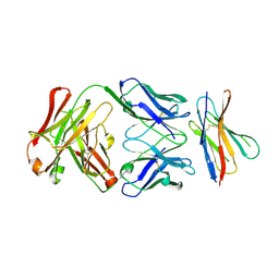
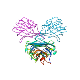
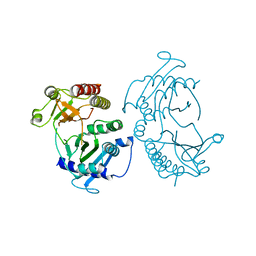
5KS8
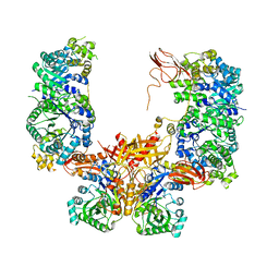 | |
2B7T
 | | Structure of ADAR2 dsRBM1 | | 分子名称: | Double-stranded RNA-specific editase 1 | | 著者 | Stefl, R, Xu, M, Skrisovska, L, Emeson, R.B, Allain, F.H.-T. | | 登録日 | 2005-10-05 | | 公開日 | 2006-03-14 | | 最終更新日 | 2024-05-22 | | 実験手法 | SOLUTION NMR | | 主引用文献 | Structure and specific RNA binding of ADAR2 double-stranded RNA binding motifs.
Structure, 14, 2006
|
|
5KPJ
 | | Mouse pgp methylated protein | | 分子名称: | Multidrug resistance protein 1A | | 著者 | Xia, D, Esser, L, Zhou, F. | | 登録日 | 2016-07-04 | | 公開日 | 2016-11-30 | | 最終更新日 | 2023-10-04 | | 実験手法 | X-RAY DIFFRACTION (3.5 Å) | | 主引用文献 | Structures of the Multidrug Transporter P-glycoprotein Reveal Asymmetric ATP Binding and the Mechanism of Polyspecificity.
J. Biol. Chem., 292, 2017
|
|
4Z2B
 | | The structure of human PDE12 residues 161-609 in complex with GSK3036342A | | 分子名称: | 1,2-ETHANEDIOL, 2',5'-phosphodiesterase 12, 2-AMINO-2-HYDROXYMETHYL-PROPANE-1,3-DIOL, ... | | 著者 | Nolte, R.T, Wisely, B, Wang, L, Wood, E.R. | | 登録日 | 2015-03-29 | | 公開日 | 2015-06-17 | | 最終更新日 | 2023-09-27 | | 実験手法 | X-RAY DIFFRACTION (1.8 Å) | | 主引用文献 | The Role of Phosphodiesterase 12 (PDE12) as a Negative Regulator of the Innate Immune Response and the Discovery of Antiviral Inhibitors.
J.Biol.Chem., 290, 2015
|
|
4TVD
 | | N-terminally truncated dextransucrase DSR-E from Leuconostoc mesenteroides NRRL B-1299 in complex with D-glucose | | 分子名称: | CALCIUM ION, DI(HYDROXYETHYL)ETHER, Dextransucrase, ... | | 著者 | Brison, Y, Remaud-Simeon, M, Mourey, L, Tranier, S. | | 登録日 | 2014-06-26 | | 公開日 | 2015-08-05 | | 最終更新日 | 2023-12-20 | | 実験手法 | X-RAY DIFFRACTION (2.3 Å) | | 主引用文献 | Structural Insights into the Carbohydrate Binding Ability of an alpha-(12) Branching Sucrase from Glycoside Hydrolase Family 70.
J.Biol.Chem., 291, 2016
|
|
4TZ7
 | | Crystal structure of type I phosphatidylinositol 4-phosphate 5-kinase alpha from Zebrafish | | 分子名称: | Phosphatidylinositol-4-phosphate 5-kinase, type I, alpha | | 著者 | Hu, J, Qin, Y, Wang, J, Li, L, Wu, D, Ha, Y. | | 登録日 | 2014-07-09 | | 公開日 | 2015-09-02 | | 最終更新日 | 2023-12-27 | | 実験手法 | X-RAY DIFFRACTION (3.31 Å) | | 主引用文献 | Resolution of structure of PIP5K1A reveals molecular mechanism for its regulation by dimerization and dishevelled.
Nat Commun, 6, 2015
|
|
4Z0W
 | | Peptaibol gichigamin isolated from Tolypocladium sup_5 | | 分子名称: | PEPTAIBOL GICHIGAMIN | | 著者 | Du, L, Risinger, A.L, Mitchell, C.A, Stamps, B.W, Pan, N, King, J.B, Motley, J.L, Thomas, L.M, Yang, Z, Stevenson, B.S, Mooberry, S.L, Cichewicz, R.H. | | 登録日 | 2015-03-26 | | 公開日 | 2016-03-30 | | 最終更新日 | 2019-12-25 | | 実験手法 | X-RAY DIFFRACTION (1.1 Å) | | 主引用文献 | Peptaibol gichigamin isolated from Tolypocladium sup_5
TO BE PUBLISHED
|
|
5OFL
 | | Crystal structure of CbXyn10C variant E140Q/E248Q complexed with cellohexaose | | 分子名称: | (4S)-2-METHYL-2,4-PENTANEDIOL, Glycoside hydrolase family 48, SULFATE ION, ... | | 著者 | Hakulinen, N, Penttinen, L, Rouvinen, J. | | 登録日 | 2017-07-11 | | 公開日 | 2017-10-04 | | 最終更新日 | 2024-01-17 | | 実験手法 | X-RAY DIFFRACTION (1.871 Å) | | 主引用文献 | Insights into the roles of non-catalytic residues in the active site of a GH10 xylanase with activity on cellulose.
J. Biol. Chem., 292, 2017
|
|
2AXF
 | | The Immunogenicity of a Viral Cytotoxic T Cell Epitope is controlled by its MHC-bound Conformation | | 分子名称: | 10-mer peptide from BZLF1 trans-activator protein, ACETIC ACID, Beta-2-microglobulin, ... | | 著者 | Tynan, F.E, Elhassen, D, Purcell, A.W, Burrows, J.M, Borg, N.A, Miles, J.J, Williamson, N.A, Green, K.J, Tellam, J, Kjer-Nielsen, L, McCluskey, J, Rossjohn, J, Burrows, S.R. | | 登録日 | 2005-09-05 | | 公開日 | 2005-11-29 | | 最終更新日 | 2017-10-11 | | 実験手法 | X-RAY DIFFRACTION (1.8 Å) | | 主引用文献 | The immunogenicity of a viral cytotoxic T cell epitope is controlled by its MHC-bound conformation
J.Exp.Med., 202, 2005
|
|
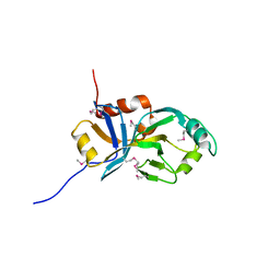
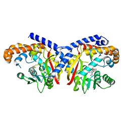
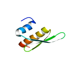
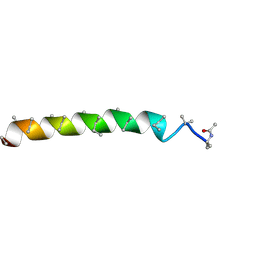
5KYE
 | |
7UOI
 | | Crystallographic structure of DapE from Enterococcus faecium | | 分子名称: | GLYCEROL, ZINC ION, succinyl-diaminopimelate desuccinylase | | 著者 | Gonzalez-Segura, L, Diaz-Vilchis, A, Terrazas-Lopez, M, Diaz-Sanchez, A.G. | | 登録日 | 2022-04-12 | | 公開日 | 2023-04-12 | | 最終更新日 | 2024-06-12 | | 実験手法 | X-RAY DIFFRACTION (1.6 Å) | | 主引用文献 | The three-dimensional structure of DapE from Enterococcus faecium reveals new insights into DapE/ArgE subfamily ligand specificity.
Int.J.Biol.Macromol., 270, 2024
|
|


