4Y9X
 
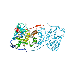 | | Crystal structure of glucosyl-3-phosphoglycerate synthase from Mycobacterium tuberculosis in complex with Mn2+, uridine-diphosphate-glucose (UDP-Glc) and phosphoglyceric acid (PGA) - GpgS Mn2+ UDP-Glc PGA-3 | | 分子名称: | 1,2-ETHANEDIOL, 3-PHOSPHOGLYCERIC ACID, CHLORIDE ION, ... | | 著者 | Albesa-Jove, D, Rodrigo-Unzueta, A, Cifuente, J.O, Urresti, S, Comino, N, Sancho-Vaello, E, Guerin, M.E. | | 登録日 | 2015-02-17 | | 公開日 | 2015-07-15 | | 最終更新日 | 2024-01-10 | | 実験手法 | X-RAY DIFFRACTION (2.637 Å) | | 主引用文献 | A Native Ternary Complex Trapped in a Crystal Reveals the Catalytic Mechanism of a Retaining Glycosyltransferase.
Angew.Chem.Int.Ed.Engl., 54, 2015
|
|
5AJC
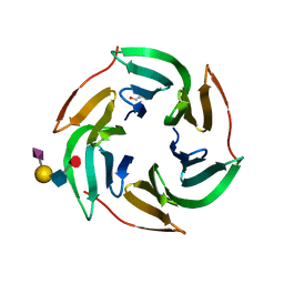 | | X-ray structure of RSL lectin in complex with sialyl lewis X tetrasaccharide | | 分子名称: | 2-AMINO-2-HYDROXYMETHYL-PROPANE-1,3-DIOL, N-acetyl-alpha-neuraminic acid-(2-3)-beta-D-galactopyranose-(1-4)-[alpha-L-fucopyranose-(1-3)]2-acetamido-2-deoxy-alpha-D-glucopyranose, PUTATIVE FUCOSE-BINDING LECTIN PROTEIN, ... | | 著者 | Topin, J, Arnaud, J, Varrot, A, Imberty, A. | | 登録日 | 2015-02-20 | | 公開日 | 2016-03-02 | | 最終更新日 | 2024-01-10 | | 実験手法 | X-RAY DIFFRACTION (1.7 Å) | | 主引用文献 | The Hidden Conformation of Lewis X, a Human Histo-Blood Group Antigen, is a Determinant for Recognition by Pathogen Lectins
Acs Chem.Biol., 11, 2016
|
|
8OO2
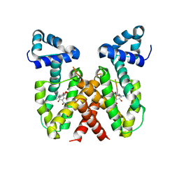 | | ChdA complex with amido-chelocardin | | 分子名称: | 2-carboxamido-2-deacetyl-chelocardin, MAGNESIUM ION, Putative transcriptional regulator | | 著者 | Koehnke, J, Sikandar, A. | | 登録日 | 2023-04-04 | | 公開日 | 2023-08-02 | | 最終更新日 | 2023-10-04 | | 実験手法 | X-RAY DIFFRACTION (1.85 Å) | | 主引用文献 | Revision of the Absolute Configurations of Chelocardin and Amidochelocardin.
Angew.Chem.Int.Ed.Engl., 62, 2023
|
|
6NPN
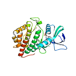 | | Crystal Structure of the Human vaccinia-related kinase bound to a N,N-dipropynyl-dihydropteridine-3-hydroxyindazole inhibitor | | 分子名称: | (7R)-7-methyl-2-[(3-oxo-2,3-dihydro-1H-indazol-6-yl)amino]-5,8-di(prop-2-yn-1-yl)-7,8-dihydropteridin-6(5H)-one, 1,2-ETHANEDIOL, DI(HYDROXYETHYL)ETHER, ... | | 著者 | Counago, R.M, dos Reis, C.V, Takarada, J.E, Azevedo, A, Guimaraes, C, Mascarello, A, Gama, F, Ferreira, M, Massirer, K.B, Arruda, P, Edwards, A.M, Elkins, J.M, Structural Genomics Consortium (SGC) | | 登録日 | 2019-01-18 | | 公開日 | 2019-02-20 | | 最終更新日 | 2023-10-11 | | 実験手法 | X-RAY DIFFRACTION (2.2 Å) | | 主引用文献 | Crystal Structure of the Human vaccinia-related kinase bound to a N,N-dipropynyl-dihydropteridine-3-hydroxyindazole inhibitor
To Be Published
|
|
1N1X
 | | Crystal Structure Analysis of the monomeric [S-carboxyamidomethyl-Cys31, S-carboxyamidomethyl-Cys32] Bovine seminal ribonuclease | | 分子名称: | Ribonuclease, seminal | | 著者 | Sica, F, Di Fiore, A, Zagari, A, Mazzarella, L. | | 登録日 | 2002-10-21 | | 公開日 | 2003-08-26 | | 最終更新日 | 2011-07-13 | | 実験手法 | X-RAY DIFFRACTION (1.45 Å) | | 主引用文献 | The unswapped chain of bovine seminal ribonuclease: Crystal structure of the free and liganded monomeric derivative
Proteins, 52, 2003
|
|
4Y9N
 | | PA3825-EAL Metal-Free-Apo Structure - Magnesium Co-crystallisation | | 分子名称: | PA3825-EAL, PHOSPHATE ION | | 著者 | Bellini, D, Horrell, S, Wagner, A, Strange, R, Walsh, M.A. | | 登録日 | 2015-02-17 | | 公開日 | 2016-03-09 | | 最終更新日 | 2024-05-08 | | 実験手法 | X-RAY DIFFRACTION (1.92 Å) | | 主引用文献 | Structure of PA3825 from P. aeruginosa bound to cyclic di-GMP and pGpG: new insights for a potential three-metal catalytic mechanism of EAL domains
To Be Published
|
|
4YFE
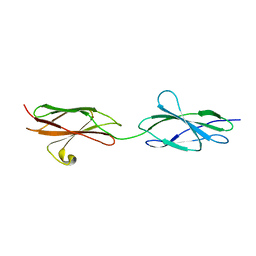 | | Crystal structure of PTP delta Fn1-Fn2 | | 分子名称: | Receptor-type tyrosine-protein phosphatase delta | | 著者 | Yamagata, A, Fukai, S. | | 登録日 | 2015-02-25 | | 公開日 | 2015-05-06 | | 最終更新日 | 2023-11-08 | | 実験手法 | X-RAY DIFFRACTION (1.972 Å) | | 主引用文献 | Mechanisms of splicing-dependent trans-synaptic adhesion by PTP delta-IL1RAPL1/IL-1RAcP for synaptic differentiation.
Nat Commun, 6, 2015
|
|
8JCS
 | |
8JCQ
 | |
8JCR
 | | Crystal structure of Procerain from Calotropis gigantea (pH 6.0) | | 分子名称: | BETA-MERCAPTOETHANOL, GLYCEROL, N-[N-[1-HYDROXYCARBOXYETHYL-CARBONYL]LEUCYLAMINO-BUTYL]-GUANIDINE, ... | | 著者 | Kumar, A, Jamdar, S.N, Srivastava, G, Makde, R.D. | | 登録日 | 2023-05-11 | | 公開日 | 2024-05-22 | | 実験手法 | X-RAY DIFFRACTION (1.4 Å) | | 主引用文献 | Crystal structure of Procerain from Calotropis gigantea
To Be Published
|
|
7QDD
 | |
3J7H
 | | Structure of beta-galactosidase at 3.2-A resolution obtained by cryo-electron microscopy | | 分子名称: | Beta-galactosidase, MAGNESIUM ION | | 著者 | Bartesaghi, A, Matthies, D, Banerjee, S, Merk, A, Subramaniam, S. | | 登録日 | 2014-06-30 | | 公開日 | 2014-07-30 | | 最終更新日 | 2024-02-21 | | 実験手法 | ELECTRON MICROSCOPY (3.2 Å) | | 主引用文献 | Structure of beta-galactosidase at 3.2- angstrom resolution obtained by cryo-electron microscopy.
Proc.Natl.Acad.Sci.USA, 111, 2014
|
|
5AJB
 | | X-ray structure of the RSL lectin in complex with Lewis X tetrasaccahride | | 分子名称: | LECTIN, alpha-L-fucopyranose-(1-3)-2-acetamido-2-deoxy-beta-D-glucopyranose, alpha-L-fucopyranose-(1-3)-[beta-D-galactopyranose-(1-4)]2-acetamido-2-deoxy-beta-D-glucopyranose, ... | | 著者 | Topin, J, Arnaud, J, Varrot, A, Imberty, A. | | 登録日 | 2015-02-20 | | 公開日 | 2016-03-02 | | 最終更新日 | 2024-01-10 | | 実験手法 | X-RAY DIFFRACTION (1.8 Å) | | 主引用文献 | The Hidden Conformation of Lewis X, a Human Histo-Blood Group Antigen, is a Determinant for Recognition by Pathogen Lectins
Acs Chem.Biol., 11, 2016
|
|
8DHK
 | |
8J3N
 | |
6NBK
 | | Crystal structure of Arginase from Bacillus cereus | | 分子名称: | Arginase, CALCIUM ION, MANGANESE (II) ION | | 著者 | Chang, C, Evdokimova, E, Mcchesney, M, Joachimiak, A, Savchenko, A, Center for Structural Genomics of Infectious Diseases (CSGID) | | 登録日 | 2018-12-07 | | 公開日 | 2018-12-19 | | 最終更新日 | 2023-10-11 | | 実験手法 | X-RAY DIFFRACTION (1.91 Å) | | 主引用文献 | Crystal structure of Arginase from Bacillus cereus
To Be Published
|
|
5JLZ
 | | Crystal structure of HLA-DRB1*04:01 in complex with modified alpha-enolase peptide 26-40 with citrulline at the position 32 | | 分子名称: | Alpha-enolase, HLA class II histocompatibility antigen, DR alpha chain, ... | | 著者 | Dubnovitsky, A, Kozhukh, G, Sandalova, T, Achour, A. | | 登録日 | 2016-04-28 | | 公開日 | 2016-12-07 | | 最終更新日 | 2024-01-10 | | 実験手法 | X-RAY DIFFRACTION (1.99 Å) | | 主引用文献 | Functional and Structural Characterization of a Novel HLA-DRB1*04:01-Restricted alpha-Enolase T Cell Epitope in Rheumatoid Arthritis.
Front Immunol, 7, 2016
|
|
8IWI
 | |
4YLJ
 | | Crystal structure of DYRK1A in complex with 10-Iodo-substituted 11H-indolo[3,2-c]quinoline-6-carboxylic acid inhibitor 5j | | 分子名称: | 10-iodo-11H-indolo[3,2-c]quinoline-6-carboxylic acid, Dual specificity tyrosine-phosphorylation-regulated kinase 1A, SULFATE ION, ... | | 著者 | Chaikuad, A, Falke, H, Nowak, R, von Delft, F, Arrowsmith, C.H, Edwards, A.M, Bountra, C, Kunick, C, Knapp, S, Structural Genomics Consortium (SGC) | | 登録日 | 2015-03-05 | | 公開日 | 2015-03-25 | | 最終更新日 | 2023-11-15 | | 実験手法 | X-RAY DIFFRACTION (2.58 Å) | | 主引用文献 | 10-Iodo-11H-indolo[3,2-c]quinoline-6-carboxylic Acids Are Selective Inhibitors of DYRK1A.
J.Med.Chem., 58, 2015
|
|
3JU0
 | | Structure of the arm-type binding domain of HAI7 integrase | | 分子名称: | Phage integrase | | 著者 | Szwagierczak, A, Antonenka, U, Popowicz, G.M, Sitar, T, Holak, T.A, Rakin, A. | | 登録日 | 2009-09-14 | | 公開日 | 2009-10-06 | | 最終更新日 | 2023-11-01 | | 実験手法 | X-RAY DIFFRACTION (1.6 Å) | | 主引用文献 | Structures of the arm-type binding domains of HPI and HAI7 integrases
J.Biol.Chem., 284, 2009
|
|
8IVK
 | |
5JJE
 | | Structure of the SRII/HtrII Complex in I212121 space group ("U" shape) | | 分子名称: | EICOSANE, RETINAL, Sensory rhodopsin II transducer, ... | | 著者 | Ishchenko, A, Round, E, Borshchevskiy, V, Grudinin, S, Gushchin, I, Klare, J, Remeeva, A, Polovinkin, V, Utrobin, P, Balandin, T, Engelhard, M, Bueldt, G, Gordeliy, V. | | 登録日 | 2016-04-23 | | 公開日 | 2017-02-15 | | 最終更新日 | 2024-01-10 | | 実験手法 | X-RAY DIFFRACTION (1.9 Å) | | 主引用文献 | New Insights on Signal Propagation by Sensory Rhodopsin II/Transducer Complex.
Sci Rep, 7, 2017
|
|
8IXS
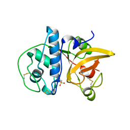
 | | Methyl and Fluorine Effects in Novel Orally Bioavailable Keap1/Nrf2 PPI Inhibitor for Treatment of Chronic Kidney Disease | | 分子名称: | (2R,3S)-3-[[(2S)-2-fluoranyl-2-(5,6,7,8-tetrahydronaphthalen-2-yl)ethanoyl]amino]-2-methyl-3-(4-methylphenyl)propanoic acid, Kelch-like ECH-associated protein 1, SULFATE ION | | 著者 | Nomura, A, Yamaguchi, K, Adachi, T. | | 登録日 | 2023-04-03 | | 公開日 | 2023-06-14 | | 最終更新日 | 2024-05-29 | | 実験手法 | X-RAY DIFFRACTION (1.48 Å) | | 主引用文献 | Methyl and Fluorine Effects in Novel Orally Bioavailable Keap1-Nrf2 PPI Inhibitor.
Acs Med.Chem.Lett., 14, 2023
|
|
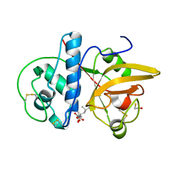
8IVR
 | | Methyl and Fluorine Effects in Novel Orally Bioavailable Keap1/Nrf2 PPI Inhibitor for Treatment of Chronic Kidney Disease | | 分子名称: | (2R,3S)-2-methyl-3-(4-methylphenyl)-3-[2-(5,6,7,8-tetrahydronaphthalen-2-yl)ethanoylamino]propanoic acid, Kelch-like ECH-associated protein 1, SULFATE ION | | 著者 | Nomura, A, Yamaguchi, K, Adachi, T. | | 登録日 | 2023-03-28 | | 公開日 | 2023-06-14 | | 最終更新日 | 2024-05-29 | | 実験手法 | X-RAY DIFFRACTION (1.5 Å) | | 主引用文献 | Methyl and Fluorine Effects in Novel Orally Bioavailable Keap1-Nrf2 PPI Inhibitor.
Acs Med.Chem.Lett., 14, 2023
|
|
8JFS
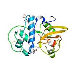
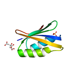 | | Phosphate bound acylphosphatase from Deinococcus radiodurans at 1 Angstrom resolution | | 分子名称: | 1,2-ETHANEDIOL, Acylphosphatase, CITRIC ACID, ... | | 著者 | Khakerwala, Z, Kumar, A, Makde, R.D. | | 登録日 | 2023-05-18 | | 公開日 | 2023-06-14 | | 最終更新日 | 2024-05-29 | | 実験手法 | X-RAY DIFFRACTION (1 Å) | | 主引用文献 | Crystal structure of phosphate bound Acyl phosphatase mini-enzyme from Deinococcus radiodurans at 1 angstrom resolution.
Biochem.Biophys.Res.Commun., 671, 2023
|
|


