3ON3
 
 | | The crystal structure of keto/oxoacid ferredoxin oxidoreductase, gamma subunit from Geobacter sulfurreducens PCA | | 分子名称: | Keto/oxoacid ferredoxin oxidoreductase, gamma subunit, SULFATE ION | | 著者 | Tan, K, Zhang, R, Hatzos, C, Buck, K, Joachimiak, A, Midwest Center for Structural Genomics (MCSG) | | 登録日 | 2010-08-27 | | 公開日 | 2010-09-22 | | 最終更新日 | 2011-07-13 | | 実験手法 | X-RAY DIFFRACTION (2.193 Å) | | 主引用文献 | The crystal structure of keto/oxoacid ferredoxin oxidoreductase, gamma subunit from Geobacter sulfurreducens PCA
To be Published
|
|
2HMC
 | | The Crystal Structure of Dihydrodipicolinate Synthase DapA from Agrobacterium tumefaciens | | 分子名称: | Dihydrodipicolinate synthase, MAGNESIUM ION | | 著者 | Kim, Y, Zhang, R, Xu, X, Zheng, H, Savchenko, A, Joachimiak, A, Midwest Center for Structural Genomics (MCSG) | | 登録日 | 2006-07-11 | | 公開日 | 2006-09-05 | | 最終更新日 | 2011-07-13 | | 実験手法 | X-RAY DIFFRACTION (1.9 Å) | | 主引用文献 | The Crystal Structure of Dihydrodipicolinate Synthase DapA from Agrobacterium tumefaciens
To be Published, 2006
|
|
4IR0
 | | Crystal Structure of Metallothiol Transferase FosB 2 from Bacillus anthracis str. Ames | | 分子名称: | 1,2-ETHANEDIOL, FOSFOMYCIN, Metallothiol transferase FosB 2, ... | | 著者 | Maltseva, N, Kim, Y, Jedrzejczak, R, Zhang, R, Anderson, W.F, Joachimiak, A, Center for Structural Genomics of Infectious Diseases (CSGID) | | 登録日 | 2013-01-14 | | 公開日 | 2013-01-23 | | 最終更新日 | 2017-11-15 | | 実験手法 | X-RAY DIFFRACTION (1.6 Å) | | 主引用文献 | Crystal Structure of Metallothiol Transferase FosB 2 from Bacillus anthracis str. Ames
To be Published
|
|
1YOZ
 | | Predicted coding region AF0941 from Archaeoglobus fulgidus | | 分子名称: | Hypothetical protein AF0941 | | 著者 | Lunin, V.V, Zhang, R, Savchenko, A, Edwards, A.M, Joachimiak, A, Midwest Center for Structural Genomics (MCSG) | | 登録日 | 2005-01-28 | | 公開日 | 2005-02-08 | | 最終更新日 | 2024-02-14 | | 実験手法 | X-RAY DIFFRACTION (2 Å) | | 主引用文献 | The crystal structure of predicted coding region AF0941 from Archaeoglobus fulgidus
To be Published
|
|
4DXA
 | | Co-crystal structure of Rap1 in complex with KRIT1 | | 分子名称: | 5'-GUANOSINE-DIPHOSPHATE-MONOTHIOPHOSPHATE, Krev interaction trapped protein 1, MAGNESIUM ION, ... | | 著者 | Li, X, Zhang, R, Boggon, T.J. | | 登録日 | 2012-02-27 | | 公開日 | 2012-05-16 | | 最終更新日 | 2023-09-13 | | 実験手法 | X-RAY DIFFRACTION (1.95 Å) | | 主引用文献 | Structural Basis for Small G Protein Effector Interaction of Ras-related Protein 1 (Rap1) and Adaptor Protein Krev Interaction Trapped 1 (KRIT1).
J.Biol.Chem., 287, 2012
|
|
3BW9
 | | Crystal Structure of HLA B*3508 in complex with a HCMV 12-mer peptide from the pp65 protein | | 分子名称: | Beta-2-microglobulin, CPS peptide from 65 kDa lower matrix phosphoprotein, HLA class I histocompatibility antigen, ... | | 著者 | Wynn, K.K, Marland, Z, Cooper, L, Silins, S.L, Gras, S, Archbold, J.K, Tynan, F.E, Miles, J.J, McCluskey, J, Burrows, S.R, Rossjohn, J, Khanna, R. | | 登録日 | 2008-01-08 | | 公開日 | 2008-04-22 | | 最終更新日 | 2023-11-01 | | 実験手法 | X-RAY DIFFRACTION (1.75 Å) | | 主引用文献 | Impact of clonal competition for peptide-MHC complexes on the CD8+ T-cell repertoire selection in a persistent viral infection
Blood, 111, 2008
|
|
3BWA
 | | Crystal Structure of HLA B*3508 in complex with a HCMV 8-mer peptide from the pp65 protein | | 分子名称: | Beta-2-microglobulin, FPT peptide from 65 kDa lower matrix phosphoprotein, HLA class I histocompatibility antigen, ... | | 著者 | Wynn, K.K, Marland, Z, Cooper, L, Silins, S.L, Gras, S, Archbold, J.K, Tynan, F.E, Miles, J.J, McCluskey, J, Burrows, S.R, Rossjohn, J, Khanna, R. | | 登録日 | 2008-01-08 | | 公開日 | 2008-04-22 | | 最終更新日 | 2023-11-01 | | 実験手法 | X-RAY DIFFRACTION (1.3 Å) | | 主引用文献 | Impact of clonal competition for peptide-MHC complexes on the CD8+ T-cell repertoire selection in a persistent viral infection
Blood, 111, 2008
|
|
4F7G
 | |
4FQN
 | | Crystal structure of the CCM2 C-terminal Harmonin Homology Domain (HHD) | | 分子名称: | Malcavernin | | 著者 | Fisher, O.S, Zhang, R, Li, X, Murphy, J.W, Boggon, T.J. | | 登録日 | 2012-06-25 | | 公開日 | 2012-12-19 | | 最終更新日 | 2024-02-28 | | 実験手法 | X-RAY DIFFRACTION (1.9 Å) | | 主引用文献 | Structural studies of cerebral cavernous malformations 2 (CCM2) reveal a folded helical domain at its C-terminus.
Febs Lett., 587, 2013
|
|
1Y89
 | | Crystal Structure of devB protein | | 分子名称: | 3,6,9,12,15,18,21,24-OCTAOXAHEXACOSAN-1-OL, DI(HYDROXYETHYL)ETHER, NONAETHYLENE GLYCOL, ... | | 著者 | Lazarski, K, Cymborowski, M, Chruszcz, M, Zheng, H, Zhang, R, Lezondra, L, Joachimiak, A, Minor, W, Midwest Center for Structural Genomics (MCSG) | | 登録日 | 2004-12-10 | | 公開日 | 2005-01-25 | | 最終更新日 | 2022-04-13 | | 実験手法 | X-RAY DIFFRACTION (2 Å) | | 主引用文献 | Crystal Structure of devB protein
To be Published
|
|
4F7H
 | | The crystal structure of kindlin-2 pleckstrin homology domain in free form | | 分子名称: | Fermitin family homolog 2, S,R MESO-TARTARIC ACID | | 著者 | Liu, Y, Zhu, Y, Qin, J, Ye, S, Zhang, R. | | 登録日 | 2012-05-16 | | 公開日 | 2012-06-13 | | 最終更新日 | 2023-09-13 | | 実験手法 | X-RAY DIFFRACTION (1.9 Å) | | 主引用文献 | Crystal structure of kindlin-2 PH domain reveals a conformational transition for its membrane anchoring and regulation of integrin activation.
Protein Cell, 3, 2012
|
|
1Y7R
 | | 1.7 A Crystal Structure of Protein of Unknown Function SA2161 from Meticillin-Resistant Staphylococcus aureus, Probable Acetyltransferase | | 分子名称: | PHOSPHATE ION, hypothetical protein SA2161 | | 著者 | Qiu, Y, Zhang, R, Collart, F, Holzle, D, Joachimiak, A, Kossiakoff, A, Midwest Center for Structural Genomics (MCSG) | | 登録日 | 2004-12-09 | | 公開日 | 2005-01-25 | | 最終更新日 | 2021-10-20 | | 実験手法 | X-RAY DIFFRACTION (1.7 Å) | | 主引用文献 | 1.7A Crystal Structure of Hypothetical Protein SA2161 from Meticillin-Resistant Staphylococcus aureus
To be Published
|
|
1P53
 | | The Crystal Structure of ICAM-1 D3-D5 fragment | | 分子名称: | 2-acetamido-2-deoxy-beta-D-glucopyranose, Intercellular adhesion molecule-1 | | 著者 | Yang, Y, Jun, C.D, Liu, J.H, Zhang, R, Jochimiak, A, Springer, T.A, Wang, J.H. | | 登録日 | 2003-04-24 | | 公開日 | 2004-05-04 | | 最終更新日 | 2020-07-29 | | 実験手法 | X-RAY DIFFRACTION (3.06 Å) | | 主引用文献 | Structural basis for dimerization of ICAM-1 on the cell surface.
Mol.Cell, 14, 2004
|
|
3B7X
 | | Crystal structure of human FK506-Binding Protein 6 | | 分子名称: | FK506-binding protein 6 | | 著者 | Walker, J.R, Davis, T, Butler-Cole, C, Paramanathan, R, Weigelt, J, Arrowsmith, C.H, Edwards, A.M, Bochkarev, A, Dhe-Paganon, S, Structural Genomics Consortium (SGC) | | 登録日 | 2007-10-31 | | 公開日 | 2007-11-27 | | 最終更新日 | 2023-08-30 | | 実験手法 | X-RAY DIFFRACTION (2.1 Å) | | 主引用文献 | Human FK506-Binding Protein 6.
To be Published
|
|
2BBJ
 | | Crystal structure of the CorA Mg2+ transporter | | 分子名称: | divalent cation transport-related protein | | 著者 | Lunin, V.V, Dobrovetsky, E, Khutoreskaya, G, Zhang, R, Joachimiak, A, Bochkarev, A, Maguire, M.E, Edwards, A.M, Koth, C.M, Structural Genomics Consortium (SGC) | | 登録日 | 2005-10-17 | | 公開日 | 2005-12-13 | | 最終更新日 | 2023-08-23 | | 実験手法 | X-RAY DIFFRACTION (3.9 Å) | | 主引用文献 | Crystal structure of the CorA Mg2+ transporter
Nature, 440, 2006
|
|
1ROC
 | | Crystal structure of the histone deposition protein Asf1 | | 分子名称: | Anti-silencing protein 1, BROMIDE ION | | 著者 | Daganzo, S.M, Erzberger, J.P, Lam, W.M, Skordalakes, E, Zhang, R, Franco, A.A, Brill, S.J, Adams, P.D, Berger, J.M, Kaufman, P.D. | | 登録日 | 2003-12-02 | | 公開日 | 2003-12-23 | | 最終更新日 | 2024-02-14 | | 実験手法 | X-RAY DIFFRACTION (1.5 Å) | | 主引用文献 | Structure and function of the conserved core of histone deposition protein Asf1.
Curr.Biol., 13, 2003
|
|
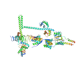
4H5Y
 | | High-resolution crystal structure of Legionella pneumophila LidA (60-594) | | 分子名称: | LidA protein, substrate of the Dot/Icm system | | 著者 | An, X, Ye, S, Liu, Y, Zheng, X, Zhang, R. | | 登録日 | 2012-09-19 | | 公開日 | 2013-09-25 | | 最終更新日 | 2024-02-28 | | 実験手法 | X-RAY DIFFRACTION (2.1 Å) | | 主引用文献 | The crystal structure of LidA, a translocated substrate of the Legionella pneumophila type IV secretion system.
Protein Cell, 4, 2013
|
|
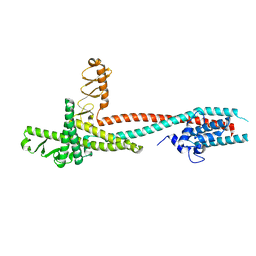
7JU4
 | | Radial spoke 2 stalk, IDAc, and N-DRC attached with doublet microtubule | | 分子名称: | 28 kDa inner dynein arm light chain, axonemal, ADENOSINE-5'-TRIPHOSPHATE, ... | | 著者 | Gui, M, Ma, M, Sze-Tu, E, Wang, X, Koh, F, Zhong, E, Berger, B, Davis, J, Dutcher, S, Zhang, R, Brown, A. | | 登録日 | 2020-08-19 | | 公開日 | 2020-12-16 | | 最終更新日 | 2024-03-06 | | 実験手法 | ELECTRON MICROSCOPY (3.4 Å) | | 主引用文献 | Structures of radial spokes and associated complexes important for ciliary motility.
Nat.Struct.Mol.Biol., 28, 2021
|
|
7JTK
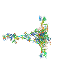 | | Radial spoke 1 isolated from Chlamydomonas reinhardtii | | 分子名称: | Cytochrome b5 heme-binding domain-containing protein, Dynein 8 kDa light chain, flagellar outer arm, ... | | 著者 | Gui, M, Ma, M, Sze-Tu, E, Wang, X, Koh, F, Zhong, E, Berger, B, Davis, J, Dutcher, S, Zhang, R, Brown, A. | | 登録日 | 2020-08-17 | | 公開日 | 2020-12-16 | | 最終更新日 | 2021-01-27 | | 実験手法 | ELECTRON MICROSCOPY (3.2 Å) | | 主引用文献 | Structures of radial spokes and associated complexes important for ciliary motility.
Nat.Struct.Mol.Biol., 28, 2021
|
|
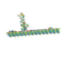
7JTS
 | | Stalk of radial spoke 1 attached with doublet microtubule from Chlamydomonas reinhardtii | | 分子名称: | Calmodulin, Dynein 8 kDa light chain, flagellar outer arm, ... | | 著者 | Gui, M, Ma, M, Sze-Tu, E, Wang, X, Koh, F, Zhong, E, Berger, B, Davis, J, Dutcher, S, Zhang, R, Brown, A. | | 登録日 | 2020-08-18 | | 公開日 | 2020-12-16 | | 最終更新日 | 2024-03-06 | | 実験手法 | ELECTRON MICROSCOPY (6.1 Å) | | 主引用文献 | Structures of radial spokes and associated complexes important for ciliary motility.
Nat.Struct.Mol.Biol., 28, 2021
|
|
1VCC
 | |
1R4V
 | | 1.9A crystal structure of protein AQ328 from Aquifex aeolicus | | 分子名称: | CACODYLATE ION, Hypothetical protein AQ_328, ZINC ION | | 著者 | Qiu, Y, Tereshko, V, Kim, Y, Zhang, R, Collart, F, Joachimiak, A, Kossiakoff, A, Midwest Center for Structural Genomics (MCSG) | | 登録日 | 2003-10-08 | | 公開日 | 2004-03-30 | | 最終更新日 | 2011-07-13 | | 実験手法 | X-RAY DIFFRACTION (1.9 Å) | | 主引用文献 | The crystal structure of Aq_328 from the hyperthermophilic bacteria Aquifex aeolicus shows an ancestral histone fold.
Proteins, 62, 2006
|
|
2MX4
 | | NMR structure of Phosphorylated 4E-BP2 | | 分子名称: | Eukaryotic translation initiation factor 4E-binding protein 2 | | 著者 | Bah, A, Forman-Kay, J, Vernon, R, Siddiqui, Z, Krzeminski, M, Muhandiram, R, Zhao, C, Sonenberg, N, Kay, L. | | 登録日 | 2014-12-10 | | 公開日 | 2015-01-07 | | 最終更新日 | 2015-03-18 | | 実験手法 | SOLUTION NMR | | 主引用文献 | Folding of an intrinsically disordered protein by phosphorylation as a regulatory switch.
Nature, 519, 2015
|
|
4DX9
 | | ICAP1 in complex with integrin beta 1 cytoplasmic tail | | 分子名称: | Integrin beta-1, Integrin beta-1-binding protein 1 | | 著者 | Liu, W, Draheim, K, Zhang, R, Calderwood, D.A, Boggon, T.J. | | 登録日 | 2012-02-27 | | 公開日 | 2013-01-09 | | 最終更新日 | 2020-09-02 | | 実験手法 | X-RAY DIFFRACTION (2.99 Å) | | 主引用文献 | Mechanism for KRIT1 Release of ICAP1-Mediated Suppression of Integrin Activation.
Mol.Cell, 49, 2013
|
|
1HO2
 | | NMR STRUCTURE OF THE POTASSIUM CHANNEL FRAGMENT L45 IN MICELLES | | 分子名称: | VOLTAGE-GATED POTASSIUM CHANNEL PROTEIN | | 著者 | Ohlenschlager, O, Hojo, H, Ramachandran, R, Gorlach, M, Haris, P.I. | | 登録日 | 2000-12-08 | | 公開日 | 2002-06-05 | | 最終更新日 | 2024-05-22 | | 実験手法 | SOLUTION NMR | | 主引用文献 | Three-dimensional structure of the S4-S5 segment of the Shaker potassium channel.
Biophys.J., 82, 2002
|
|


