8DEA
 
 | |
8DG6
 | |
3IUF
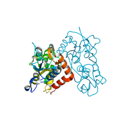
 | | Crystal structure of the C2H2-type zinc finger domain of human ubi-d4 | | 分子名称: | ZINC ION, Zinc finger protein ubi-d4 | | 著者 | Tempel, W, Xu, C, Bian, C, Adams-Cioaba, M, Eryilmaz, J, Bountra, C, Weigelt, J, Arrowsmith, C.H, Edwards, A.M, Bochkarev, A, Min, J, Structural Genomics Consortium (SGC) | | 登録日 | 2009-08-31 | | 公開日 | 2009-11-03 | | 最終更新日 | 2024-02-21 | | 実験手法 | X-RAY DIFFRACTION (1.8 Å) | | 主引用文献 | Crystal structure of the Cys2His2-type zinc finger domain of human DPF2.
Biochem.Biophys.Res.Commun., 413, 2011
|
|
2FZN
 | |
2FZM
 | |
5C7J
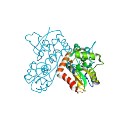
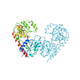
 | | CRYSTAL STRUCTURE OF NEDD4 WITH A UB VARIANT | | 分子名称: | E3 ubiquitin-protein ligase NEDD4, Polyubiquitin-C | | 著者 | Walker, J.R, Hu, J, Dong, A, Bountra, C, Edwards, A.M, Arrowsmith, C.H, Tong, Y, Structural Genomics Consortium (SGC) | | 登録日 | 2015-06-24 | | 公開日 | 2016-03-16 | | 最終更新日 | 2023-09-27 | | 実験手法 | X-RAY DIFFRACTION (3 Å) | | 主引用文献 | System-Wide Modulation of HECT E3 Ligases with Selective Ubiquitin Variant Probes.
Mol.Cell, 62, 2016
|
|
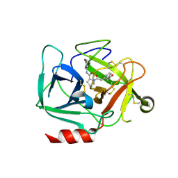
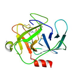
3B6T
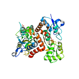
 | | Crystal Structure of the GLUR2 Ligand Binding Core (S1S2J) T686A Mutant in Complex with Quisqualate at 2.1 Resolution | | 分子名称: | (S)-2-AMINO-3-(3,5-DIOXO-[1,2,4]OXADIAZOLIDIN-2-YL)-PROPIONIC ACID, Glutamate receptor 2, SULFATE ION | | 著者 | Cho, Y, Lolis, E, Howe, J.R. | | 登録日 | 2007-10-29 | | 公開日 | 2008-02-05 | | 最終更新日 | 2024-11-06 | | 実験手法 | X-RAY DIFFRACTION (2.1 Å) | | 主引用文献 | Structural and single-channel results indicate that the rates of ligand binding domain closing and opening directly impact AMPA receptor gating.
J.Neurosci., 28, 2008
|
|
3B6Q
 | |
3B6W
 | |
5HPK
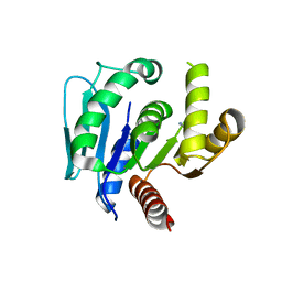
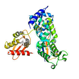 | | System-wide modulation of HECT E3 ligases with selective ubiquitin variant probes: NEDD4L and UbV NL.1 | | 分子名称: | E3 ubiquitin-protein ligase NEDD4-like, Ubiquitin variant NL.1 | | 著者 | Wu, K.-P, Mukherjee, M, Mercredi, P.Y, Schulman, B.A. | | 登録日 | 2016-01-20 | | 公開日 | 2016-03-16 | | 最終更新日 | 2023-09-27 | | 実験手法 | X-RAY DIFFRACTION (2.431 Å) | | 主引用文献 | System-Wide Modulation of HECT E3 Ligases with Selective Ubiquitin Variant Probes.
Mol.Cell, 62, 2016
|
|
5HPT
 | |
5HPL
 | |
5HPS
 | |
5NS9
 | |
8JNJ
 | |
8JNI
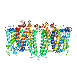 | | Structure of AE2 in complex with PIP2 | | 分子名称: | Anion exchange protein 2, CHLORIDE ION, [(2R)-1-octadecanoyloxy-3-[oxidanyl-[(1R,2R,3S,4R,5R,6S)-2,3,6-tris(oxidanyl)-4,5-diphosphonooxy-cyclohexyl]oxy-phospho ryl]oxy-propan-2-yl] (8Z)-icosa-5,8,11,14-tetraenoate | | 著者 | Yin, Y.X, Ding, D. | | 登録日 | 2023-06-06 | | 公開日 | 2024-02-07 | | 最終更新日 | 2024-11-06 | | 実験手法 | ELECTRON MICROSCOPY (3.2 Å) | | 主引用文献 | Structural and functional insights into the lipid regulation of human anion exchanger 2.
Nat Commun, 15, 2024
|
|
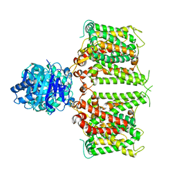
9LGW
 | | Cryo-EM structure of SiAgo-Aga1 complex | | 分子名称: | Piwi domain-containing protein, SiAgo-associated protein1, SiAga1 | | 著者 | Dai, Z.K, Guan, Z.Y, Zou, T.T. | | 登録日 | 2025-01-10 | | 公開日 | 2025-02-19 | | 実験手法 | ELECTRON MICROSCOPY (2.7 Å) | | 主引用文献 | Structural and mechanistic insights into the activation of a short prokaryotic argonaute system from archaeon Sulfolobus islandicus.
Nucleic Acids Res., 53, 2025
|
|
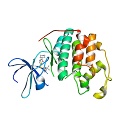
6LCY
 | | Crystal structure of Synaptotagmin-7 C2B in complex with IP6 | | 分子名称: | INOSITOL HEXAKISPHOSPHATE, Synaptotagmin-7 | | 著者 | Zhang, Y, Zhang, X, Rao, F, Wang, C. | | 登録日 | 2019-11-20 | | 公開日 | 2021-03-03 | | 最終更新日 | 2023-11-22 | | 実験手法 | X-RAY DIFFRACTION (2.301 Å) | | 主引用文献 | 5-IP 7 is a GPCR messenger mediating neural control of synaptotagmin-dependent insulin exocytosis and glucose homeostasis.
Nat Metab, 3, 2021
|
|
5D1J
 | |
7FG9
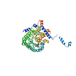
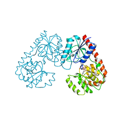 | | Alpha-1,2-glucosyltransferase_UDP_tll1591 | | 分子名称: | Glycosyl transferase, URIDINE-5'-DIPHOSPHATE | | 著者 | Su, J.Y. | | 登録日 | 2021-07-26 | | 公開日 | 2022-06-01 | | 最終更新日 | 2023-11-29 | | 実験手法 | X-RAY DIFFRACTION (2.66 Å) | | 主引用文献 | Structural basis for glucosylsucrose synthesis by a member of the alpha-1,2-glucosyltransferase family
Acta Biochim.Biophys.Sin., 54, 2022
|
|
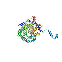
7FGA
 | | Alpha-1,2-glucosyltransferase_UDP_sucrose_tll1591 | | 分子名称: | Glycosyl transferase, URIDINE-5'-DIPHOSPHATE, alpha-D-glucopyranose-(1-1)-alpha-D-fructofuranose | | 著者 | Su, J.Y. | | 登録日 | 2021-07-26 | | 公開日 | 2022-06-01 | | 最終更新日 | 2023-11-29 | | 実験手法 | X-RAY DIFFRACTION (3.2 Å) | | 主引用文献 | Structural basis for glucosylsucrose synthesis by a member of the alpha-1,2-glucosyltransferase family
Acta Biochim.Biophys.Sin., 54, 2022
|
|
6LDQ
 | |
6LJP
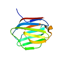 | | Crystal structure of human galectin-16 | | 分子名称: | Galectin-16 | | 著者 | Su, J. | | 登録日 | 2019-12-17 | | 公開日 | 2020-10-14 | | 最終更新日 | 2023-11-22 | | 実験手法 | X-RAY DIFFRACTION (2 Å) | | 主引用文献 | Human galectin-16 has a pseudo ligand binding site and plays a role in regulating c-Rel-mediated lymphocyte activity.
Biochim Biophys Acta Gen Subj, 1865, 2020
|
|
6LJQ
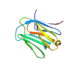 | | human galectin-16 R55N | | 分子名称: | Galectin-16, beta-D-galactopyranose-(1-4)-beta-D-glucopyranose | | 著者 | Su, J. | | 登録日 | 2019-12-17 | | 公開日 | 2020-10-14 | | 最終更新日 | 2023-11-22 | | 実験手法 | X-RAY DIFFRACTION (1.49 Å) | | 主引用文献 | Human galectin-16 has a pseudo ligand binding site and plays a role in regulating c-Rel-mediated lymphocyte activity.
Biochim Biophys Acta Gen Subj, 1865, 2020
|
|
6LJR
 | | human galectin-16 R55N/H57R | | 分子名称: | Galectin-16, beta-D-galactopyranose-(1-4)-beta-D-glucopyranose | | 著者 | Su, J. | | 登録日 | 2019-12-17 | | 公開日 | 2020-10-14 | | 最終更新日 | 2023-11-22 | | 実験手法 | X-RAY DIFFRACTION (2 Å) | | 主引用文献 | Human galectin-16 has a pseudo ligand binding site and plays a role in regulating c-Rel-mediated lymphocyte activity.
Biochim Biophys Acta Gen Subj, 1865, 2020
|
|


