Loading
PDBj
メニューPDBj@FacebookPDBj@TwitterPDBj@YouTubewwPDB FoundationwwPDB
RCSB PDBPDBeBMRBAdv. SearchSearch help
Search by PDB author
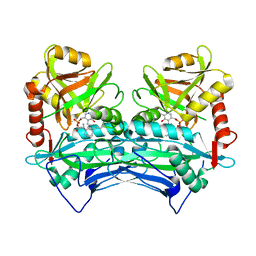
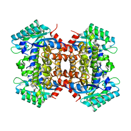
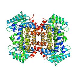
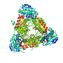
7Y05
DownloadVisualize
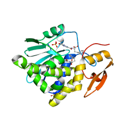
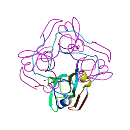
BU of 7y05 by Molmil
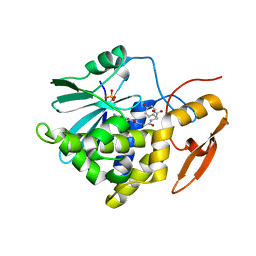
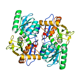
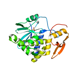
Crystal structure of Ricin A chain bound with (R)-2-(2-amino-4-oxo-3,4-dihydropteridine-7-carboxamido)-2-phenylacetic acid
分子名称: (2R)-2-[(2-azanyl-4-oxidanylidene-3H-pteridin-7-yl)carbonylamino]-2-phenyl-ethanoic acid, Ricin A chain, SULFATE ION
著者Goto, M, Higashi, S, Ohba, T, Kawata, R, Nagatsu, K, Suzuki, S, Saito, R.
登録日2022-06-03
公開日2022-12-21
最終更新日2023-11-29
実験手法X-RAY DIFFRACTION (1.6 Å)
主引用文献Conformational change in ricin toxin A-Chain: A critical factor for inhibitor binding to the secondary pocket.
Biochem.Biophys.Res.Commun., 627, 2022
7Y06
DownloadVisualize
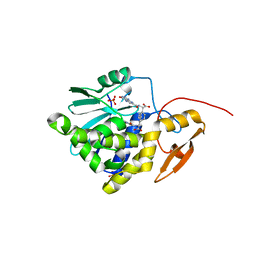
BU of 7y06 by Molmil
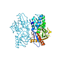
Crystal structure of Ricin A chain bound with (S)-2-(2-amino-4-oxo-3,4-dihydropteridine-7-carboxamido)-4-phenylbutanoic acid
分子名称: (2S)-2-[(2-azanyl-4-oxidanylidene-3H-pteridin-7-yl)carbonylamino]-4-phenyl-butanoic acid, Ricin A chain, SULFATE ION
著者Goto, M, Higashi, S, Ohba, T, Kawata, R, Nagatsu, K, Suzuki, S, Saito, R.
登録日2022-06-03
公開日2022-12-21
最終更新日2023-11-29
実験手法X-RAY DIFFRACTION (1.35 Å)
主引用文献Conformational change in ricin toxin A-Chain: A critical factor for inhibitor binding to the secondary pocket.
Biochem.Biophys.Res.Commun., 627, 2022
7Y03
DownloadVisualize
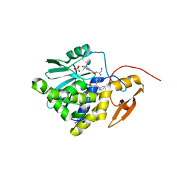
BU of 7y03 by Molmil
Crystal structure of Ricin A chain bound with (S)-2-(2-amino-4-oxo-3,4-dihydropteridine-7-carboxamido)-2-phenylacetic acid
分子名称: (2S)-2-[(2-azanyl-4-oxidanylidene-3H-pteridin-7-yl)carbonylamino]-2-phenyl-ethanoic acid, Ricin A chain, SULFATE ION
著者Goto, M, Higashi, S, Ohba, T, Kawata, R, Nagatsu, K, Suzuki, S, Saito, R.
登録日2022-06-03
公開日2022-12-21
最終更新日2023-11-29
実験手法X-RAY DIFFRACTION (1.35 Å)
主引用文献Conformational change in ricin toxin A-Chain: A critical factor for inhibitor binding to the secondary pocket.
Biochem.Biophys.Res.Commun., 627, 2022
7Y07
DownloadVisualize
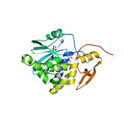
BU of 7y07 by Molmil
Crystal structure of Ricin A chain bound with (S)-2-amino-N-(1-hydroxy-3-phenylpropan-2-yl)-4-oxo-3,4-dihydropteridine-7-carboxamide
分子名称: 2-azanyl-4-oxidanylidene-N-[(2S)-1-oxidanyl-3-phenyl-propan-2-yl]-3H-pteridine-7-carboxamide, Ricin A chain, SULFATE ION
著者Goto, M, Higashi, S, Ohba, T, Kawata, R, Nagatsu, K, Suzuki, S, Saito, R.
登録日2022-06-03
公開日2022-12-21
最終更新日2023-11-29
実験手法X-RAY DIFFRACTION (1.25 Å)
主引用文献Conformational change in ricin toxin A-Chain: A critical factor for inhibitor binding to the secondary pocket.
Biochem.Biophys.Res.Commun., 627, 2022
7Y08
DownloadVisualize
BU of 7y08 by Molmil
Crystal structure of Ricin A chain bound with (2-amino-4-oxo-3,4-dihydropteridine-7-carbonyl)glycyl-L-phenylalanine
分子名称: N-[(2-amino-4-oxo-1,4-dihydropteridin-7-yl)carbonyl]glycyl-L-phenylalanine, Ricin A chain, SULFATE ION
著者Goto, M, Higashi, S, Ohba, T, Kawata, R, Nagatsu, K, Suzuki, S, Saito, R.
登録日2022-06-03
公開日2022-12-21
最終更新日2023-11-29
実験手法X-RAY DIFFRACTION (1.25 Å)
主引用文献Conformational change in ricin toxin A-Chain: A critical factor for inhibitor binding to the secondary pocket.
Biochem.Biophys.Res.Commun., 627, 2022
8I7P
DownloadVisualize
BU of 8i7p by Molmil
Crystal structure of Ricin A chain bound with N2-(2-amino-4-oxo-3,4-dihydropteridine-7-carbonyl)glycyl-L-tyrosine
分子名称: 6-(2-ethyl-4-hydroxyphenyl)-1H-indazole-3-carboxamide, Ricin A chain, SULFATE ION
著者Goto, M, Sakamoto, N, Higashi, S, Kawata, R, Nagatsu, K, Saito, R.
登録日2023-02-01
公開日2023-09-20
最終更新日2024-10-09
実験手法X-RAY DIFFRACTION (1.6 Å)
主引用文献Crystal structure of ricin toxin A chain complexed with a highly potent pterin-based small-molecular inhibitor.
J Enzyme Inhib Med Chem, 38, 2023
2COG
DownloadVisualize
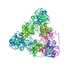
BU of 2cog by Molmil
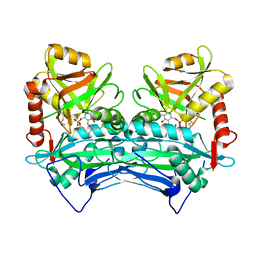
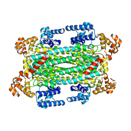
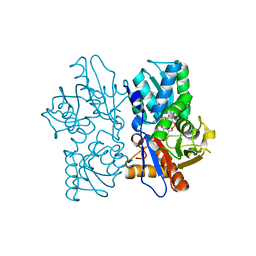
Crystal structure of oxidized human cytosolic branched-chain aminotransferase complexed with 4-methylvalerate
分子名称: 4-METHYL VALERIC ACID, MAGNESIUM ION, PYRIDOXAL-5'-PHOSPHATE, ...
著者Goto, M.
登録日2005-05-17
公開日2005-08-30
最終更新日2021-11-10
実験手法X-RAY DIFFRACTION (2.1 Å)
主引用文献Structural determinants for branched-chain aminotransferase isozyme-specific inhibition by the anticonvulsant drug gabapentin
J.Biol.Chem., 280, 2005
2COI
DownloadVisualize
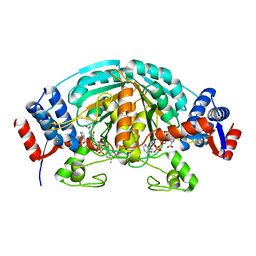
BU of 2coi by Molmil
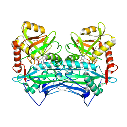
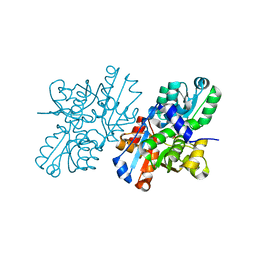
Crystal structure of oxidized human cytosolic branched-chain aminotransferase complexed with gabapentin
分子名称: MAGNESIUM ION, PYRIDOXAL-5'-PHOSPHATE, [1-(AMINOMETHYL)CYCLOHEXYL]ACETIC ACID, ...
著者Goto, M.
登録日2005-05-17
公開日2005-08-30
最終更新日2021-11-10
実験手法X-RAY DIFFRACTION (1.9 Å)
主引用文献Structural determinants for branched-chain aminotransferase isozyme-specific inhibition by the anticonvulsant drug gabapentin
J.Biol.Chem., 280, 2005
2DVX
DownloadVisualize
BU of 2dvx by Molmil
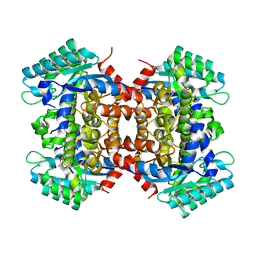
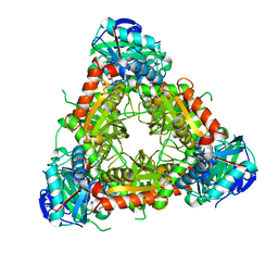
Crystal Structure of 2,6-Dihydroxybenzoate Decarboxylase Complexed with inhibitor 2,3-dihydroxybenzaldehyde
分子名称: 2,3-DIHYDROXYBENZALDEHYDE, Thermophilic reversible gamma-resorcylate decarboxylase, ZINC ION
著者Goto, M.
登録日2006-08-01
公開日2006-09-19
最終更新日2023-10-25
実験手法X-RAY DIFFRACTION (1.7 Å)
主引用文献Crystal Structures of Nonoxidative Zn-Dependent 2,6-Dihydroxybenzoate (gamma-Resorcylate) Decarboxylase from Rhizobium sp. Strain MTP-10005
To be Published
2COJ
DownloadVisualize
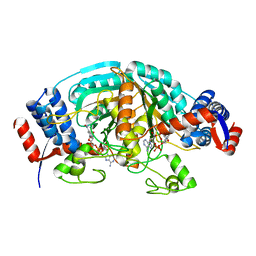
BU of 2coj by Molmil
Crystal structure of reduced human cytosolic branched-chain aminotransferase complexed with gabapentin
分子名称: MAGNESIUM ION, PYRIDOXAL-5'-PHOSPHATE, [1-(AMINOMETHYL)CYCLOHEXYL]ACETIC ACID, ...
著者Goto, M.
登録日2005-05-17
公開日2005-08-30
最終更新日2021-11-10
実験手法X-RAY DIFFRACTION (2.4 Å)
主引用文献Structural determinants for branched-chain aminotransferase isozyme-specific inhibition by the anticonvulsant drug gabapentin
J.Biol.Chem., 280, 2005
2DVT
DownloadVisualize
BU of 2dvt by Molmil
Crystal Structure of 2,6-Dihydroxybenzoate Decarboxylase from Rhizobium
分子名称: Thermophilic reversible gamma-resorcylate decarboxylase, ZINC ION
著者Goto, M.
登録日2006-08-01
公開日2006-09-19
最終更新日2024-03-13
実験手法X-RAY DIFFRACTION (1.7 Å)
主引用文献Crystal Structures of Nonoxidative Zn-Dependent 2,6-Dihydroxybenzoate (gamma-Resorcylate) Decarboxylase from Rhizobium sp. Strain MTP-10005
To be Published
2DVU
DownloadVisualize
BU of 2dvu by Molmil
Crystal Structure of 2,6-Dihydroxybenzoate Decarboxylase Complexed with 2,6-Dihydroxybenzoate
分子名称: 2,6-DIHYDROXYBENZOIC ACID, Thermophilic reversible gamma-resorcylate decarboxylase, ZINC ION
著者Goto, M.
登録日2006-08-01
公開日2006-09-19
最終更新日2023-10-25
実験手法X-RAY DIFFRACTION (1.9 Å)
主引用文献Crystal Structures of Nonoxidative Zn-Dependent 2,6-Dihydroxybenzoate (gamma-Resorcylate) Decarboxylase from Rhizobium sp. Strain MTP-10005
To be Published
2EJ0
DownloadVisualize
BU of 2ej0 by Molmil
Crystal Structure of T.th.HB8 Branched-Chain Amino Acid Aminotransferase with Pyridoxamine 5'-phosphate
分子名称: (4S)-2-METHYL-2,4-PENTANEDIOL, 4'-DEOXY-4'-AMINOPYRIDOXAL-5'-PHOSPHATE, Branched-chain amino acid aminotransferase
著者Goto, M.
登録日2007-03-14
公開日2008-03-18
最終更新日2023-10-25
実験手法X-RAY DIFFRACTION (1.6 Å)
主引用文献Crystal Structure of T.th.HB8 Branched-Chain Amino Acid Aminotransferase with Pyridoxamine 5'-phosphate
To be published
2E9F
DownloadVisualize
BU of 2e9f by Molmil
Crystal Structure of T.th.HB8 Argininosuccinate lyase complexed with L-Arginine
分子名称: ARGININE, Argininosuccinate lyase
著者Goto, M.
登録日2007-01-25
公開日2007-07-31
最終更新日2023-10-25
実験手法X-RAY DIFFRACTION (2.8 Å)
主引用文献Crystal Structure of T.th.HB8 Argininosuccinate lyase complexed with L-Arginine
To be published
2EJ2
DownloadVisualize
BU of 2ej2 by Molmil
Crystal Structure of T.th.HB8 Branched-Chain Amino Acid Aminotransferase Complexed with N-(5'-Phosphopyridoxyl)-L-Glutamate
分子名称: (4S)-2-METHYL-2,4-PENTANEDIOL, 4-[(1,3-DICARBOXY-PROPYLAMINO)-METHYL]-3-HYDROXY-2-METHYL-5-PHOSPHONOOXYMETHYL-PYRIDINIUM, Branched-chain amino acid aminotransferase
著者Goto, M.
登録日2007-03-14
公開日2008-03-18
最終更新日2023-10-25
実験手法X-RAY DIFFRACTION (2 Å)
主引用文献Crystal Structure of T.th.HB8 Branched-Chain Amino Acid Aminotransferase Complexed with N-(5'-Phosphopyridoxyl)-L-Glutamate
TO BE PUBLISHED
2EFY
DownloadVisualize
BU of 2efy by Molmil
Crystal Structure of T.th. HB8 O-acetylserine sulfhydrylase Complexed with 4-Acetylbutyric acid
分子名称: 5-OXOHEXANOIC ACID, O-acetylserine (Thiol)-lyase, PYRIDOXAL-5'-PHOSPHATE
著者Goto, M.
登録日2007-02-26
公開日2008-02-26
最終更新日2023-10-25
実験手法X-RAY DIFFRACTION (2.35 Å)
主引用文献Crystal Structure of T.th. HB8 O-acetylserine sulfhydrylase Complexed with 4-Acetylbutyric acid
to be published
2ECQ
DownloadVisualize
BU of 2ecq by Molmil
Crystal Structure of T.th. HB8 O-acetylserine sulfhydrylase Complexed with 3-Hydroxylactate
分子名称: (3S)-3-HYDROXYBUTANOIC ACID, O-acetylserine (Thiol)-lyase, PYRIDOXAL-5'-PHOSPHATE
著者Goto, M.
登録日2007-02-13
公開日2008-02-19
最終更新日2023-10-25
実験手法X-RAY DIFFRACTION (1.9 Å)
主引用文献Crystal Structure of T.th. HB8 O-acetylserine sulfhydrylase Complexed with 3-Hydroxylactate
to be published
2EEO
DownloadVisualize
BU of 2eeo by Molmil
Crystal Structure of T.th. HB8 L-Aspartate-alpha-Decarboxylase Complexed with Fumarate
分子名称: Aspartate 1-decarboxylase, FUMARIC ACID, PYRUVIC ACID
著者Goto, M.
登録日2007-02-16
公開日2008-02-19
最終更新日2023-11-15
実験手法X-RAY DIFFRACTION (1.6 Å)
主引用文献Crystal Structure of T.th. HB8 L-Aspartate-alpha-Decarboxylase
To be Published
2EIY
DownloadVisualize
BU of 2eiy by Molmil
Crystal Structure of T.th.HB8 Branched-Chain Amino Acid Aminotransferase Complexed with 4-Methylvaleric Acid
分子名称: (4S)-2-METHYL-2,4-PENTANEDIOL, 4-METHYL VALERIC ACID, Branched-chain amino acid aminotransferase, ...
著者Goto, M.
登録日2007-03-14
公開日2008-03-18
最終更新日2023-10-25
実験手法X-RAY DIFFRACTION (1.35 Å)
主引用文献Crystal Structure of T.th.HB8 Branched-Chain Amino Acid Aminotransferase Complexed with 4-Methylvaleric Acid
To be published
2CWH
DownloadVisualize
BU of 2cwh by Molmil
Crystal Structure of delta1-piperideine-2-carboxylate reductase from Pseudomonas syringae complexed with NADPH and pyrrole-2-carboxylate
分子名称: NADPH DIHYDRO-NICOTINAMIDE-ADENINE-DINUCLEOTIDE PHOSPHATE, PYRROLE-2-CARBOXYLATE, delta1-piperideine-2-carboxylate reductase
著者Goto, M, Muramatsu, H, Mihara, H, Kurihara, T, Esaki, N, Omi, R, Miyahara, I, Hirotsu, K.
登録日2005-06-20
公開日2005-10-04
最終更新日2024-03-13
実験手法X-RAY DIFFRACTION (1.7 Å)
主引用文献Crystal structures of Delta1-piperideine-2-carboxylate/Delta1-pyrroline-2-carboxylate reductase belonging to a new family of NAD(P)H-dependent oxidoreductases: conformational change, substrate recognition, and stereochemistry of the reaction
J.Biol.Chem., 280, 2005
2CWF
DownloadVisualize
BU of 2cwf by Molmil
Crystal Structure of delta1-piperideine-2-carboxylate reductase from Pseudomonas syringae complexed with NADPH
分子名称: NADPH DIHYDRO-NICOTINAMIDE-ADENINE-DINUCLEOTIDE PHOSPHATE, delta1-piperideine-2-carboxylate reductase
著者Goto, M, Muramatsu, H, Mihara, H, Kurihara, T, Esaki, N, Omi, R, Miyahara, I, Hirotsu, K.
登録日2005-06-20
公開日2005-10-04
最終更新日2024-03-13
実験手法X-RAY DIFFRACTION (1.8 Å)
主引用文献Crystal structures of Delta1-piperideine-2-carboxylate/Delta1-pyrroline-2-carboxylate reductase belonging to a new family of NAD(P)H-dependent oxidoreductases: conformational change, substrate recognition, and stereochemistry of the reaction
J.Biol.Chem., 280, 2005
2ECO
DownloadVisualize
BU of 2eco by Molmil
Crystal Structure of T.th. HB8 O-acetylserine sulfhydrylase Complexed with 4-methylvalerate
分子名称: 4-METHYL VALERIC ACID, O-acetylserine (Thiol)-lyase, PYRIDOXAL-5'-PHOSPHATE
著者Goto, M.
登録日2007-02-13
公開日2008-02-19
最終更新日2023-10-25
実験手法X-RAY DIFFRACTION (1.9 Å)
主引用文献Crystal Structure of T.th. HB8 O-acetylserine sulfhydrylase Complexed with 4-methylvalerate
to be published
2EJ3
DownloadVisualize
BU of 2ej3 by Molmil
Crystal Structure of T.th.HB8 Branched-Chain Amino Acid Aminotransferase Complexed with Gabapentin
分子名称: (4S)-2-METHYL-2,4-PENTANEDIOL, Branched-chain amino acid aminotransferase, PYRIDOXAL-5'-PHOSPHATE, ...
著者Goto, M.
登録日2007-03-14
公開日2008-03-18
最終更新日2023-10-25
実験手法X-RAY DIFFRACTION (2.2 Å)
主引用文献Crystal Structure of T.th.HB8 Branched-Chain Amino Acid Aminotransferase Complexed with Gabapentin
To be Published
2ZR8
DownloadVisualize
BU of 2zr8 by Molmil
Crystal Structure of Modified Serine Racemase complexed with Serine
分子名称: MAGNESIUM ION, N-(5'-PHOSPHOPYRIDOXYL)-D-ALANINE, SERINE, ...
著者Goto, M.
登録日2008-08-25
公開日2009-07-28
最終更新日2023-11-01
実験手法X-RAY DIFFRACTION (2.2 Å)
主引用文献Crystal structure of a homolog of mammalian serine racemase from Schizosaccharomyces pombe
J.Biol.Chem., 284, 2009
4TV7
DownloadVisualize
BU of 4tv7 by Molmil
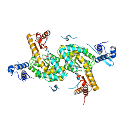
Crystal structure of Bacillus subtilis GabR at 2.05 Angstroms resolution
分子名称: HTH-type transcriptional regulatory protein GabR
著者Goto, M, Okuda, K, Yoshimura, T.
登録日2014-06-26
公開日2014-12-24
最終更新日2020-01-29
実験手法X-RAY DIFFRACTION (2.05 Å)
主引用文献Role of the aminotransferase domain in Bacillus subtilis GabR, a pyridoxal 5'-phosphate-dependent transcriptional regulator
Mol. Microbiol., 95, 2015

225946

件を2024-10-09に公開中

PDB statisticsPDBj update infoContact PDBjnumon