2R68
 
 | |
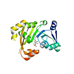
3P2K
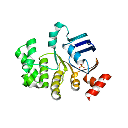
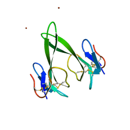
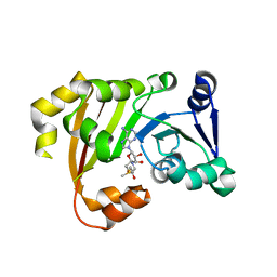 | | Structure of an antibiotic related Methyltransferase | | 分子名称: | 16S rRNA methylase, S-ADENOSYLMETHIONINE | | 著者 | Sivaraman, J, Husain, N. | | 登録日 | 2010-10-02 | | 公開日 | 2010-11-17 | | 最終更新日 | 2023-11-01 | | 実験手法 | X-RAY DIFFRACTION (2.7 Å) | | 主引用文献 | Structural basis for the methylation of A1408 in 16S rRNA by a panaminoglycoside resistance methyltransferase NpmA from a clinical isolate and analysis of the NpmA interactions with the 30S ribosomal subunit.
Nucleic Acids Res., 39, 2011
|
|
3P2E
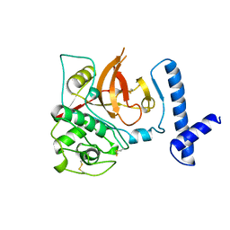
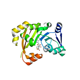 | | Structure of an antibiotic related Methyltransferase | | 分子名称: | 16S rRNA methylase, S-ADENOSYL-L-HOMOCYSTEINE | | 著者 | Sivaraman, J, Husain, N. | | 登録日 | 2010-10-02 | | 公開日 | 2010-11-17 | | 最終更新日 | 2023-11-01 | | 実験手法 | X-RAY DIFFRACTION (1.68 Å) | | 主引用文献 | Structural basis for the methylation of A1408 in 16S rRNA by a panaminoglycoside resistance methyltransferase NpmA from a clinical isolate and analysis of the NpmA interactions with the 30S ribosomal subunit.
Nucleic Acids Res., 2010
|
|
3P2I
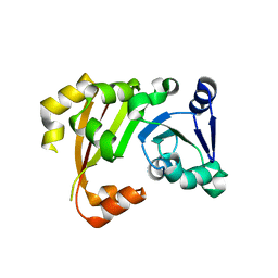 | | Structure of an antibiotic related Methyltransferase | | 分子名称: | 16S rRNA methylase | | 著者 | Sivaraman, J, Husain, N. | | 登録日 | 2010-10-02 | | 公開日 | 2010-11-17 | | 最終更新日 | 2023-11-01 | | 実験手法 | X-RAY DIFFRACTION (2.4 Å) | | 主引用文献 | Structural basis for the methylation of A1408 in 16S rRNA by a panaminoglycoside resistance methyltransferase NpmA from a clinical isolate and analysis of the NpmA interactions with the 30S ribosomal subunit.
Nucleic Acids Res., 39, 2011
|
|
3PB3
 | | Structure of an Antibiotic Related Methyltransferase | | 分子名称: | 16S rRNA methylase, S-ADENOSYL-L-HOMOCYSTEINE | | 著者 | Sivaraman, J, Husain, N. | | 登録日 | 2010-10-20 | | 公開日 | 2010-11-24 | | 最終更新日 | 2024-03-20 | | 実験手法 | X-RAY DIFFRACTION (1.9 Å) | | 主引用文献 | Structural basis for the methylation of A1408 in 16S rRNA by a panaminoglycoside resistance methyltransferase NpmA from a clinical isolate and analysis of the NpmA interactions with the 30S ribosomal subunit.
Nucleic Acids Res., 2010
|
|
8K70
 | |
8JB0
 | |
8JAX
 | |
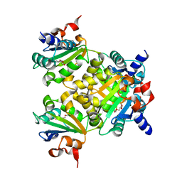
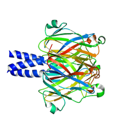
4U00
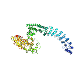
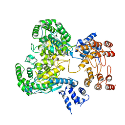
 | | Crystal structure of TTHA1159 in complex with ADP | | 分子名称: | ADENOSINE-5'-DIPHOSPHATE, Amino acid ABC transporter, ATP-binding protein, ... | | 著者 | Karthiga Devi, S, Chichili, V.P.R, Velmurugan, D, Sivaraman, J. | | 登録日 | 2014-07-11 | | 公開日 | 2015-05-13 | | 最終更新日 | 2021-09-15 | | 実験手法 | X-RAY DIFFRACTION (2.1 Å) | | 主引用文献 | Structural basis for the hydrolysis of ATP by a nucleotide binding subunit of an amino acid ABC transporter from Thermus thermophilus
J.Struct.Biol., 190, 2015
|
|
4U02
 | | Crystal structure of apo-TTHA1159 | | 分子名称: | Amino acid ABC transporter, ATP-binding protein, SULFATE ION | | 著者 | Karthiga Devi, S, Chichili, V.P.R, Velmurugan, D, Sivaraman, J. | | 登録日 | 2014-07-11 | | 公開日 | 2015-05-13 | | 最終更新日 | 2024-03-20 | | 実験手法 | X-RAY DIFFRACTION (2.399 Å) | | 主引用文献 | Structural basis for the hydrolysis of ATP by a nucleotide binding subunit of an amino acid ABC transporter from Thermus thermophilus
J.Struct.Biol., 190, 2015
|
|
1CJL
 | |
7C28
 | | Unusual quaternary structure of a homodimeric synergistic toxin from mamba snake venom | | 分子名称: | SULFATE ION, Synergistic-type venom protein S2C4 | | 著者 | Jobichen, C, Narumi, A, Sivaraman, J, Kini, R.M. | | 登録日 | 2020-05-07 | | 公開日 | 2020-10-14 | | 最終更新日 | 2023-11-29 | | 実験手法 | X-RAY DIFFRACTION (2.4 Å) | | 主引用文献 | Unusual quaternary structure of a homodimeric synergistic-type toxin from mamba snake venom defines its molecular evolution.
Biochem.J., 477, 2020
|
|
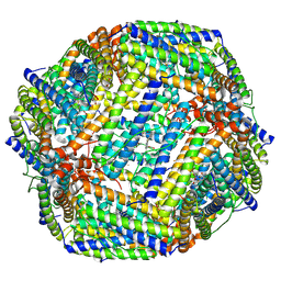
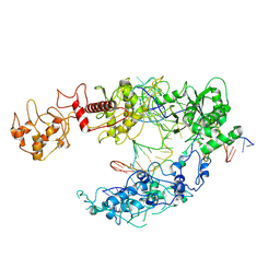
8H8X
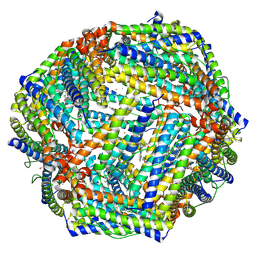
 | | Cryo-EM structure of HACE1 monomer | | 分子名称: | E3 ubiquitin-protein ligase HACE1 | | 著者 | Singh, S, Machida, S, Tulsian, N.K, Choong, Y.K, Ng, J, Shanker, S, Yaochen, L.D, Shi, J, Sivaraman, J. | | 登録日 | 2022-10-24 | | 公開日 | 2023-06-28 | | 最終更新日 | 2024-01-10 | | 実験手法 | ELECTRON MICROSCOPY (3.92 Å) | | 主引用文献 | Structural Basis for the Enzymatic Activity of the HACE1 HECT-Type E3 Ligase Through N-Terminal Helix Dimerization.
Adv Sci, 10, 2023
|
|
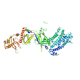
8HAE
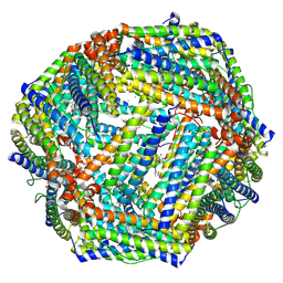
 | | Cryo-EM structure of HACE1 dimer | | 分子名称: | E3 ubiquitin-protein ligase HACE1 | | 著者 | Singh, S, Machida, S, Tulsian, N.K, Choong, Y.K, Ng, J, Shanker, S, Yaochen, L.D, Shi, J, Sivaraman, J, Machida, S. | | 登録日 | 2022-10-26 | | 公開日 | 2023-06-28 | | 最終更新日 | 2023-10-04 | | 実験手法 | ELECTRON MICROSCOPY (4.55 Å) | | 主引用文献 | Structural Basis for the Enzymatic Activity of the HACE1 HECT-Type E3 Ligase Through N-Terminal Helix Dimerization.
Adv Sci, 10, 2023
|
|
4RUD
 | |
8GWR
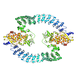
 | | Near full length Kidney type Glutaminase in complex with 2,2-Dimethyl-2,3-Dihydrobenzo[a] Phenanthridin-4(1H)-one (DDP) | | 分子名称: | 2,2-dimethyl-1,3-dihydrobenzo[a]phenanthridin-4-one, Glutaminase kidney isoform, mitochondrial | | 著者 | Shankar, S, Jobichen, C, Sivaraman, J. | | 登録日 | 2022-09-17 | | 公開日 | 2022-12-21 | | 最終更新日 | 2023-11-29 | | 実験手法 | X-RAY DIFFRACTION (2.801 Å) | | 主引用文献 | A novel allosteric site employs a conserved inhibition mechanism in human kidney-type glutaminase.
Febs J., 290, 2023
|
|
7Y6G
 | |
7Y6P
 | |
7Y6F
 | |
8I54
 | | Lb2Cas12a RNA DNA complex | | 分子名称: | DNA (25-MER), DNA (5'-D(*AP*GP*TP*GP*CP*TP*TP*TP*A)-3'), Lb2Cas12a, ... | | 著者 | Li, J, Sivaraman, J, Satoru, M. | | 登録日 | 2023-01-24 | | 公開日 | 2023-02-22 | | 最終更新日 | 2023-05-03 | | 実験手法 | ELECTRON MICROSCOPY (3.95 Å) | | 主引用文献 | Structures of apo Cas12a and its complex with crRNA and DNA reveal the dynamics of ternary complex formation and target DNA cleavage.
Plos Biol., 21, 2023
|
|
2ED6
 | |
8H9D
 | | Crystal structure of Cas12a protein | | 分子名称: | Cas12A, MAGNESIUM ION, RNA (5'-R(P*AP*AP*UP*UP*UP*CP*UP*AP*CP*UP*AP*AP*GP*UP*GP*UP*AP*GP*AP*UP*C)-3'), ... | | 著者 | Jianwei, L, Jobichen, C, Sivaraman, J. | | 登録日 | 2022-10-25 | | 公開日 | 2023-02-08 | | 最終更新日 | 2023-11-29 | | 実験手法 | X-RAY DIFFRACTION (3.1 Å) | | 主引用文献 | Structures of apo Cas12a and its complex with crRNA and DNA reveal the dynamics of ternary complex formation and target DNA cleavage.
Plos Biol., 21, 2023
|
|
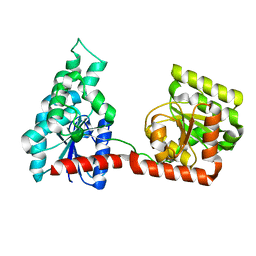
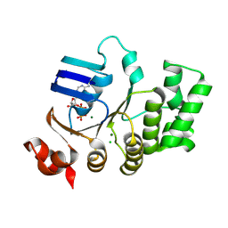
4E53
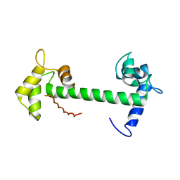 | | Calmodulin and Nm peptide complex | | 分子名称: | Calmodulin, Linker, IQ motif of Neuromodulin | | 著者 | Kumar, V, Sivaraman, J. | | 登録日 | 2012-03-13 | | 公開日 | 2013-03-20 | | 最終更新日 | 2024-03-20 | | 実験手法 | X-RAY DIFFRACTION (2.69 Å) | | 主引用文献 | Structural basis for the interaction of unstructured neuron specific substrates neuromodulin and neurogranin with calmodulin
Sci Rep, 3, 2013
|
|
4E50
 | | Calmodulin and Ng peptide complex | | 分子名称: | Calmodulin, Linker, IQ motif of Neurogranin | | 著者 | Kumar, V, Sivaraman, J. | | 登録日 | 2012-03-13 | | 公開日 | 2013-03-20 | | 最終更新日 | 2024-03-20 | | 実験手法 | X-RAY DIFFRACTION (2.7 Å) | | 主引用文献 | Structural basis for the interaction of unstructured neuron specific substrates neuromodulin and neurogranin with calmodulin
Sci Rep, 3, 2013
|
|
2EDM
 | |


