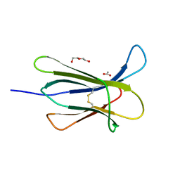
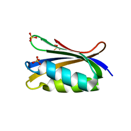
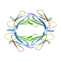
5CSB
 
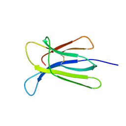 | | The crystal structure of beta2-microglobulin D76N mutant at room temperature | | 分子名称: | Beta-2-microglobulin | | 著者 | de Rosa, M, Mota, C.S, de Sanctis, D, Bolognesi, M, Ricagno, S. | | 登録日 | 2015-07-23 | | 公開日 | 2016-08-10 | | 最終更新日 | 2024-01-10 | | 実験手法 | X-RAY DIFFRACTION (1.719 Å) | | 主引用文献 | Conformational dynamics in crystals reveal the molecular bases for D76N beta-2 microglobulin aggregation propensity.
Nat Commun, 9, 2018
|
|
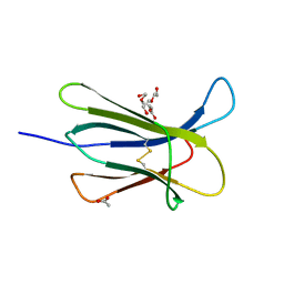
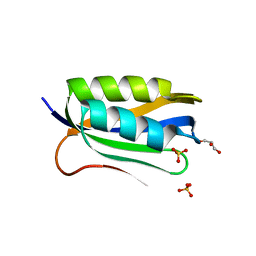
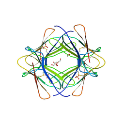
5CS7
 | | The crystal structure of wt beta2-microglobulin at room temperature | | 分子名称: | Beta-2-microglobulin | | 著者 | de Rosa, M, Mota, C.S, de Sanctis, D, Bolognesi, M, Ricagno, S. | | 登録日 | 2015-07-23 | | 公開日 | 2016-08-10 | | 最終更新日 | 2024-01-10 | | 実験手法 | X-RAY DIFFRACTION (2.1 Å) | | 主引用文献 | Conformational dynamics in crystals reveal the molecular bases for D76N beta-2 microglobulin aggregation propensity.
Nat Commun, 9, 2018
|
|
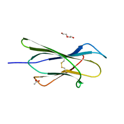
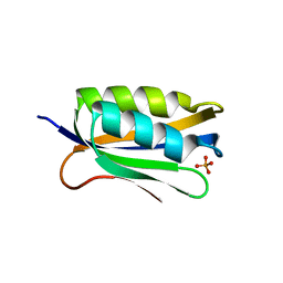
3DHJ
 | | Beta 2 microglobulin mutant W60C | | 分子名称: | Beta-2-microglobulin | | 著者 | Ricagno, S, Colombo, M, de Rosa, M, Bolognesi, M, Giorgetti, S, Bellotti, V. | | 登録日 | 2008-06-18 | | 公開日 | 2008-11-18 | | 最終更新日 | 2023-11-01 | | 実験手法 | X-RAY DIFFRACTION (2 Å) | | 主引用文献 | DE loop mutations affect beta2-microglobulin stability and amyloid aggregation
Biochem.Biophys.Res.Commun., 377, 2008
|
|
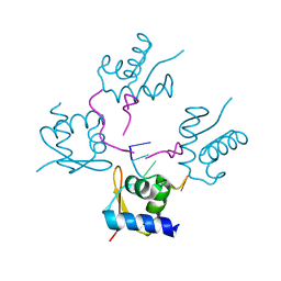
4HOB
 | | The crystal structure of the Zalpha domain from Cyprinid Herpes virus 3 | | 分子名称: | Putative uncharacterized protein, SULFATE ION | | 著者 | Tome, A.R, Kus, K, de Rosa, M, Paulo, L.M, Figueiredo, D, Athanasiadis, A. | | 登録日 | 2012-10-22 | | 公開日 | 2013-09-11 | | 最終更新日 | 2023-11-08 | | 実験手法 | X-RAY DIFFRACTION (1.76 Å) | | 主引用文献 | Crystal structure of a poxvirus-like zalpha domain from cyprinid herpesvirus 3
J.Virol., 87, 2013
|
|
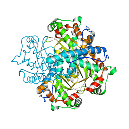
2C7G
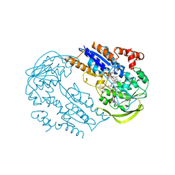 | | FprA from Mycobacterium tuberculosis: His57Gln mutant | | 分子名称: | 4-OXO-NICOTINAMIDE-ADENINE DINUCLEOTIDE PHOSPHATE, FLAVIN-ADENINE DINUCLEOTIDE, NADPH-FERREDOXIN REDUCTASE FPRA, ... | | 著者 | Pennati, A, Razeto, A, De Rosa, M, Pandini, V, Vanoni, M.A, Aliverti, A, Mattevi, A, Coda, A, Zanetti, G. | | 登録日 | 2005-11-24 | | 公開日 | 2006-07-26 | | 最終更新日 | 2023-12-13 | | 実験手法 | X-RAY DIFFRACTION (1.8 Å) | | 主引用文献 | Role of the His57-Glu214 Ionic Couple Located in the Active Site of Mycobacterium Tuberculosis Fpra.
Biochemistry, 45, 2006
|
|
4RMW
 | |
4RMU
 | |
4RMV
 | |
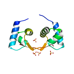
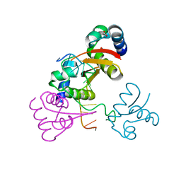
4BND
 | | Structure of an atypical alpha-phosphoglucomutase similar to eukaryotic phosphomannomutases | | 分子名称: | ALPHA-PHOSPHOGLUCOMUTASE, GLYCEROL, SULFATE ION | | 著者 | Nogly, P, Matias, P.M, De Rosa, M, Castro, R, Santos, H, Neves, A.R, Archer, M. | | 登録日 | 2013-05-14 | | 公開日 | 2013-10-02 | | 最終更新日 | 2024-05-08 | | 実験手法 | X-RAY DIFFRACTION (1.5 Å) | | 主引用文献 | High-Resolution Structure of an Atypical [Alpha]-Phosphoglucomutase Related to Eukaryotic Phosphomannomutases
Acta Crystallogr.,Sect.D, 69, 2013
|
|
4RMS
 | |
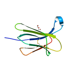
4RMT
 | | Crystal structure of the D98N Beta-2 Microglobulin mutant | | 分子名称: | ACETATE ION, Beta-2-microglobulin, DI(HYDROXYETHYL)ETHER, ... | | 著者 | de Rosa, M, Bolognesi, M, Ricagno, S. | | 登録日 | 2014-10-22 | | 公開日 | 2015-11-18 | | 最終更新日 | 2024-10-09 | | 実験手法 | X-RAY DIFFRACTION (1.242 Å) | | 主引用文献 | Decoding the Structural Bases of D76N 2-Microglobulin High Amyloidogenicity through Crystallography and Asn-Scan Mutagenesis.
Plos One, 10, 2015
|
|
4RMR
 | |
4RMQ
 | |
4OJH
 | |
4OJ3
 | |
4OJG
 | |
4KA4
 | |
4LB6
 | |
4LB5
 | |
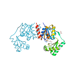
6H1F
 | | Structure of the nanobody-stabilized gelsolin D187N variant (second domain) | | 分子名称: | Gelsolin, THIOCYANATE ION, gelsolin nanobody, ... | | 著者 | Hassan, A, Milani, M, Mastrangelo, E, de Rosa, M. | | 登録日 | 2018-07-11 | | 公開日 | 2019-01-23 | | 最終更新日 | 2024-01-17 | | 実験手法 | X-RAY DIFFRACTION (1.9 Å) | | 主引用文献 | Nanobody interaction unveils structure, dynamics and proteotoxicity of the Finnish-type amyloidogenic gelsolin variant.
Biochim Biophys Acta Mol Basis Dis, 1865, 2019
|
|
4C7U
 | | Crystal structure of manganese superoxide dismutase from Arabidopsis thaliana | | 分子名称: | MANGANESE (II) ION, SUPEROXIDE DISMUTASE [MN] 1, MITOCHONDRIAL | | 著者 | Marques, A, Santos, S.P, Rosa, M, Carrondo, M.A, Abreu, I.A, Romao, C.V, Frazao, C. | | 登録日 | 2013-09-25 | | 公開日 | 2014-10-15 | | 最終更新日 | 2023-12-20 | | 実験手法 | X-RAY DIFFRACTION (1.951 Å) | | 主引用文献 | Crystal Structure of the Arabidopsis Thaliana Manganese Superoxide Dismutase at 1.95 A Resolution
To be Published
|
|
5KK3
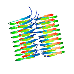 | | Atomic Resolution Structure of Monomorphic AB42 Amyloid Fibrils | | 分子名称: | Beta-amyloid protein 42 | | 著者 | Colvin, M.T, Silvers, R, Zhe Ni, Q, Can, T.V, Sergeyev, I, Rosay, M, Donovan, K.J, Michael, B, Wall, J, Linse, S, Griffin, R.G. | | 登録日 | 2016-06-20 | | 公開日 | 2016-07-13 | | 最終更新日 | 2024-05-01 | | 実験手法 | SOLID-STATE NMR | | 主引用文献 | Atomic Resolution Structure of Monomorphic A beta 42 Amyloid Fibrils.
J.Am.Chem.Soc., 138, 2016
|
|
3TLR
 | |
3TM6
 | |
6EY2
 | | Crystal structure of XIAP-BIR3 in complex with a cIAP1-selective SM | | 分子名称: | (3~{S},6~{S},7~{S},9~{a}~{S})-~{N}-[(4-~{tert}-butylphenyl)methyl]-7-(hydroxymethyl)-6-[[(2~{S})-2-(methylamino)butanoyl]amino]-5-oxidanylidene-1,2,3,6,7,8,9,9~{a}-octahydropyrrolo[1,2-a]azepine-3-carboxamide, E3 ubiquitin-protein ligase XIAP, ZINC ION | | 著者 | Cossu, F, Corti, A, Milani, M, Mastrangelo, E. | | 登録日 | 2017-11-10 | | 公開日 | 2018-08-08 | | 最終更新日 | 2024-01-17 | | 実験手法 | X-RAY DIFFRACTION (2.7 Å) | | 主引用文献 | Structure-based design and molecular profiling of Smac-mimetics selective for cellular IAPs.
FEBS J., 285, 2018
|
|


