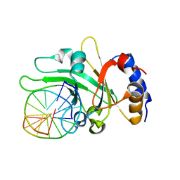
2OQA
 
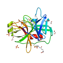
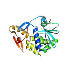 | | X-ray Sequence and Crystal Structure of Luffaculin 1, a Novel Type 1 Ribosome-inactivating Protein | | 分子名称: | 2-acetamido-2-deoxy-beta-D-glucopyranose, DI(HYDROXYETHYL)ETHER, Luffaculin 1, ... | | 著者 | Hou, X, Huang, M. | | 登録日 | 2007-01-31 | | 公開日 | 2007-05-29 | | 最終更新日 | 2024-12-25 | | 実験手法 | X-RAY DIFFRACTION (1.4 Å) | | 主引用文献 | X-ray sequence and crystal structure of luffaculin 1, a novel type 1 ribosome-inactivating protein
Bmc Struct.Biol., 7, 2007
|
|
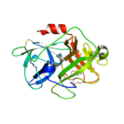
2FAT
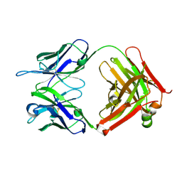
 | | An anti-urokinase plasminogen activator receptor (UPAR) antibody: Crystal structure and binding epitope | | 分子名称: | FAB ATN-615, heavy chain, light chain | | 著者 | Li, Y, Parry, G, Shi, X, Chen, L, Callahan, J.A, Mazar, A.P, Huang, M. | | 登録日 | 2005-12-07 | | 公開日 | 2006-11-14 | | 最終更新日 | 2024-10-16 | | 実験手法 | X-RAY DIFFRACTION (1.77 Å) | | 主引用文献 | An anti-urokinase plasminogen activator receptor (uPAR) antibody: crystal structure and binding epitope
J.Mol.Biol., 365, 2007
|
|
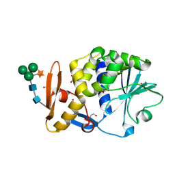
3MHW
 | | The complex crystal Structure of Urokianse and 2-Aminobenzothiazole | | 分子名称: | 1,3-benzothiazol-2-amine, SULFATE ION, Urokinase-type plasminogen activator | | 著者 | Jiang, L.-G, Yuan, C, Chen, L.-Q, Huang, M.-D. | | 登録日 | 2010-04-09 | | 公開日 | 2010-04-21 | | 最終更新日 | 2024-10-30 | | 実験手法 | X-RAY DIFFRACTION (1.45 Å) | | 主引用文献 | Crystal Structures of 2-Aminobenzothiazole-based Inhibitors in Complexes with Urokinase-type Plasminogen Activator
CHIN.J.STRUCT.CHEM., 28, 2009
|
|
3BWH
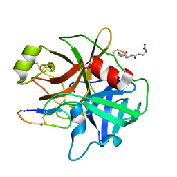
 | | Atomic resolution structure of cucurmosin, a novel type 1 RIP from the sarcocarp of Cucurbita moschata | | 分子名称: | 1,2-ETHANEDIOL, PHOSPHATE ION, beta-D-xylopyranose-(1-2)-[alpha-D-mannopyranose-(1-3)][alpha-D-mannopyranose-(1-6)]beta-D-mannopyranose-(1-4)-2-acetamido-2-deoxy-beta-D-glucopyranose-(1-4)-2-acetamido-2-deoxy-beta-D-glucopyranose, ... | | 著者 | Chen, L. | | 登録日 | 2008-01-09 | | 公開日 | 2008-10-07 | | 最終更新日 | 2024-11-06 | | 実験手法 | X-RAY DIFFRACTION (1 Å) | | 主引用文献 | Atomic resolution structure of cucurmosin, a novel type 1 ribosome-inactivating protein from the sarcocarp of Cucurbita moschata.
J.Struct.Biol., 164, 2008
|
|
3CX9
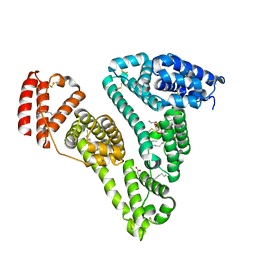 | | Crystal Structure of Human serum albumin complexed with Myristic acid and lysophosphatidylethanolamine | | 分子名称: | (2S)-3-{[(R)-(2-aminoethoxy)(hydroxy)phosphoryl]oxy}-2-hydroxypropyl hexadecanoate, MYRISTIC ACID, Serum albumin | | 著者 | Guo, S, Yang, F, Chen, L, Bian, C, Huang, M. | | 登録日 | 2008-04-24 | | 公開日 | 2009-04-28 | | 最終更新日 | 2024-11-13 | | 実験手法 | X-RAY DIFFRACTION (2.8 Å) | | 主引用文献 | Structural basis of transport of lysophospholipids by human serum albumin.
Biochem.J., 423, 2009
|
|
4K23
 | | Structure of anti-uPAR Fab ATN-658 | | 分子名称: | anti-uPAR antibody, heavy chain, light chain | | 著者 | Yuan, C, Huang, M, Chen, L. | | 登録日 | 2013-04-08 | | 公開日 | 2014-02-26 | | 最終更新日 | 2024-10-30 | | 実験手法 | X-RAY DIFFRACTION (1.6 Å) | | 主引用文献 | Identification of a New Epitope in uPAR as a Target for the Cancer Therapeutic Monoclonal Antibody ATN-658, a Structural Homolog of the uPAR Binding Integrin CD11b ( alpha M)
Plos One, 9, 2014
|
|
4K24
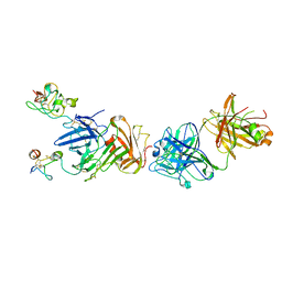 | | Structure of anti-uPAR Fab ATN-658 in complex with uPAR | | 分子名称: | 2-acetamido-2-deoxy-beta-D-glucopyranose, 2-acetamido-2-deoxy-beta-D-glucopyranose-(1-4)-2-acetamido-2-deoxy-beta-D-glucopyranose, Urokinase plasminogen activator surface receptor, ... | | 著者 | Huang, M.D, Xu, X, Yuan, C. | | 登録日 | 2013-04-08 | | 公開日 | 2014-02-26 | | 最終更新日 | 2024-11-20 | | 実験手法 | X-RAY DIFFRACTION (4.5 Å) | | 主引用文献 | Identification of a New Epitope in uPAR as a Target for the Cancer Therapeutic Monoclonal Antibody ATN-658, a Structural Homolog of the uPAR Binding Integrin CD11b ( alpha M)
Plos One, 9, 2014
|
|
3HOB
 | |
3G7N
 | | Crystal Structure of a Triacylglycerol Lipase from Penicillium Expansum at 1.3 | | 分子名称: | DI(HYDROXYETHYL)ETHER, Lipase, PENTAETHYLENE GLYCOL, ... | | 著者 | Bian, C.B, Yuan, C, Chen, L.Q, Edward, J.M, Lin, L, Jiang, L.G, Huang, Z.X, Huang, M.D. | | 登録日 | 2009-02-10 | | 公開日 | 2010-02-23 | | 最終更新日 | 2024-10-30 | | 実験手法 | X-RAY DIFFRACTION (1.3 Å) | | 主引用文献 | Crystal structure of a triacylglycerol lipase from Penicillium expansum at 1.3 A determined by sulfur SAD
Proteins, 78, 2010
|
|
3HNB
 | |
3HNY
 | |
3P8F
 | |
3P8G
 | | Crystal Structure of MT-SP1 in complex with benzamidine | | 分子名称: | 1,2-ETHANEDIOL, BENZAMIDINE, GLUTATHIONE, ... | | 著者 | Yuan, C, Huang, M, Chen, L. | | 登録日 | 2010-10-13 | | 公開日 | 2011-08-03 | | 最終更新日 | 2023-11-01 | | 実験手法 | X-RAY DIFFRACTION (1.2 Å) | | 主引用文献 | Structure of catalytic domain of Matriptase in complex with Sunflower trypsin inhibitor-1.
Bmc Struct.Biol., 11, 2011
|
|
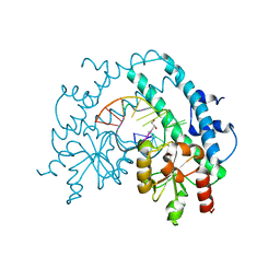
3HQF
 | | Crystal structure of restriction endonuclease EcoRII N-terminal effector-binding domain in complex with cognate DNA | | 分子名称: | 5'-D(*CP*GP*CP*CP*AP*GP*GP*GP*C)-3', 5'-D(*GP*CP*CP*CP*TP*GP*GP*CP*G)-3', Restriction endonuclease | | 著者 | Golovenko, D, Manakova, E, Grazulis, S, Tamulaitiene, G, Siksnys, V. | | 登録日 | 2009-06-06 | | 公開日 | 2009-09-22 | | 最終更新日 | 2023-09-06 | | 実験手法 | X-RAY DIFFRACTION (2.51 Å) | | 主引用文献 | Structural mechanisms for the 5'-CCWGG sequence recognition by the N- and C-terminal domains of EcoRII.
Nucleic Acids Res., 37, 2009
|
|
3HQG
 | | Crystal structure of restriction endonuclease EcoRII catalytic C-terminal domain in complex with cognate DNA | | 分子名称: | 5'-D(*TP*AP*GP*CP*CP*TP*GP*GP*TP*CP*GP*A)-3', 5'-D(*TP*CP*GP*AP*CP*CP*AP*GP*GP*CP*TP*A)-3', GLYCEROL, ... | | 著者 | Golovenko, D, Manakova, E, Grazulis, S, Tamulaitiene, G, Siksnys, V. | | 登録日 | 2009-06-06 | | 公開日 | 2009-09-22 | | 最終更新日 | 2023-09-06 | | 実験手法 | X-RAY DIFFRACTION (2.6 Å) | | 主引用文献 | Structural mechanisms for the 5'-CCWGG sequence recognition by the N- and C-terminal domains of EcoRII.
Nucleic Acids Res., 37, 2009
|
|
3Q4W
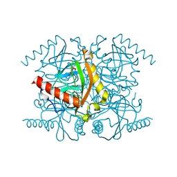 | | The structure of archaeal inorganic pyrophosphatase in complex with substrate | | 分子名称: | BROMIDE ION, CALCIUM ION, PYROPHOSPHATE 2-, ... | | 著者 | Hughes, R.C, Meehan, E.J, Coates, L, Ng, J.D. | | 登録日 | 2010-12-24 | | 公開日 | 2012-01-04 | | 最終更新日 | 2023-09-13 | | 実験手法 | X-RAY DIFFRACTION (1.441 Å) | | 主引用文献 | Inorganic pyrophosphatase crystals from Thermococcus thioreducens for X-ray and neutron diffraction.
Acta Crystallogr.,Sect.F, 68, 2012
|
|
3Q46
 | | Magnesium activated Inorganic pyrophosphatase from Thermococcus thioreducens bound to hydrolyzed product at 0.99 Angstrom resolution | | 分子名称: | 4-(2-HYDROXYETHYL)-1-PIPERAZINE ETHANESULFONIC ACID, CHLORIDE ION, MAGNESIUM ION, ... | | 著者 | Hughes, R.C, Coates, L, Meehan, E.J, Ng, J.D. | | 登録日 | 2010-12-23 | | 公開日 | 2012-01-04 | | 最終更新日 | 2023-09-13 | | 実験手法 | X-RAY DIFFRACTION (0.99 Å) | | 主引用文献 | Inorganic pyrophosphatase crystals from Thermococcus thioreducens for X-ray and neutron diffraction.
Acta Crystallogr.,Sect.F, 68, 2012
|
|
3Q3L
 | | The neutron crystallographic structure of inorganic pyrophosphatase from Thermococcus thioreducens | | 分子名称: | CALCIUM ION, Tt-IPPase | | 著者 | Hughes, R.C, Coates, L, Blakeley, M.P, Tomanicek, S.J, Meehan, E.J, Garcia-Ruiz, J.M, Ng, J.D. | | 登録日 | 2010-12-22 | | 公開日 | 2012-02-08 | | 最終更新日 | 2023-09-13 | | 実験手法 | NEUTRON DIFFRACTION (2.5 Å) | | 主引用文献 | Inorganic pyrophosphatase crystals from Thermococcus thioreducens for X-ray and neutron diffraction.
Acta Crystallogr.,Sect.F, 68, 2012
|
|
2LTN
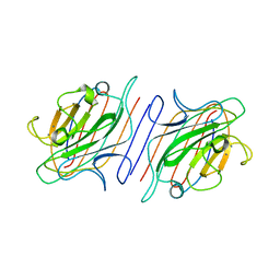 | | DESIGN, EXPRESSION, AND CRYSTALLIZATION OF RECOMBINANT LECTIN FROM THE GARDEN PEA (PISUM SATIVUM) | | 分子名称: | CALCIUM ION, MANGANESE (II) ION, PEA LECTIN, ... | | 著者 | Suddath, F.L, Phillips, S.R, Einspahr, H. | | 登録日 | 1990-06-26 | | 公開日 | 1990-07-15 | | 最終更新日 | 2024-02-21 | | 実験手法 | X-RAY DIFFRACTION (1.7 Å) | | 主引用文献 | Design, expression, and crystallization of recombinant lectin from the garden pea (Pisum sativum).
J.Biol.Chem., 264, 1989
|
|


