7D9T
 
 | |
7VSQ
 | |
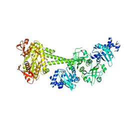
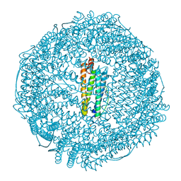
4DE6
 | | Horse spleen apo-ferritin complex with arachidonic acid | | 分子名称: | ARACHIDONIC ACID, CADMIUM ION, Ferritin light chain, ... | | 著者 | Bu, W, Liu, R, Dmochowski, I.J, Loll, P.J, Eckenhoff, R.G. | | 登録日 | 2012-01-19 | | 公開日 | 2012-03-07 | | 最終更新日 | 2023-09-13 | | 実験手法 | X-RAY DIFFRACTION (2.18 Å) | | 主引用文献 | Ferritin couples iron and fatty acid metabolism.
Faseb J., 26, 2012
|
|
8ZWN
 | |
7ARO
 | | Crystal structure of the non-ribose partial agonist LUF5833 bound to the adenosine A2A receptor | | 分子名称: | 2-azanyl-6-(1~{H}-imidazol-2-ylmethylsulfanyl)-4-phenyl-pyridine-3,5-dicarbonitrile, Adenosine receptor A2a,Soluble cytochrome b562,Adenosine receptor A2a, CHOLESTEROL, ... | | 著者 | Verdon, G, Amelia, T, van Veldhoven, J, Falsini, M, Liu, R, Heitman, L, van Westen, G, Segala, E, Cheng, R, Cooke, R, van der Es, D, Ijzerman, A. | | 登録日 | 2020-10-25 | | 公開日 | 2021-04-07 | | 最終更新日 | 2024-11-13 | | 実験手法 | X-RAY DIFFRACTION (3.119 Å) | | 主引用文献 | Crystal Structure and Subsequent Ligand Design of a Nonriboside Partial Agonist Bound to the Adenosine A 2A Receptor.
J.Med.Chem., 64, 2021
|
|
8IM0
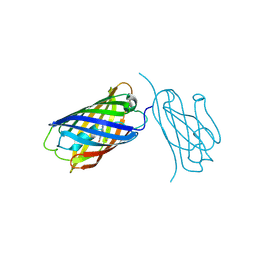 | | mCherry-LaM8 complex | | 分子名称: | LaM8, MCherry fluorescent protein | | 著者 | Liang, H, Liu, R, Ding, Y. | | 登録日 | 2023-03-05 | | 公開日 | 2023-06-21 | | 最終更新日 | 2024-11-13 | | 実験手法 | X-RAY DIFFRACTION (1.31 Å) | | 主引用文献 | Structural Insights into the Binding of Red Fluorescent Protein mCherry-Specific Nanobodies.
Int J Mol Sci, 24, 2023
|
|
8ILX
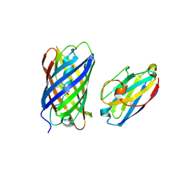 | | mCherry-LaM3 complex | | 分子名称: | LAM3, MCherry fluorescent protein | | 著者 | Liang, H, Liu, R, Ding, Y. | | 登録日 | 2023-03-05 | | 公開日 | 2023-06-21 | | 最終更新日 | 2024-10-16 | | 実験手法 | X-RAY DIFFRACTION (3.29 Å) | | 主引用文献 | Structural Insights into the Binding of Red Fluorescent Protein mCherry-Specific Nanobodies.
Int J Mol Sci, 24, 2023
|
|
8IM1
 | | mCherry-LaM1 complex | | 分子名称: | LaM1, MCherry fluorescent protein, SULFATE ION | | 著者 | Liang, H, Liu, R, Ding, Y. | | 登録日 | 2023-03-05 | | 公開日 | 2023-06-21 | | 最終更新日 | 2024-10-23 | | 実験手法 | X-RAY DIFFRACTION (2.05 Å) | | 主引用文献 | Structural Insights into the Binding of Red Fluorescent Protein mCherry-Specific Nanobodies.
Int J Mol Sci, 24, 2023
|
|
9ILW
 | |
4WOY
 | | Crystal structure and functional analysis of MiD49, a receptor for the mitochondrial fission protein Drp1 | | 分子名称: | Mitochondrial dynamics protein MID49 | | 著者 | Loson, O.C, Meng, S, Ngo, H.B, Liu, R, Kaiser, J.T, Chan, D.C. | | 登録日 | 2014-10-17 | | 公開日 | 2015-01-28 | | 最終更新日 | 2023-12-27 | | 実験手法 | X-RAY DIFFRACTION (2.4 Å) | | 主引用文献 | Crystal structure and functional analysis of MiD49, a receptor for the mitochondrial fission protein Drp1.
Protein Sci., 24, 2015
|
|
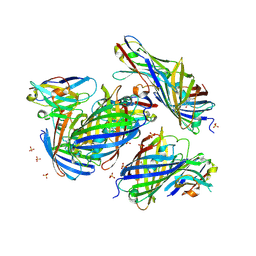
8YBN
 | | Crystal structure of nanobody SEB-Nb8 bound to staphylococcal enterotoxin B (SEB) | | 分子名称: | Enterotoxin type B, nanobody SEB-Nb8 | | 著者 | Ding, Y, Zong, X, Liu, R, Liu, P. | | 登録日 | 2024-02-15 | | 公開日 | 2024-12-25 | | 実験手法 | X-RAY DIFFRACTION (2.18 Å) | | 主引用文献 | Structural insights into the binding of nanobodies to the Staphylococcal enterotoxin B.
Int.J.Biol.Macromol., 276, 2024
|
|
8YBO
 | | Crystal structure of nanobody SEB-Nb11 bound to staphylococcal enterotoxin B (SEB) | | 分子名称: | Enterotoxin type B, nanobody SEB-Nb11 | | 著者 | Ding, Y, Zong, X, Liu, R, Liu, P. | | 登録日 | 2024-02-15 | | 公開日 | 2024-12-25 | | 最終更新日 | 2025-01-01 | | 実験手法 | X-RAY DIFFRACTION (1.59 Å) | | 主引用文献 | Structural insights into the binding of nanobodies to the Staphylococcal enterotoxin B.
Int.J.Biol.Macromol., 276, 2024
|
|
8YBM
 | | Crystal structure of nanobody SEB-Nb6 bound to staphylococcal enterotoxin B (SEB) | | 分子名称: | Enterotoxin type B, nanobody SEB-Nb6 | | 著者 | Ding, Y, Zong, X, Liu, R, Liu, P. | | 登録日 | 2024-02-15 | | 公開日 | 2024-12-25 | | 最終更新日 | 2025-01-01 | | 実験手法 | X-RAY DIFFRACTION (2.04 Å) | | 主引用文献 | Structural insights into the binding of nanobodies to the Staphylococcal enterotoxin B.
Int.J.Biol.Macromol., 276, 2024
|
|
8YBL
 | | Crystal structure of nanobody SEB-Nb3 bound to staphylococcal enterotoxin B (SEB) | | 分子名称: | Enterotoxin type B, nanobody SEB-Nb3 | | 著者 | Ding, Y, Zong, X, Liu, R, Liu, P. | | 登録日 | 2024-02-15 | | 公開日 | 2024-12-25 | | 実験手法 | X-RAY DIFFRACTION (2.33 Å) | | 主引用文献 | Structural insights into the binding of nanobodies to the Staphylococcal enterotoxin B.
Int.J.Biol.Macromol., 276, 2024
|
|
8YBP
 | | Crystal structure of nanobody SEB-Nb20 bound to staphylococcal enterotoxin B (SEB) | | 分子名称: | Enterotoxin type B, nanobody SEB-Nb20 | | 著者 | Ding, Y, Zong, X, Liu, R, Liu, P. | | 登録日 | 2024-02-15 | | 公開日 | 2024-12-25 | | 実験手法 | X-RAY DIFFRACTION (2.08 Å) | | 主引用文献 | Structural insights into the binding of nanobodies to the Staphylococcal enterotoxin B.
Int.J.Biol.Macromol., 276, 2024
|
|
2F9V
 | | HCV NS3 protease domain with NS4a peptide and a ketoamide inhibitor with P1 and P2 cyclopropylalannines | | 分子名称: | (2S,8R,9S,15S)-15-CYCLOHEXYL-9,12-BIS(CYCLOPROPYLMETHYL)-8-HYDROXY-20-METHYL-4,7,11,14,17-PENTAOXO-2-PHENYL-18-OXA-3,6,10,12,13,16-HEXAAZAHENICOSAN-1-OIC ACID, NS3 protease/helicase, ZINC ION, ... | | 著者 | Bogen, S.L, Ruan, S, Liu, R, Agrawal, S, Pichardo, J, Prongay, A, Baroudy, B, Saksena, A, Girijavallabhan, V, Njoroge, F.G. | | 登録日 | 2005-12-06 | | 公開日 | 2007-01-09 | | 最終更新日 | 2024-10-16 | | 実験手法 | X-RAY DIFFRACTION (2.6 Å) | | 主引用文献 | Depeptidization efforts on P3-P2 a-ketoamide inhibitors of HCV NS3-4A serine protease: Effect on HCV replicon activity.
Bioorg.Med.Chem.Lett., 16, 2006
|
|
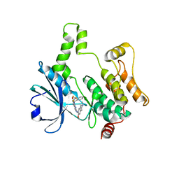
2R13
 | | Crystal structure of human mitoNEET reveals a novel [2Fe-2S] cluster coordination | | 分子名称: | CHLORIDE ION, FE2/S2 (INORGANIC) CLUSTER, Zinc finger CDGSH domain-containing protein 1 | | 著者 | Hou, X, Liu, R, Ross, S, Smart, E.J, Zhu, H, Gong, W. | | 登録日 | 2007-08-22 | | 公開日 | 2007-09-11 | | 最終更新日 | 2024-03-13 | | 実験手法 | X-RAY DIFFRACTION (1.8 Å) | | 主引用文献 | Crystallographic studies of human MitoNEET
J.Biol.Chem., 282, 2007
|
|
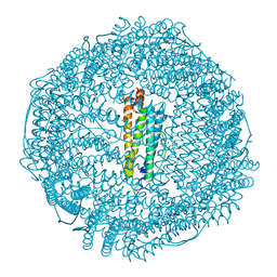
3RAV
 | | Horse spleen apo-ferritin with bound Pentobarbital | | 分子名称: | 5-ethyl-5-[(2S)-pentan-2-yl]pyrimidine-2,4,6(1H,3H,5H)-trione, CADMIUM ION, Ferritin light chain, ... | | 著者 | Oakley, S.H, Vedula, L.S, Xi, J, Liu, R, Eckenhoff, R.G, Loll, P.J. | | 登録日 | 2011-03-28 | | 公開日 | 2011-05-11 | | 最終更新日 | 2024-02-21 | | 実験手法 | X-RAY DIFFRACTION (1.9 Å) | | 主引用文献 | High resolution view of barbiturate recognition by a protein binding site
to be published
|
|
3RD0
 | | Horse spleen apo-ferritin with bound thiopental | | 分子名称: | 5-ethyl-5-[(2R)-pentan-2-yl]-2-thioxodihydropyrimidine-4,6(1H,5H)-dione, CADMIUM ION, Ferritin light chain, ... | | 著者 | Oakley, S.H, Vedula, L.S, Xi, J, Liu, R, Eckenhoff, R.G, Loll, P.J. | | 登録日 | 2011-03-31 | | 公開日 | 2011-05-11 | | 最終更新日 | 2023-09-13 | | 実験手法 | X-RAY DIFFRACTION (2 Å) | | 主引用文献 | High resolution view of barbiturate recognition by a protein binding site
to be published
|
|
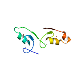
2LSP
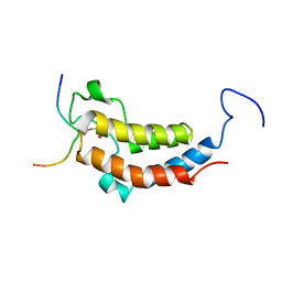 | | solution structures of BRD4 second bromodomain with NF-kB-K310ac peptide | | 分子名称: | Bromodomain-containing protein 4, NF-kB-K310ac peptide | | 著者 | Zhang, G, Liu, R, Zhong, Y, Plotnikov, A.N, Zhang, W, Rusinova, E, Gerona-Nevarro, G, Moshkina, N, Joshua, J, Chuang, P.Y, Ohlmeyer, M, He, J, Zhou, M.-M. | | 登録日 | 2012-05-03 | | 公開日 | 2012-07-18 | | 最終更新日 | 2024-11-06 | | 実験手法 | SOLUTION NMR | | 主引用文献 | Down-regulation of NF-kappa B transcriptional activity in HIV-associated kidney disease by BRD4 inhibition.
J.Biol.Chem., 287, 2012
|
|
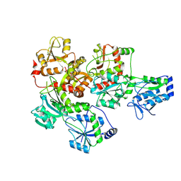
6JYV
 | | Structure of an isopenicillin N synthase from Pseudomonas aeruginosa PAO1 | | 分子名称: | Probable iron/ascorbate oxidoreductase, SODIUM ION | | 著者 | Hao, Z, Che, S, Wang, R, Liu, R, Zhang, Q, Bartlam, M. | | 登録日 | 2019-04-28 | | 公開日 | 2019-05-22 | | 最終更新日 | 2023-11-22 | | 実験手法 | X-RAY DIFFRACTION (1.651 Å) | | 主引用文献 | Structural characterization of an isopenicillin N synthase family oxygenase from Pseudomonas aeruginosa PAO1.
Biochem.Biophys.Res.Commun., 514, 2019
|
|
8XN7
 | | Crystal structure of HPK1 kinase domain T165E,S171E phosphomimetic mutant in complex with compound 9f | | 分子名称: | 5-amino-2-((6-methoxy-2-methyl-1,2,3,4-tetrahydroisoquinolin-7-yl)amino)-8-(2-(trifluoromethyl)benzyl)pyrido[2,3-d]pyrimidin-7(8H)-one, Mitogen-activated protein kinase kinase kinase kinase 1 | | 著者 | Huang, W.X, Liu, R, Ding, K. | | 登録日 | 2023-12-29 | | 公開日 | 2024-04-10 | | 実験手法 | X-RAY DIFFRACTION (2.65 Å) | | 主引用文献 | Discovery of 5-aminopyrido[2,3-d]pyrimidin-7(8H)-one derivatives as new hematopoietic progenitor kinase 1 (HPK1) inhibitors.
Eur.J.Med.Chem., 269, 2024
|
|
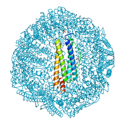
6JT0
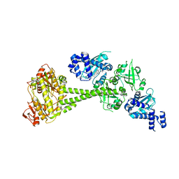 | | Structure of human soluble guanylate cyclase in the unliganded state | | 分子名称: | Guanylate cyclase soluble subunit alpha-1, Guanylate cyclase soluble subunit beta-1, PROTOPORPHYRIN IX CONTAINING FE | | 著者 | Chen, L, Kang, Y, Liu, R, Wu, J.-X. | | 登録日 | 2019-04-08 | | 公開日 | 2019-08-28 | | 最終更新日 | 2024-03-27 | | 実験手法 | ELECTRON MICROSCOPY (4 Å) | | 主引用文献 | Structural insights into the mechanism of human soluble guanylate cyclase.
Nature, 574, 2019
|
|
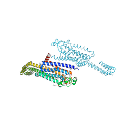
6JT1
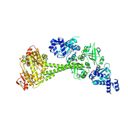 | | Structure of human soluble guanylate cyclase in the heme oxidised state | | 分子名称: | Guanylate cyclase soluble subunit alpha-1, Guanylate cyclase soluble subunit beta-1, PROTOPORPHYRIN IX CONTAINING FE | | 著者 | Chen, L, Kang, Y, Liu, R, Wu, J.-X. | | 登録日 | 2019-04-08 | | 公開日 | 2019-08-28 | | 最終更新日 | 2024-03-27 | | 実験手法 | ELECTRON MICROSCOPY (3.9 Å) | | 主引用文献 | Structural insights into the mechanism of human soluble guanylate cyclase.
Nature, 574, 2019
|
|
6JT2
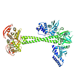 | | Structure of human soluble guanylate cyclase in the NO activated state | | 分子名称: | Guanylate cyclase soluble subunit alpha-1, Guanylate cyclase soluble subunit beta-1, MAGNESIUM ION, ... | | 著者 | Chen, L, Kang, Y, Liu, R, Wu, J.-X. | | 登録日 | 2019-04-08 | | 公開日 | 2019-09-04 | | 最終更新日 | 2024-03-27 | | 実験手法 | ELECTRON MICROSCOPY (3.8 Å) | | 主引用文献 | Structural insights into the mechanism of human soluble guanylate cyclase.
Nature, 574, 2019
|
|


