6BDD
 
 | | Crystal structure of Fe(II) unliganded H-NOX protein from K. algicida | | 分子名称: | 2,4-dihydroxyhept-2-ene-1,7-dioic acid aldolase, PROTOPORPHYRIN IX CONTAINING FE | | 著者 | Bruegger, J.J, Hespen, C.W, Marletta, M.A. | | 登録日 | 2017-10-22 | | 公開日 | 2018-05-30 | | 最終更新日 | 2023-10-04 | | 実験手法 | X-RAY DIFFRACTION (1.901 Å) | | 主引用文献 | Native Alanine Substitution in the Glycine Hinge Modulates Conformational Flexibility of Heme Nitric Oxide/Oxygen (H-NOX) Sensing Proteins.
ACS Chem. Biol., 13, 2018
|
|
7CPV
 | | Cryo-EM structure of 80S ribosome from mouse testis | | 分子名称: | 40S ribosomal protein S10, 40S ribosomal protein S11, 40S ribosomal protein S13, ... | | 著者 | Huo, Y.G, He, X, Jiang, T, Qin, Y, Guo, X.J, Sha, J.H. | | 登録日 | 2020-08-08 | | 公開日 | 2022-02-02 | | 最終更新日 | 2024-05-29 | | 実験手法 | ELECTRON MICROSCOPY (3.03 Å) | | 主引用文献 | A male germ-cell-specific ribosome controls male fertility.
Nature, 612, 2022
|
|
2OSL
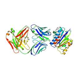
 | | Crystal structure of Rituximab Fab in complex with an epitope peptide | | 分子名称: | B-lymphocyte antigen CD20, heavy chain of the Rituximab Fab fragment,heavy chain of the Rituximab Fab fragment, light chain of the Rituximab Fab fragment,light chain of the Rituximab Fab fragment | | 著者 | Du, J, Zhong, C, Ding, J. | | 登録日 | 2007-02-06 | | 公開日 | 2007-04-10 | | 最終更新日 | 2024-04-10 | | 実験手法 | X-RAY DIFFRACTION (2.6 Å) | | 主引用文献 | Structural basis for recognition of CD20 by therapeutic antibody Rituximab
J.Biol.Chem., 282, 2007
|
|
2PVX
 | | NMR and X-ray Analysis of Structural Additivity in Metal Binding Site-Swapped Hybrids of Rubredoxin | | 分子名称: | Rubredoxin, ZINC ION | | 著者 | Wang, L, LeMaster, D.M, Hernandez, G, Li, H. | | 登録日 | 2007-05-10 | | 公開日 | 2007-12-18 | | 最終更新日 | 2023-08-30 | | 実験手法 | X-RAY DIFFRACTION (1.04 Å) | | 主引用文献 | NMR and X-ray analysis of structural additivity in metal binding site-swapped hybrids of rubredoxin
Bmc Struct.Biol., 7, 2007
|
|
3EOB
 | |
3EOA
 | |
3EO9
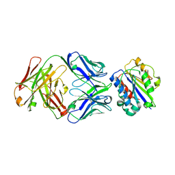
 | | Crystal structure the Fab fragment of Efalizumab | | 分子名称: | Efalizumab Fab fragment, heavy chain, light chain | | 著者 | Li, S, Ding, J. | | 登録日 | 2008-09-26 | | 公開日 | 2009-04-14 | | 最終更新日 | 2023-11-01 | | 実験手法 | X-RAY DIFFRACTION (1.8 Å) | | 主引用文献 | Efalizumab binding to the LFA-1 alphaL I domain blocks ICAM-1 binding via steric hindrance.
Proc.Natl.Acad.Sci.USA, 106, 2009
|
|
4HUN
 | |
4HPT
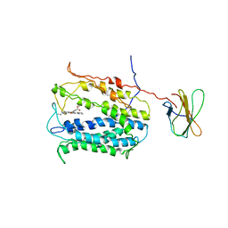
 | | Crystal structure of the catalytic subunit of cAMP-dependent protein kinase displaying complete phosphoryl transfer of AMP-PNP onto a substrate peptide | | 分子名称: | MAGNESIUM ION, PHOSPHOAMINOPHOSPHONIC ACID-ADENYLATE ESTER, cAMP-dependent protein kinase catalytic subunit alpha, ... | | 著者 | Bastidas, A.C, Steichen, J.M, Wu, J, Taylor, S.S. | | 登録日 | 2012-10-24 | | 公開日 | 2013-03-20 | | 最終更新日 | 2017-11-15 | | 実験手法 | X-RAY DIFFRACTION (2.15 Å) | | 主引用文献 | Phosphoryl transfer by protein kinase a is captured in a crystal lattice.
J.Am.Chem.Soc., 135, 2013
|
|
4HUL
 | |
4HPU
 | | Crystal structure of the catalytic subunit of cAMP-dependent protein kinase displaying partial phosphoryl transfer of AMP-PNP onto a substrate peptide | | 分子名称: | MAGNESIUM ION, MYRISTIC ACID, PHOSPHOAMINOPHOSPHONIC ACID-ADENYLATE ESTER, ... | | 著者 | Bastidas, A.C, Steichen, J.M, Wu, J, Taylor, S.S. | | 登録日 | 2012-10-24 | | 公開日 | 2013-03-20 | | 最終更新日 | 2017-11-15 | | 実験手法 | X-RAY DIFFRACTION (1.55 Å) | | 主引用文献 | Phosphoryl transfer by protein kinase a is captured in a crystal lattice.
J.Am.Chem.Soc., 135, 2013
|
|
4HUK
 | |
4HUM
 | |
3CXD
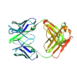
 | | Crystal structure of anti-osteopontin antibody 23C3 in complex with its epitope peptide | | 分子名称: | Fab fragment of anti-osteopontin antibody 23C3, Heavy chain, Light chain, ... | | 著者 | Du, J, Yang, H, Zhong, C, Ding, J. | | 登録日 | 2008-04-24 | | 公開日 | 2008-10-14 | | 最終更新日 | 2023-11-01 | | 実験手法 | X-RAY DIFFRACTION (2.8 Å) | | 主引用文献 | Molecular basis of recognition of human osteopontin by 23C3, a potential therapeutic antibody for treatment of rheumatoid arthritis
J.Mol.Biol., 382, 2008
|
|
3GIZ
 | | Crystal structure of the Fab fragment of anti-CD20 antibody Ofatumumab | | 分子名称: | Fab fragment of anti-CD20 antibody Ofatumumab, heavy chain, light chain, ... | | 著者 | Du, J, Yang, H, Ding, J. | | 登録日 | 2009-03-07 | | 公開日 | 2009-05-26 | | 最終更新日 | 2023-11-01 | | 実験手法 | X-RAY DIFFRACTION (2.2 Å) | | 主引用文献 | Structure of the Fab fragment of therapeutic antibody Ofatumumab provides insights into the recognition mechanism with CD20
Mol.Immunol., 46, 2009
|
|
3DSF
 | | Crystal structure of anti-osteopontin antibody 23C3 in complex with W43A mutated epitope peptide | | 分子名称: | Fab fragment of anti-osteopontin antibody 23C3, Heavy chain, Light chain, ... | | 著者 | Du, J, Zhong, C, Yang, H, Ding, J. | | 登録日 | 2008-07-12 | | 公開日 | 2008-10-14 | | 最終更新日 | 2023-11-01 | | 実験手法 | X-RAY DIFFRACTION (2.8 Å) | | 主引用文献 | Molecular basis of recognition of human osteopontin by 23C3, a potential therapeutic antibody for treatment of rheumatoid arthritis
J.Mol.Biol., 382, 2008
|
|
8JD9
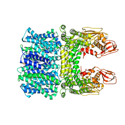 | | Cyro-EM structure of the Na+/H+ antipoter SOS1 from Arabidopsis thaliana,class1 | | 分子名称: | 1,2-DIACYL-SN-GLYCERO-3-PHOSPHOCHOLINE, Sodium/hydrogen exchanger 7 | | 著者 | Yang, G.H, Zhang, Y.M, Zhou, J.Q, Jia, Y.T, Xu, X, Fu, P, Wu, H.Y. | | 登録日 | 2023-05-13 | | 公開日 | 2023-11-08 | | 最終更新日 | 2023-11-29 | | 実験手法 | ELECTRON MICROSCOPY (2.87 Å) | | 主引用文献 | Structural basis for the activity regulation of Salt Overly Sensitive 1 in Arabidopsis salt tolerance.
Nat.Plants, 9, 2023
|
|
8JDA
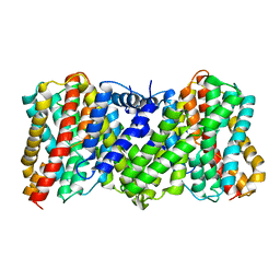 | | Cyro-EM structure of the Na+/H+ antipoter SOS1 from Arabidopsis thaliana,class2 | | 分子名称: | Sodium/hydrogen exchanger 7 | | 著者 | Yang, G.H, Zhang, Y.M, Zhou, J.Q, Jia, Y.T, Xu, X, Fu, P, Wu, H.Y. | | 登録日 | 2023-05-13 | | 公開日 | 2023-11-08 | | 最終更新日 | 2023-11-29 | | 実験手法 | ELECTRON MICROSCOPY (3.67 Å) | | 主引用文献 | Structural basis for the activity regulation of Salt Overly Sensitive 1 in Arabidopsis salt tolerance.
Nat.Plants, 9, 2023
|
|
3STU
 | |
3STT
 | |
3STV
 | |
3STW
 | |
3STX
 | |
3STY
 | |
4K7E
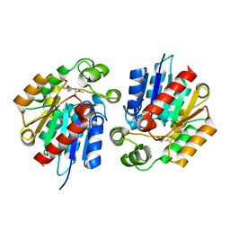
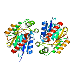
 | | Crystal structure of Junin virus nucleoprotein | | 分子名称: | Nucleoprotein | | 著者 | Zhang, Y.J, Li, L, Liu, X, Dong, S.S, Wang, W.M, Huo, T, Rao, Z.H, Yang, C. | | 登録日 | 2013-04-17 | | 公開日 | 2013-08-07 | | 最終更新日 | 2013-10-16 | | 実験手法 | X-RAY DIFFRACTION (2.2 Å) | | 主引用文献 | Crystal structure of Junin virus nucleoprotein
J.Gen.Virol., 94, 2013
|
|


