1G83
 
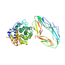
 | | CRYSTAL STRUCTURE OF FYN SH3-SH2 | | 分子名称: | PROTO-ONCOGENE TYROSINE-PROTEIN KINASE FYN | | 著者 | Arold, S.T, Ulmer, T.S, Mulhern, T.D, Werner, J.M, Ladbury, J.E, Campbell, I.D, Noble, M.E.M. | | 登録日 | 2000-11-16 | | 公開日 | 2001-05-30 | | 最終更新日 | 2024-02-07 | | 実験手法 | X-RAY DIFFRACTION (2.6 Å) | | 主引用文献 | The role of the Src homology 3-Src homology 2 interface in the regulation of Src kinases.
J.Biol.Chem., 276, 2001
|
|
2JL9
 | | Structural explanation for the role of Mn in the activity of phi6 RNA- dependent RNA polymerase | | 分子名称: | RNA-DIRECTED RNA POLYMERASE | | 著者 | Poranen, M.M, Salgado, P.S, Koivunen, M.R.L, Wright, S, Bamford, D.H, Stuart, D.I, Grimes, J.M. | | 登録日 | 2008-09-05 | | 公開日 | 2008-11-04 | | 最終更新日 | 2024-05-08 | | 実験手法 | X-RAY DIFFRACTION (3.2 Å) | | 主引用文献 | Structural Explanation for the Role of Mn2+ in the Activity of {Phi}6 RNA-Dependent RNA Polymerase.
Nucleic Acids Res., 36, 2008
|
|
2JLN
 | | Structure of Mhp1, a nucleobase-cation-symport-1 family transporter | | 分子名称: | MERCURY (II) ION, MHP1, SODIUM ION | | 著者 | Weyand, S, Shimamura, T, Yajima, S, Suzuki, S, Mirza, O, Krusong, K, Carpenter, E.P, Rutherford, N.G, Hadden, J.M, O'Reilly, J, Ma, P, Saidijam, M, Patching, S.G, Hope, R.J, Norbertczak, H.T, Roach, P.C.J, Iwata, S, Henderson, P.J.F, Cameron, A.D. | | 登録日 | 2008-09-11 | | 公開日 | 2008-10-28 | | 最終更新日 | 2024-05-08 | | 実験手法 | X-RAY DIFFRACTION (2.85 Å) | | 主引用文献 | Structure and Molecular Mechanism of a Nucleobase-Cation-Symport-1 Family Transporter.
Science, 322, 2008
|
|
1M9O
 | |
2JHA
 | | The structure of bluetongue virus VP4 reveals a multifunctional RNA- capping production-line | | 分子名称: | DIGUANOSINE-5'-TRIPHOSPHATE, GUANINE, VP4 CORE PROTEIN | | 著者 | Sutton, G, Grimes, J.M, Stuart, D.I, Roy, P. | | 登録日 | 2007-02-21 | | 公開日 | 2007-04-10 | | 最終更新日 | 2024-05-08 | | 実験手法 | X-RAY DIFFRACTION (3.4 Å) | | 主引用文献 | Bluetongue Virus Vp4 is an RNA-Capping Assembly Line.
Nat.Struct.Mol.Biol., 14, 2007
|
|
2K6R
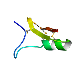
 | | Protein folding on a highly rugged landscape: Experimental observation of glassy dynamics and structural frustration | | 分子名称: | Full Sequence Design 1 Synthetic Superstable | | 著者 | Sadqi, M, de Alba, E, Perez-Jimenez, R, Sanchez-Ruiz, J.M, Munoz, V. | | 登録日 | 2008-07-18 | | 公開日 | 2009-06-16 | | 最終更新日 | 2016-06-08 | | 実験手法 | SOLUTION NMR | | 主引用文献 | A designed protein as experimental model of primordial folding
Proc.Natl.Acad.Sci.USA, 106, 2009
|
|
1M1J
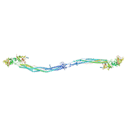 | | Crystal structure of native chicken fibrinogen with two different bound ligands | | 分子名称: | 2-acetamido-2-deoxy-alpha-D-glucopyranose, 2-acetamido-2-deoxy-beta-D-glucopyranose, CALCIUM ION, ... | | 著者 | Yang, Z, Kollman, J.M, Pandi, L, Doolittle, R.F. | | 登録日 | 2002-06-19 | | 公開日 | 2002-06-26 | | 最終更新日 | 2020-07-29 | | 実験手法 | X-RAY DIFFRACTION (2.7 Å) | | 主引用文献 | Crystal Structure of Native Chicken Fibrinogen at 2.7 A Resolution
Biochemistry, 40, 2001
|
|
1M7T
 | |
1G1Z
 | | NMR Solution Structures of delta-Conotoxin EVIA from Conus ermineus that Selectively Acts on Vertebrate Neuronal Na+ Channels, LEU12-PRO13 Cis isomer | | 分子名称: | CONOTOXIN EVIA | | 著者 | Volpon, L, Lamthanh, H, Le Gall, F, Menez, A, Lancelin, J.M. | | 登録日 | 2000-10-16 | | 公開日 | 2000-11-01 | | 最終更新日 | 2022-02-23 | | 実験手法 | SOLUTION NMR | | 主引用文献 | NMR Solution Structures of delta-Conotoxin EVIA from Conus ermineus That Selectively Acts on Vertebrate Neuronal Na+ Channels.
J.Biol.Chem., 279, 2004
|
|
1G4D
 | |
2N34
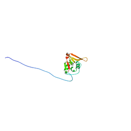
 | | NMR assignments and solution structure of the JAK interaction region of SOCS5 | | 分子名称: | Suppressor of cytokine signaling 5 | | 著者 | Chandrashekaran, I.R, Mohanty, B, Linossi, E.M, Nicholson, S.E, Babon, J, Norton, R.S, Dagley, L.F, Leung, E.W.W, Murphy, J.M. | | 登録日 | 2015-05-21 | | 公開日 | 2015-07-29 | | 最終更新日 | 2024-05-15 | | 実験手法 | SOLUTION NMR | | 主引用文献 | Structure and Functional Characterization of the Conserved JAK Interaction Region in the Intrinsically Disordered N-Terminus of SOCS5.
Biochemistry, 54, 2015
|
|
1G2A
 | | THE CRYSTAL STRUCTURE OF E.COLI PEPTIDE DEFORMYLASE COMPLEXED WITH ACTINONIN | | 分子名称: | ACTINONIN, NICKEL (II) ION, POLYPEPTIDE DEFORMYLASE | | 著者 | Clements, J.M, Beckett, P, Brown, A, Catlin, C, Lobell, M, Palan, S, Thomas, W, Whittaker, M, Baker, P.J, Rodgers, H.F, Barynin, V, Rice, D.W, Hunter, M.G. | | 登録日 | 2000-10-18 | | 公開日 | 2001-10-17 | | 最終更新日 | 2024-02-07 | | 実験手法 | X-RAY DIFFRACTION (1.75 Å) | | 主引用文献 | Antibiotic activity and characterization of BB-3497, a novel peptide deformylase inhibitor.
Antimicrob.Agents Chemother., 45, 2001
|
|
2MPC
 | |
1G25
 | | SOLUTION STRUCTURE OF THE N-TERMINAL DOMAIN OF THE HUMAN TFIIH MAT1 SUBUNIT | | 分子名称: | CDK-ACTIVATING KINASE ASSEMBLY FACTOR MAT1, ZINC ION | | 著者 | Gervais, V, Wasielewski, E, Busso, D, Poterszman, A, Egly, J.M, Thierry, J.C, Kieffer, B. | | 登録日 | 2000-10-17 | | 公開日 | 2000-11-01 | | 最終更新日 | 2024-05-22 | | 実験手法 | SOLUTION NMR | | 主引用文献 | Solution Structure of the N-terminal Domain of the Human TFIIH MAT1 Subunit: New Insights into the RING Finger Family
J.Biol.Chem., 276, 2001
|
|
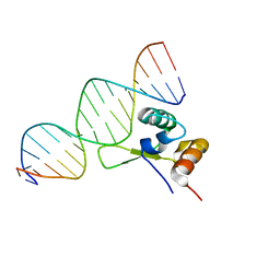
2JX1
 | | Structure of the fifth zinc finger of Myelin Transcription Factor 1 in complex with RARE DNA | | 分子名称: | DNA (5'-D(*DAP*DCP*DCP*DGP*DAP*DAP*DAP*DGP*DTP*DTP*DCP*DAP*DC)-3'), DNA (5'-D(*DGP*DTP*DGP*DAP*DAP*DCP*DTP*DTP*DTP*DCP*DGP*DGP*DT)-3'), Myelin transcription factor 1 | | 著者 | Gamsjaeger, R, Swanton, M.K, Kobus, F.J, Lehtomaki, E, Lowry, J.A, Kwan, A.H, Matthews, J.M, Mackay, J.P. | | 登録日 | 2007-11-01 | | 公開日 | 2007-12-11 | | 最終更新日 | 2024-05-29 | | 実験手法 | SOLUTION NMR | | 主引用文献 | Structure of the fifth zinc finger of Myelin Transcription Factor 1 in complex with RARE DNA
To be Published
|
|
1GHQ
 | | CR2-C3D COMPLEX STRUCTURE | | 分子名称: | 2-acetamido-2-deoxy-alpha-D-glucopyranose, COMPLEMENT C3, CR2/CD121/C3D/EPSTEIN-BARR VIRUS RECEPTOR, ... | | 著者 | Szakonyi, G, Guthridge, J.M, Li, D, Holers, V.M, Chen, X.S. | | 登録日 | 2001-01-11 | | 公開日 | 2001-06-13 | | 最終更新日 | 2023-12-27 | | 実験手法 | X-RAY DIFFRACTION (2.04 Å) | | 主引用文献 | Structure of complement receptor 2 in complex with its C3d ligand.
Science, 292, 2001
|
|
1ICA
 | |
2K3A
 | | NMR solution structure of Staphylococcus saprophyticus CHAP (cysteine, histidine-dependent amidohydrolases/peptidases) domain protein. Northeast Structural Genomics Consortium target SyR11 | | 分子名称: | CHAP domain protein | | 著者 | Rossi, P, Aramini, J.M, Chen, C.X, Nwosu, C, Cunningham, K.C, Owens, L.A, Xiao, R, Liu, J, Baran, M.C, Swapna, G, Acton, T.B, Rost, B, Montelione, G.T, Northeast Structural Genomics Consortium (NESG) | | 登録日 | 2008-04-29 | | 公開日 | 2008-05-13 | | 最終更新日 | 2024-05-29 | | 実験手法 | SOLUTION NMR | | 主引用文献 | Structural elucidation of the Cys-His-Glu-Asn proteolytic relay in the secreted CHAP domain enzyme from the human pathogen Staphylococcus saprophyticus.
Proteins, 74, 2008
|
|
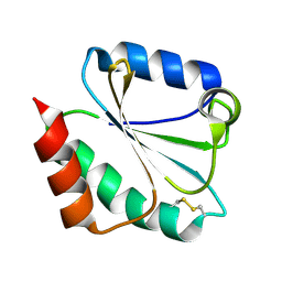
1I1J
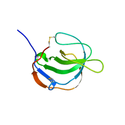 | | STRUCTURE OF MELANOMA INHIBITORY ACTIVITY PROTEIN: A MEMBER OF A NEW FAMILY OF SECRETED PROTEINS | | 分子名称: | MELANOMA DERIVED GROWTH REGULATORY PROTEIN | | 著者 | Lougheed, J.C, Holton, J.M, Alber, T, Bazan, J.F, Handel, T.M. | | 登録日 | 2001-02-02 | | 公開日 | 2001-05-16 | | 最終更新日 | 2011-07-13 | | 実験手法 | X-RAY DIFFRACTION (1.39 Å) | | 主引用文献 | Structure of melanoma inhibitory activity protein, a member of a recently identified family of secreted proteins.
Proc.Natl.Acad.Sci.USA, 98, 2001
|
|
1KSI
 | | CRYSTAL STRUCTURE OF A EUKARYOTIC (PEA SEEDLING) COPPER-CONTAINING AMINE OXIDASE AT 2.2A RESOLUTION | | 分子名称: | 2-acetamido-2-deoxy-beta-D-glucopyranose, 2-acetamido-2-deoxy-beta-D-glucopyranose-(1-4)-2-acetamido-2-deoxy-beta-D-glucopyranose, COPPER (II) ION, ... | | 著者 | Wilce, M.C.J, Kumar, V, Freeman, H.C, Guss, J.M. | | 登録日 | 1996-07-20 | | 公開日 | 1997-12-24 | | 最終更新日 | 2020-07-29 | | 実験手法 | X-RAY DIFFRACTION (2.2 Å) | | 主引用文献 | Crystal structure of a eukaryotic (pea seedling) copper-containing amine oxidase at 2.2 A resolution.
Structure, 4, 1996
|
|
1KJ5
 | | Solution Structure of Human beta-defensin 1 | | 分子名称: | BETA-DEFENSIN 1 | | 著者 | Schibli, D.J, Hunter, H.N, Aseyev, V, Starner, T.D, Wiencek, J.M, McCray Jr, P.B, Tack, B.F, Vogel, H.J. | | 登録日 | 2001-12-04 | | 公開日 | 2002-03-20 | | 最終更新日 | 2022-02-23 | | 実験手法 | SOLUTION NMR | | 主引用文献 | The solution structures of the human beta-defensins lead to a better understanding of the potent bactericidal activity of HBD3 against Staphylococcus aureus.
J.Biol.Chem., 277, 2002
|
|
1KWA
 | | HUMAN CASK/LIN-2 PDZ DOMAIN | | 分子名称: | HCASK/LIN-2 PROTEIN, SULFATE ION | | 著者 | Daniels, D.L, Cohen, A.R, Anderson, J.M, Brunger, A.T. | | 登録日 | 1998-01-16 | | 公開日 | 1998-05-27 | | 最終更新日 | 2024-02-14 | | 実験手法 | X-RAY DIFFRACTION (1.93 Å) | | 主引用文献 | Crystal structure of the hCASK PDZ domain reveals the structural basis of class II PDZ domain target recognition
Nat.Struct.Biol., 5, 1998
|
|
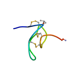
2JP8
 | | Angiotensin 1-7 | | 分子名称: | Angiotensin-(1-7) | | 著者 | Lula, I, Denadai, A.L, Resende, J.M, de Souza, F.B, de Lima, G.F, Pilo-Veloso, D, Heine, T, Duarte, H.A, Santos, R.A.S, Sinesterra, R.D. | | 登録日 | 2007-04-27 | | 公開日 | 2007-10-30 | | 最終更新日 | 2023-12-20 | | 実験手法 | SOLUTION NMR | | 主引用文献 | Study of angiotensin-(1-7) vasoactive peptide and its beta-cyclodextrin inclusion complexes: complete sequence-specific NMR assignments and structural studies
Peptides, 28, 2007
|
|
1I86
 | |
1I89
 | | Chalcone synthase (G256L) | | 分子名称: | CHALCONE SYNTHASE 2 | | 著者 | Jez, J.M, Bowman, M.E, Noel, J.P. | | 登録日 | 2001-03-12 | | 公開日 | 2001-12-12 | | 最終更新日 | 2024-04-03 | | 実験手法 | X-RAY DIFFRACTION (1.86 Å) | | 主引用文献 | Structure-guided programming of polyketide chain-length determination in chalcone synthase.
Biochemistry, 40, 2001
|
|


