2YVJ
 
 | |
2YVF
 | |
3KCJ
 | |
3KBJ
 | |
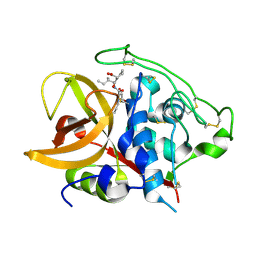
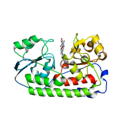
7XLP
 | | MEK1 bound to DS03090629 | | 分子名称: | (1~{R},3~{S})-3-[[6-[2-chloranyl-4-(4-methylpyrimidin-2-yl)oxy-phenyl]-3-methyl-1~{H}-indazol-4-yl]oxy]cyclohexan-1-amine, CALCIUM ION, DIMETHYL SULFOXIDE, ... | | 著者 | Kishikawa, S, Takano, K, Ubukata, O, Hanzawa, H. | | 登録日 | 2022-04-22 | | 公開日 | 2023-03-01 | | 最終更新日 | 2023-11-29 | | 実験手法 | X-RAY DIFFRACTION (2.1 Å) | | 主引用文献 | Discovery of a Novel ATP-Competitive MEK Inhibitor DS03090629 that Overcomes Resistance Conferred by BRAF Overexpression in BRAF-Mutated Melanoma.
Mol.Cancer Ther., 22, 2023
|
|
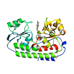
7XNC
 | | MEK1 bound to DS94070624 | | 分子名称: | CALCIUM ION, DIMETHYL SULFOXIDE, Dual specificity mitogen-activated protein kinase kinase 1, ... | | 著者 | Kishikawa, S, Takano, K, Ubukata, O, Hanzawa, H. | | 登録日 | 2022-04-28 | | 公開日 | 2023-03-01 | | 最終更新日 | 2023-11-29 | | 実験手法 | X-RAY DIFFRACTION (2.1 Å) | | 主引用文献 | Discovery of a Novel ATP-Competitive MEK Inhibitor DS03090629 that Overcomes Resistance Conferred by BRAF Overexpression in BRAF-Mutated Melanoma.
Mol.Cancer Ther., 22, 2023
|
|
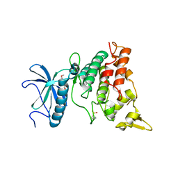
5Z5C
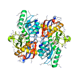 | | Crystal structure of hydrogen sulfide-producing enzyme (Fn1055) from Fusobacterium nucleatum: lysine-dimethylated form | | 分子名称: | CHLORIDE ION, Cysteine synthase, PYRIDOXAL-5'-PHOSPHATE | | 著者 | Kezuka, Y, Yoshida, Y, Nonaka, T. | | 登録日 | 2018-01-17 | | 公開日 | 2018-02-14 | | 最終更新日 | 2018-03-07 | | 実験手法 | X-RAY DIFFRACTION (2.07 Å) | | 主引用文献 | Structural insights into the catalytic mechanism of cysteine (hydroxyl) lyase from the hydrogen sulfide-producing oral pathogen,
Biochem. J., 475, 2018
|
|
5CZL
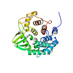 | | Crystal structure of a novel GH8 endo-beta-1,4-glucanase from an Achatina fulica gut metagenomic library | | 分子名称: | Glucanase, PHOSPHATE ION | | 著者 | Scapin, S.M.N, Souza, F.H.M, Zanphorlin, L.M, Almeida, T.S, Sade, Y.B, Cardoso, A.M, Pinheiro, G.L, Murakami, M.T. | | 登録日 | 2015-07-31 | | 公開日 | 2016-08-03 | | 最終更新日 | 2024-03-06 | | 実験手法 | X-RAY DIFFRACTION (2.391 Å) | | 主引用文献 | Crystal structure of a novel GH8 endo-beta-1,4-glucanase from an Achatina fulica gut metagenomic library
To Be Published
|
|
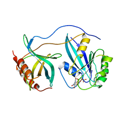
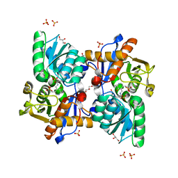
5B1S
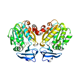 | | Crystal structure of Trypanosoma cruzi spermidine synthase in complex with 2-(2-fluorophenyl)ethanamine | | 分子名称: | 2-(2-fluorophenyl)ethanamine, 5'-[(S)-(3-AMINOPROPYL)(METHYL)-LAMBDA~4~-SULFANYL]-5'-DEOXYADENOSINE, Spermidine synthase, ... | | 著者 | Amano, Y, Tateishi, Y. | | 登録日 | 2015-12-17 | | 公開日 | 2016-12-21 | | 最終更新日 | 2023-11-08 | | 実験手法 | X-RAY DIFFRACTION (1.58 Å) | | 主引用文献 | In silico, in vitro, X-ray crystallography, and integrated strategies for discovering spermidine synthase inhibitors for Chagas disease
Sci Rep, 7, 2017
|
|
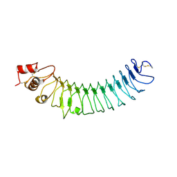
6KD5
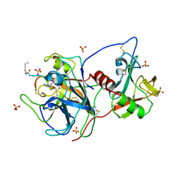 | | Crystal structure of the extracellular domain of MSPL/TMPRSS13 in complex with dec-RVKR-cmk inhibitor | | 分子名称: | 2-acetamido-2-deoxy-beta-D-glucopyranose-(1-4)-2-acetamido-2-deoxy-beta-D-glucopyranose, CALCIUM ION, DECANOYL-ARG-VAL-LYS-ARG-CHLOROMETHYLKETONE INHIBITOR, ... | | 著者 | Ohno, A, Maita, N, Okumura, Y, Nikawa, T. | | 登録日 | 2019-06-30 | | 公開日 | 2020-06-24 | | 最終更新日 | 2023-11-22 | | 実験手法 | X-RAY DIFFRACTION (2.6 Å) | | 主引用文献 | Crystal structure of inhibitor-bound human MSPL that can activate high pathogenic avian influenza.
Life Sci Alliance, 4, 2021
|
|
7XVX
 | |
3KRD
 | |
6KIP
 | |
1GCU
 | |
1QDQ
 | | X-RAY CRYSTAL STRUCTURE OF BOVINE CATHEPSIN B-CA074 COMPLEX | | 分子名称: | CATHEPSIN B, [PROPYLAMINO-3-HYDROXY-BUTAN-1,4-DIONYL]-ISOLEUCYL-PROLINE | | 著者 | Yamamoto, A. | | 登録日 | 1999-07-10 | | 公開日 | 2000-07-10 | | 最終更新日 | 2018-01-31 | | 実験手法 | X-RAY DIFFRACTION (2.18 Å) | | 主引用文献 | Substrate specificity of bovine cathepsin B and its inhibition by CA074, based on crystal structure refinement of the complex.
J.Biochem.(Tokyo), 127, 2000
|
|
7FHT
 | | Crystal structure of DYRK1A in complex with RD0448 | | 分子名称: | (5~{Z})-5-[(3-ethynyl-4-methoxy-phenyl)methylidene]-2-sulfanylidene-1,3-thiazolidin-4-one, Dual specificity tyrosine-phosphorylation-regulated kinase 1A | | 著者 | Kikuchi, M, Sumida, Y, Hosoya, T, Kii, I, Umehara, T. | | 登録日 | 2021-07-30 | | 公開日 | 2022-03-23 | | 最終更新日 | 2023-11-29 | | 実験手法 | X-RAY DIFFRACTION (2.68 Å) | | 主引用文献 | Structure-activity relationship for the folding intermediate-selective inhibition of DYRK1A.
Eur.J.Med.Chem., 227, 2022
|
|
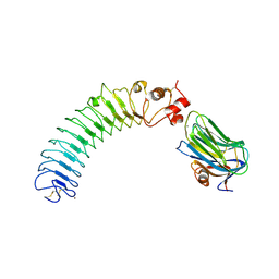
1RRP
 | | STRUCTURE OF THE RAN-GPPNHP-RANBD1 COMPLEX | | 分子名称: | MAGNESIUM ION, NUCLEAR PORE COMPLEX PROTEIN NUP358, PHOSPHOAMINOPHOSPHONIC ACID-GUANYLATE ESTER, ... | | 著者 | Vetter, I.R, Nowak, C, Nishimoto, T, Kuhlmann, J, Wittinghofer, A. | | 登録日 | 1999-01-15 | | 公開日 | 1999-05-18 | | 最終更新日 | 2024-04-03 | | 実験手法 | X-RAY DIFFRACTION (2.96 Å) | | 主引用文献 | Structure of a Ran-binding domain complexed with Ran bound to a GTP analogue: implications for nuclear transport.
Nature, 398, 1999
|
|
5Z8X
 | | Crystal structure of human LRRTM2 | | 分子名称: | 2-acetamido-2-deoxy-beta-D-glucopyranose, Leucine-rich repeat transmembrane neuronal protein 2 | | 著者 | Yamagata, A, Fukai, S. | | 登録日 | 2018-02-01 | | 公開日 | 2018-10-10 | | 最終更新日 | 2023-11-22 | | 実験手法 | X-RAY DIFFRACTION (3.15 Å) | | 主引用文献 | Structural insights into modulation and selectivity of transsynaptic neurexin-LRRTM interaction.
Nat Commun, 9, 2018
|
|
5Z8Y
 | | Crystal structure of human LRRTM2 in complex with Neurexin 1beta | | 分子名称: | 2-acetamido-2-deoxy-beta-D-glucopyranose, 2-acetamido-2-deoxy-beta-D-glucopyranose-(1-4)-[alpha-L-fucopyranose-(1-6)]2-acetamido-2-deoxy-beta-D-glucopyranose, CALCIUM ION, ... | | 著者 | Yamagata, A, Fukai, S. | | 登録日 | 2018-02-01 | | 公開日 | 2018-10-10 | | 最終更新日 | 2023-11-22 | | 実験手法 | X-RAY DIFFRACTION (3.4 Å) | | 主引用文献 | Structural insights into modulation and selectivity of transsynaptic neurexin-LRRTM interaction.
Nat Commun, 9, 2018
|
|
1QQL
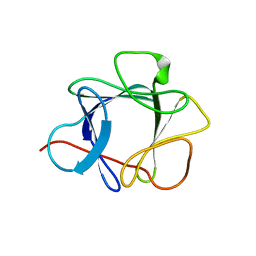 | | THE CRYSTAL STRUCTURE OF FIBROBLAST GROWTH FACTOR 7/1 CHIMERA | | 分子名称: | FIBROBLAST GROWTH FACTOR 7/1 CHIMERA | | 著者 | Ye, S, Luo, Y, Pelletier, H, McKeehan, W.L. | | 登録日 | 1999-06-07 | | 公開日 | 2000-01-14 | | 最終更新日 | 2024-02-14 | | 実験手法 | X-RAY DIFFRACTION (2.3 Å) | | 主引用文献 | Structural basis for interaction of FGF-1, FGF-2, and FGF-7 with different heparan sulfate motifs.
Biochemistry, 40, 2001
|
|
1QQK
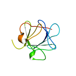 | | THE CRYSTAL STRUCTURE OF FIBROBLAST GROWTH FACTOR 7 (KERATINOCYTE GROWTH FACTOR) | | 分子名称: | FIBROBLAST GROWTH FACTOR 7 | | 著者 | Ye, S, Luo, Y, Pelletier, H, McKeehan, W.L. | | 登録日 | 1999-06-07 | | 公開日 | 2000-01-14 | | 最終更新日 | 2024-02-14 | | 実験手法 | X-RAY DIFFRACTION (3.1 Å) | | 主引用文献 | Structural basis for interaction of FGF-1, FGF-2, and FGF-7 with different heparan sulfate motifs.
Biochemistry, 40, 2001
|
|
5B1H
 | |
5BNQ
 | | Crystal structure of hRANKL-mRANK complex | | 分子名称: | CHLORIDE ION, PHOSPHATE ION, SODIUM ION, ... | | 著者 | Ren, J. | | 登録日 | 2015-05-26 | | 公開日 | 2015-10-14 | | 最終更新日 | 2024-01-10 | | 実験手法 | X-RAY DIFFRACTION (2.8 Å) | | 主引用文献 | A RANKL mutant used as an inter-species vaccine for efficient immunotherapy of osteoporosis.
Sci Rep, 5, 2015
|
|
5B50
 | | Crystal structure of heme binding protein HmuT Y240A | | 分子名称: | ABC-type transporter, periplasmic component, PROTOPORPHYRIN IX CONTAINING FE | | 著者 | Muraki, N, Aono, S. | | 登録日 | 2016-04-20 | | 公開日 | 2017-03-01 | | 最終更新日 | 2023-11-08 | | 実験手法 | X-RAY DIFFRACTION (1.65 Å) | | 主引用文献 | Structural Characterization of Heme Environmental Mutants of CgHmuT that Shuttles Heme Molecules to Heme Transporters
Int J Mol Sci, 17, 2016
|
|
5B51
 | |


