1YDS
 
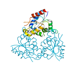
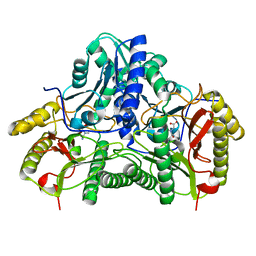
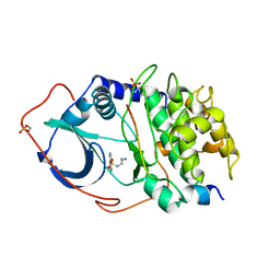 | | Structure of CAMP-dependent protein kinase, alpha-catalytic subunit in complex with H8 protein kinase inhibitor [N-(2-methylamino)ethyl]-5-isoquinolinesulfonamide | | 分子名称: | C-AMP-DEPENDENT PROTEIN KINASE, N-[2-(METHYLAMINO)ETHYL]-5-ISOQUINOLINESULFONAMIDE, PROTEIN KINASE INHIBITOR PEPTIDE | | 著者 | Engh, R.A, Girod, A, Kinzel, V, Huber, R, Bossemeyer, D. | | 登録日 | 1996-07-24 | | 公開日 | 1997-04-01 | | 最終更新日 | 2024-06-05 | | 実験手法 | X-RAY DIFFRACTION (2.2 Å) | | 主引用文献 | Crystal structures of catalytic subunit of cAMP-dependent protein kinase in complex with isoquinolinesulfonyl protein kinase inhibitors H7, H8, and H89. Structural implications for selectivity.
J.Biol.Chem., 271, 1996
|
|
1PPE
 | |
1MVN
 | | PPC decarboxylase mutant C175S complexed with pantothenoylaminoethenethiol | | 分子名称: | 2,4-DIHYDROXY-N-[2-(2-MERCAPTO-VINYLCARBAMOYL)-ETHYL]-3,3-DIMETHYL-BUTYRAMIDE, FLAVIN MONONUCLEOTIDE, PPC decarboxylase AtHAL3a | | 著者 | Steinbacher, S, Hernandez-Acosta, P, Bieseler, B, Blaesse, M, Huber, R, Culianez-Macia, F.A, Kupke, T. | | 登録日 | 2002-09-26 | | 公開日 | 2003-03-04 | | 最終更新日 | 2023-10-25 | | 実験手法 | X-RAY DIFFRACTION (2.21 Å) | | 主引用文献 | Crystal Structure of the Plant PPC Decarboxylase AtHAL3a Complexed with an Ene-thiol Reaction Intermediate
J.Mol.Biol., 327, 2003
|
|
1CTS
 | |
1N6E
 | | tricorn protease in complex with a tridecapeptide chloromethyl ketone derivative | | 分子名称: | DQTQKAAAELTFF, Tricorn protease | | 著者 | Kim, J.-S, Groll, M, Huber, R, Brandstetter, H. | | 登録日 | 2002-11-10 | | 公開日 | 2002-12-30 | | 最終更新日 | 2024-01-31 | | 実験手法 | X-RAY DIFFRACTION (2.6 Å) | | 主引用文献 | Navigation Inside a Protease: Substrate Selection and Product Exit in the Tricorn Protease
from Thermoplasma acidophilum
J.Mol.Biol., 324, 2002
|
|
1ILU
 | | X-RAY CRYSTAL STRUCTURE THE TWO SITE-SPECIFIC MUTANTS ILE7SER AND PHE110SER OF AZURIN FROM PSEUDOMONAS AERUGINOSA | | 分子名称: | AZURIN, COPPER (II) ION | | 著者 | Hammann, C, Nar, H, Huber, R, Messerschmidt, A. | | 登録日 | 1995-10-12 | | 公開日 | 1996-03-08 | | 最終更新日 | 2021-11-03 | | 実験手法 | X-RAY DIFFRACTION (2.3 Å) | | 主引用文献 | X-ray crystal structure of the two site-specific mutants Ile7Ser and Phe110Ser of azurin from Pseudomonas aeruginosa.
J.Mol.Biol., 255, 1996
|
|
1N6D
 | | Tricorn protease in complex with tetrapeptide chloromethyl ketone derivative | | 分子名称: | DECANE, RVRK, Tricorn protease | | 著者 | Kim, J.-S, Groll, M, Huber, R, Brandstetter, H. | | 登録日 | 2002-11-10 | | 公開日 | 2002-12-30 | | 最終更新日 | 2024-01-31 | | 実験手法 | X-RAY DIFFRACTION (2.8 Å) | | 主引用文献 | Navigation Inside a Protease: Substrate Selection and Product Exit in the Tricorn Protease
from Thermoplasma acidophilum
J.Mol.Biol., 324, 2002
|
|
1ILS
 | | X-RAY CRYSTAL STRUCTURE THE TWO SITE-SPECIFIC MUTANTS ILE7SER AND PHE110SER OF AZURIN FROM PSEUDOMONAS AERUGINOSA | | 分子名称: | AZURIN, COPPER (II) ION, NITRATE ION | | 著者 | Hammann, C, Nar, H, Huber, R, Messerschmidt, A. | | 登録日 | 1995-10-12 | | 公開日 | 1996-03-08 | | 最終更新日 | 2021-11-03 | | 実験手法 | X-RAY DIFFRACTION (2.2 Å) | | 主引用文献 | X-ray crystal structure of the two site-specific mutants Ile7Ser and Phe110Ser of azurin from Pseudomonas aeruginosa.
J.Mol.Biol., 255, 1996
|
|
1JDX
 | |
1MVL
 | | PPC decarboxylase mutant C175S | | 分子名称: | FLAVIN MONONUCLEOTIDE, PPC decarboxylase AtHAL3a | | 著者 | Steinbacher, S, Hernandez-Acosta, P, Bieseler, B, Blaesse, M, Huber, R, Culianez-Macia, F.A, Kupke, T. | | 登録日 | 2002-09-26 | | 公開日 | 2003-03-04 | | 最終更新日 | 2023-10-25 | | 実験手法 | X-RAY DIFFRACTION (2 Å) | | 主引用文献 | Crystal Structure of the Plant PPC Decarboxylase AtHAL3a Complexed with an Ene-thiol Reaction Intermediate
J.Mol.Biol., 327, 2003
|
|
1DX5
 | | Crystal structure of the thrombin-thrombomodulin complex | | 分子名称: | 2-acetamido-2-deoxy-beta-D-glucopyranose, CALCIUM ION, FORMIC ACID, ... | | 著者 | Fuentes-Prior, P, Iwanaga, Y, Huber, R, Pagila, R, Rumennik, G, Seto, M, Morser, J, Light, D.R, Bode, W. | | 登録日 | 1999-12-20 | | 公開日 | 2000-04-10 | | 最終更新日 | 2024-05-01 | | 実験手法 | X-RAY DIFFRACTION (2.3 Å) | | 主引用文献 | Structural Basis for the Anticoagulant Activity of the Thrombin-Thrombomodulin Complex
Nature, 404, 2000
|
|
1CL1
 | |
1CLW
 | | TAILSPIKE PROTEIN FROM PHAGE P22, V331A MUTANT | | 分子名称: | TAILSPIKE PROTEIN | | 著者 | Steinbacher, S, Baxa, U, Weintraub, A, Huber, R, Seckler, R. | | 登録日 | 1999-05-04 | | 公開日 | 1999-11-05 | | 最終更新日 | 2023-08-09 | | 実験手法 | X-RAY DIFFRACTION (2 Å) | | 主引用文献 | Mutations improving the folding of phage P22 tailspike protein affect its receptor binding activity.
J.Mol.Biol., 293, 1999
|
|
1DHP
 | | DIHYDRODIPICOLINATE SYNTHASE | | 分子名称: | DIHYDRODIPICOLINATE SYNTHASE, POTASSIUM ION | | 著者 | Mirwaldt, C, Korndoerfer, I, Huber, R. | | 登録日 | 1995-02-09 | | 公開日 | 1997-02-12 | | 最終更新日 | 2024-02-07 | | 実験手法 | X-RAY DIFFRACTION (2.3 Å) | | 主引用文献 | The crystal structure of dihydrodipicolinate synthase from Escherichia coli at 2.5 A resolution.
J.Mol.Biol., 246, 1995
|
|
2H6T
 | | Secreted aspartic proteinase (Sap) 3 from Candida albicans complexed with pepstatin A | | 分子名称: | Candidapepsin-3, ZINC ION, pepstatin A | | 著者 | Ruge, E, Borelli, C, Maskos, K, Huber, R. | | 登録日 | 2006-06-01 | | 公開日 | 2007-06-12 | | 最終更新日 | 2017-10-18 | | 実験手法 | X-RAY DIFFRACTION (1.9 Å) | | 主引用文献 | The crystal structure of the secreted aspartic proteinase 3 from Candida albicans and its complex with pepstatin A.
Proteins, 68, 2007
|
|
2H6S
 | | Secreted aspartic proteinase (Sap) 3 from Candida albicans | | 分子名称: | Candidapepsin-3, ZINC ION | | 著者 | Ruge, E, Borelli, C, Maskos, K, Huber, R. | | 登録日 | 2006-06-01 | | 公開日 | 2007-06-12 | | 最終更新日 | 2017-10-18 | | 実験手法 | X-RAY DIFFRACTION (2.2 Å) | | 主引用文献 | The crystal structure of the secreted aspartic proteinase 3 from Candida albicans and its complex with pepstatin A.
Proteins, 68, 2007
|
|
1U9L
 | | Structural basis for a NusA- protein N interaction | | 分子名称: | GOLD ION, Lambda N, Transcription elongation protein nusA | | 著者 | Bonin, I, Muehlberger, R, Bourenkov, G.P, Huber, R, Bacher, A, Richter, G, Wahl, M.C. | | 登録日 | 2004-08-10 | | 公開日 | 2004-08-31 | | 最終更新日 | 2024-03-13 | | 実験手法 | X-RAY DIFFRACTION (1.9 Å) | | 主引用文献 | Structural basis for the interaction of Escherichia coli NusA with protein N of phage lambda
Proc.Natl.Acad.Sci.Usa, 101, 2004
|
|
1CHM
 | | ENZYMATIC MECHANISM OF CREATINE AMIDINOHYDROLASE AS DEDUCED FROM CRYSTAL STRUCTURES | | 分子名称: | CARBAMOYL SARCOSINE, CREATINE AMIDINOHYDROLASE | | 著者 | Hoeffken, H.W, Knof, S.H, Bartlett, P.A, Huber, R, Moellering, H, Schumacher, G. | | 登録日 | 1993-07-19 | | 公開日 | 1994-04-30 | | 最終更新日 | 2024-02-07 | | 実験手法 | X-RAY DIFFRACTION (1.9 Å) | | 主引用文献 | Enzymatic mechanism of creatine amidinohydrolase as deduced from crystal structures.
J.Mol.Biol., 214, 1990
|
|
1CL2
 | |
1RVV
 | | SYNTHASE/RIBOFLAVIN SYNTHASE COMPLEX OF BACILLUS SUBTILIS | | 分子名称: | 5-NITRO-6-RIBITYL-AMINO-2,4(1H,3H)-PYRIMIDINEDIONE, PHOSPHATE ION, RIBOFLAVIN SYNTHASE | | 著者 | Ritsert, K, Huber, R, Turk, D, Ladenstein, R, Schmidt-Baese, K, Bacher, A. | | 登録日 | 1995-10-25 | | 公開日 | 1996-12-07 | | 最終更新日 | 2024-02-14 | | 実験手法 | X-RAY DIFFRACTION (2.4 Å) | | 主引用文献 | Studies on the lumazine synthase/riboflavin synthase complex of Bacillus subtilis: crystal structure analysis of reconstituted, icosahedral beta-subunit capsids with bound substrate analogue inhibitor at 2.4 A resolution.
J.Mol.Biol., 253, 1995
|
|
1N6F
 | | tricorn protease in complex with Z-Phe-diketo-Arg-Glu-Phe | | 分子名称: | 4-[2-(3-BENZYLOXYCARBONYLAMINO-4-CYCLOHEXYL-1-HYDROXY-2-OXO-BUTYLAMINO)-5-GUANIDINO-PENTANOYLAMINO]-4-(1-CARBOXY-2-CYCLOHEXYL-ETHYLCARBAMOYL)-BUTYRIC ACID, tricorn protease | | 著者 | Kim, J.-S, Groll, M, Huber, R, Brandstetter, H. | | 登録日 | 2002-11-10 | | 公開日 | 2002-12-30 | | 最終更新日 | 2023-10-25 | | 実験手法 | X-RAY DIFFRACTION (2.7 Å) | | 主引用文献 | Navigation Inside a Protease: Substrate Selection and Product Exit in the Tricorn Protease
from Thermoplasma acidophilum
J.Mol.Biol., 324, 2002
|
|
1TSP
 | | CRYSTAL STRUCTURE OF P22 TAILSPIKE PROTEIN: INTERDIGITATED SUBUNITS IN A THERMOSTABLE TRIMER | | 分子名称: | TAILSPIKE-PROTEIN | | 著者 | Steinbacher, S, Seckler, R, Miller, S, Steipe, B, Huber, R, Reinemer, P. | | 登録日 | 1994-06-16 | | 公開日 | 1995-09-15 | | 最終更新日 | 2024-02-14 | | 実験手法 | X-RAY DIFFRACTION (2 Å) | | 主引用文献 | Crystal structure of P22 tailspike protein: interdigitated subunits in a thermostable trimer.
Science, 265, 1994
|
|
1NPS
 | | CRYSTAL STRUCTURE OF N-TERMINAL DOMAIN OF PROTEIN S | | 分子名称: | CALCIUM ION, DEVELOPMENT-SPECIFIC PROTEIN S | | 著者 | Wenk, M, Baumgartner, R, Mayer, E.M, Huber, R, Holak, T.A, Jaenicke, R. | | 登録日 | 1999-02-01 | | 公開日 | 2000-02-04 | | 最終更新日 | 2023-12-27 | | 実験手法 | X-RAY DIFFRACTION (1.8 Å) | | 主引用文献 | The domains of protein S from Myxococcus xanthus: structure, stability and interactions.
J.Mol.Biol., 286, 1999
|
|
2F9N
 | | Crystal Structure of the Recombinant Human Alpha I Tryptase Mutant K192Q/D216G in Complex with Leupeptin | | 分子名称: | (R,R)-2,3-BUTANEDIOL, 2-acetamido-2-deoxy-beta-D-glucopyranose, Leupeptin, ... | | 著者 | Rohr, K.B, Selwood, T, Marquardt, U, Huber, R, Schechter, N.M, Bode, W, Than, M.E. | | 登録日 | 2005-12-06 | | 公開日 | 2006-01-31 | | 最終更新日 | 2023-08-30 | | 実験手法 | X-RAY DIFFRACTION (1.6 Å) | | 主引用文献 | X-ray Structures of Free and Leupeptin-complexed Human alpha I-Tryptase Mutants: Indication for an alpha to beta-Tryptase Transition
J.Mol.Biol., 357, 2005
|
|
1BLI
 | | BACILLUS LICHENIFORMIS ALPHA-AMYLASE | | 分子名称: | ALPHA-AMYLASE, CALCIUM ION, SODIUM ION | | 著者 | Machius, M, Declerck, N, Huber, R, Wiegand, G. | | 登録日 | 1998-01-07 | | 公開日 | 1999-03-23 | | 最終更新日 | 2024-05-22 | | 実験手法 | X-RAY DIFFRACTION (1.9 Å) | | 主引用文献 | Activation of Bacillus licheniformis alpha-amylase through a disorder-->order transition of the substrate-binding site mediated by a calcium-sodium-calcium metal triad.
Structure, 6, 1998
|
|


