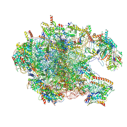
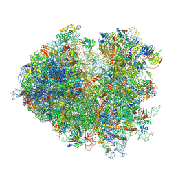
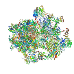
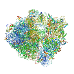
8XT0
 
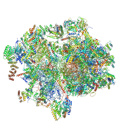 | | Cryo-EM structure of the human 55S mitoribosome with 5um Tigecycline | | 分子名称: | 12s rRNA, 16s rRNA, 39S ribosomal protein L22, ... | | 著者 | Li, X, Wang, M, Cheng, J. | | 登録日 | 2024-01-10 | | 公開日 | 2024-07-10 | | 実験手法 | ELECTRON MICROSCOPY (3.2 Å) | | 主引用文献 | Structural basis for differential inhibition of eukaryotic ribosomes by tigecycline.
Nat Commun, 15, 2024
|
|
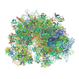
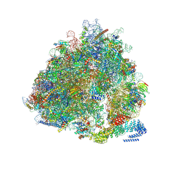
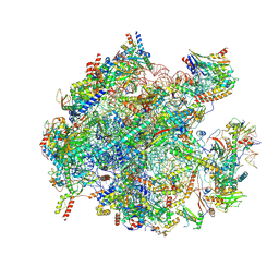
8YOO
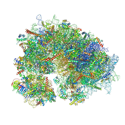 | | Cryo-EM structure of the human 80S ribosome with 100 um Tigecycline | | 分子名称: | 18S rRNA, 28S rRNA, 40S ribosomal protein S10, ... | | 著者 | Li, X, Wang, M, Denk, T, Cheng, J. | | 登録日 | 2024-03-13 | | 公開日 | 2024-07-10 | | 実験手法 | ELECTRON MICROSCOPY (2 Å) | | 主引用文献 | Structural basis for differential inhibition of eukaryotic ribosomes by tigecycline.
Nat Commun, 15, 2024
|
|
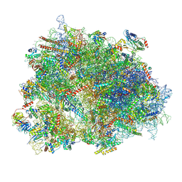
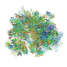
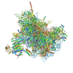
8XSX
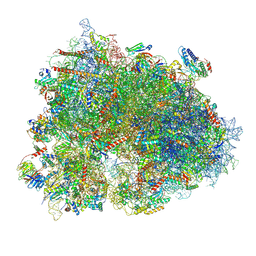 | | Cryo-EM structure of the human 80S ribosome with Tigecycline, E-tRNA, SERBP1 and eEF2 | | 分子名称: | 18S rRNA, 28S rRNA, 40S ribosomal protein S10, ... | | 著者 | Li, X, Wang, M, Cheng, J. | | 登録日 | 2024-01-10 | | 公開日 | 2024-07-10 | | 実験手法 | ELECTRON MICROSCOPY (2.4 Å) | | 主引用文献 | Structural basis for differential inhibition of eukaryotic ribosomes by tigecycline.
Nat Commun, 15, 2024
|
|
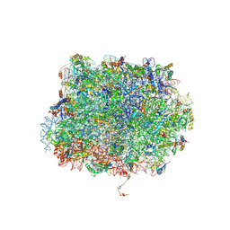
8YOP
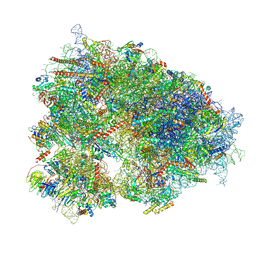 | | Cryo-EM structure of the human 80S ribosome with 4 um Tigecycline | | 分子名称: | 18S rRNA, 28S rRNA, 40S ribosomal protein S10, ... | | 著者 | Li, X, Wang, M, Denk, T, Cheng, J. | | 登録日 | 2024-03-13 | | 公開日 | 2024-07-10 | | 実験手法 | ELECTRON MICROSCOPY (2.2 Å) | | 主引用文献 | Structural basis for differential inhibition of eukaryotic ribosomes by tigecycline.
Nat Commun, 15, 2024
|
|
8XT3
 | | Cryo-EM structure of the human 39S mitoribosome with 10uM Tigecycline | | 分子名称: | 16s rRNA, 39S ribosomal protein L22, mitochondrial, ... | | 著者 | Li, X, Wang, M, Cheng, J. | | 登録日 | 2024-01-10 | | 公開日 | 2024-07-10 | | 実験手法 | ELECTRON MICROSCOPY (3.1 Å) | | 主引用文献 | Structural basis for differential inhibition of eukaryotic ribosomes by tigecycline.
Nat Commun, 15, 2024
|
|
8XSZ
 | | Cryo-EM structure of the human 80S ribosome with Tigecycline, E-tRNA and P-tRNA | | 分子名称: | 18S rRNA, 28S rRNA, 40S ribosomal protein S10, ... | | 著者 | Li, X, Wang, M, Cheng, J. | | 登録日 | 2024-01-10 | | 公開日 | 2024-07-10 | | 実験手法 | ELECTRON MICROSCOPY (3.2 Å) | | 主引用文献 | Structural basis for differential inhibition of eukaryotic ribosomes by tigecycline.
Nat Commun, 15, 2024
|
|
8XSY
 | | Cryo-EM structure of the human 80S ribosome with Tigecycline, e-tRNA and CCDC124 (40S head Swivelled) | | 分子名称: | 18S rRNA, 28S rRNA, 40S ribosomal protein S10, ... | | 著者 | Li, X, Wang, M, Cheng, J. | | 登録日 | 2024-01-10 | | 公開日 | 2024-07-10 | | 実験手法 | ELECTRON MICROSCOPY (3 Å) | | 主引用文献 | Structural basis for differential inhibition of eukaryotic ribosomes by tigecycline.
Nat Commun, 15, 2024
|
|
5GAK
 | | Yeast 60S ribosomal subunit with A-site tRNA, P-site tRNA and eIF-5A | | 分子名称: | 25S rRNA, 4-{(2R)-2-[(1S,3S,5S)-3,5-dimethyl-2-oxocyclohexyl]-2-hydroxyethyl}piperidine-2,6-dione, 5.8S rRNA, ... | | 著者 | Schmidt, C, Becker, T. | | 登録日 | 2015-12-09 | | 公開日 | 2016-02-24 | | 最終更新日 | 2019-12-11 | | 実験手法 | ELECTRON MICROSCOPY (3.88 Å) | | 主引用文献 | Structure of the hypusinylated eukaryotic translation factor eIF-5A bound to the ribosome.
Nucleic Acids Res., 44, 2016
|
|
7NRD
 | | Structure of the yeast Gcn1 bound to a colliding stalled 80S ribosome with MBF1, A/P-tRNA and P/E-tRNA | | 分子名称: | 25S rRNA (3184-MER), 40S ribosomal protein S0-A, 40S ribosomal protein S1-A, ... | | 著者 | Pochopien, A.A, Beckert, B, Wilson, D.N. | | 登録日 | 2021-03-03 | | 公開日 | 2021-04-14 | | 実験手法 | ELECTRON MICROSCOPY (4.36 Å) | | 主引用文献 | Structure of Gcn1 bound to stalled and colliding 80S ribosomes.
Proc.Natl.Acad.Sci.USA, 118, 2021
|
|
7NRC
 | | Structure of the yeast Gcn1 bound to a leading stalled 80S ribosome with Rbg2, Gir2, A- and P-tRNA and eIF5A | | 分子名称: | 18S rRNA (1771-MER), 25S rRNA (3184-MER), 40S ribosomal protein S0-A, ... | | 著者 | Pochopien, A.A, Beckert, B, Wilson, D.N. | | 登録日 | 2021-03-03 | | 公開日 | 2021-05-05 | | 実験手法 | ELECTRON MICROSCOPY (3.9 Å) | | 主引用文献 | Structure of Gcn1 bound to stalled and colliding 80S ribosomes.
Proc.Natl.Acad.Sci.USA, 118, 2021
|
|
6YXJ
 | |
8K2C
 | | Cryo-EM structure of the human 80S ribosome with Tigecycline | | 分子名称: | 18S rRNA, 28S rRNA, 40S ribosomal protein S10, ... | | 著者 | Li, X, Wang, M, Cheng, J. | | 登録日 | 2023-07-12 | | 公開日 | 2024-07-10 | | 実験手法 | ELECTRON MICROSCOPY (2.4 Å) | | 主引用文献 | Structural basis for differential inhibition of eukaryotic ribosomes by tigecycline.
Nat Commun, 15, 2024
|
|
8K2A
 | | Cryo-EM structure of the human 55S mitoribosome with Tigecycline | | 分子名称: | 12S rRNA, 16S rRNA, 39S ribosomal protein L22, ... | | 著者 | Li, X, Wang, M, Cheng, J. | | 登録日 | 2023-07-12 | | 公開日 | 2024-07-10 | | 実験手法 | ELECTRON MICROSCOPY (2.9 Å) | | 主引用文献 | Structural basis for differential inhibition of eukaryotic ribosomes by tigecycline.
Nat Commun, 15, 2024
|
|
8K2B
 | | Cryo-EM structure of the human 39S mitoribosome with Tigecycline | | 分子名称: | 16s rRNA, 39S ribosomal protein L22, mitochondrial, ... | | 著者 | Li, X, Wang, M, Cheng, J. | | 登録日 | 2023-07-12 | | 公開日 | 2024-07-10 | | 実験手法 | ELECTRON MICROSCOPY (3.4 Å) | | 主引用文献 | Structural basis for differential inhibition of eukaryotic ribosomes by tigecycline.
Nat Commun, 15, 2024
|
|
6FTG
 | |
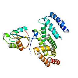
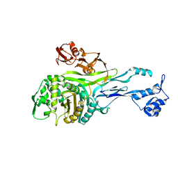
3KL4
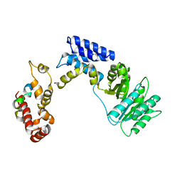 | | Recognition of a signal peptide by the signal recognition particle | | 分子名称: | Signal peptide of yeast dipeptidyl aminopeptidase B, Signal recognition 54 kDa protein | | 著者 | Janda, C.Y, Nagai, K, Li, J, Oubridge, C. | | 登録日 | 2009-11-06 | | 公開日 | 2010-03-31 | | 最終更新日 | 2024-02-21 | | 実験手法 | X-RAY DIFFRACTION (3.5 Å) | | 主引用文献 | Recognition of a signal peptide by the signal recognition particle.
Nature, 465, 2010
|
|
6Y69
 | |
1PMD
 | |
3OCI
 | | Crystal structure of TBP (TATA box binding protein) | | 分子名称: | 1,2-ETHANEDIOL, TRANSCRIPTION INITIATION FACTOR TFIID (TFIID-1) | | 著者 | Cui, S, Wollmann, P, Moldt, M, Hopfner, K.-P. | | 登録日 | 2010-08-10 | | 公開日 | 2011-07-13 | | 最終更新日 | 2024-03-20 | | 実験手法 | X-RAY DIFFRACTION (1.899 Å) | | 主引用文献 | Structure and mechanism of the Swi2/Snf2 remodeller Mot1 in complex with its substrate TBP.
Nature, 475, 2011
|
|
2XL1
 | |
1QMF
 | | PENICILLIN-BINDING PROTEIN 2X (PBP-2X) ACYL-ENZYME COMPLEX | | 分子名称: | 2-[CARBOXY-(2-FURAN-2-YL-2-METHOXYIMINO-ACETYLAMINO)-METHYL]-5-METHYL-3,6-DIHYDRO-2H-[1,3]THIAZINE-4-CARBOXYLIC ACID, CEFUROXIME (OCT-3-ENE FORM), PENICILLIN-BINDING PROTEIN 2X | | 著者 | Gordon, E.J, Mouz, N, Duee, E, Dideberg, O. | | 登録日 | 1999-09-28 | | 公開日 | 2000-05-25 | | 最終更新日 | 2024-05-01 | | 実験手法 | X-RAY DIFFRACTION (2.8 Å) | | 主引用文献 | The Crystal Structure of the Penicillin Binding Protein 2X from Streptococcus Pneumoniae and its Acyl-Enzyme Form: Implication in Drug Resistance
J.Mol.Biol., 299, 2000
|
|
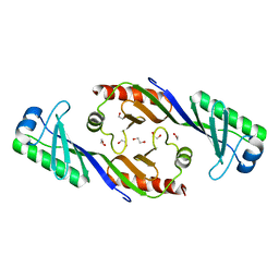
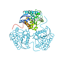
4K68
 | | Structure of a novel GH10 endoxylanase retrieved from sugarcane soil metagenome | | 分子名称: | GH10 xylanase, GLYCEROL | | 著者 | Santos, C.R, Polo, C.C, Alvarez, T.M, Paixao, D.A.A, Almeida, R.F, Pereira, I.O, Squina, F.M, Murakami, M.T. | | 登録日 | 2013-04-15 | | 公開日 | 2013-10-23 | | 最終更新日 | 2024-02-28 | | 実験手法 | X-RAY DIFFRACTION (2.74 Å) | | 主引用文献 | Development and biotechnological application of a novel endoxylanase family GH10 identified from sugarcane soil metagenome.
Plos One, 8, 2013
|
|
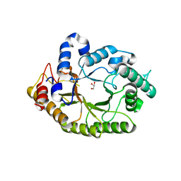
4IE3
 | | Crystal structure of human Arginase-2 complexed with inhbitor 1o | | 分子名称: | Arginase-2, mitochondrial, BENZAMIDINE, ... | | 著者 | Cousido-Siah, A, Mitschler, A, Ruiz, F.X, Beckett, P, Van Zandt, M.C, Ji, M.K, Whitehouse, D, Ryder, T, Jagdmann, E, Andreoli, M, Mazur, A, Padmanilayam, M, Schroeter, H, Golebiowski, A, Podjarny, A. | | 登録日 | 2012-12-13 | | 公開日 | 2013-03-20 | | 最終更新日 | 2023-09-20 | | 実験手法 | X-RAY DIFFRACTION (2.3522 Å) | | 主引用文献 | 2-Substituted-2-amino-6-boronohexanoic acids as arginase inhibitors.
Bioorg.Med.Chem.Lett., 23, 2013
|
|
4IE2
 | | Crystal structure of human Arginase-2 complexed with inhibitor 1h | | 分子名称: | Arginase-2, mitochondrial, BENZAMIDINE, ... | | 著者 | Cousido-Siah, A, Mitschler, A, Ruiz, F.X, Beckett, P, Van Zandt, M.C, Ji, M.K, Whitehouse, D, Ryder, T, Jagdmann, E, Andreoli, M, Mazur, A, Padmanilayam, M, Schroeter, H, Golebiowski, A, Podjarny, A. | | 登録日 | 2012-12-13 | | 公開日 | 2013-03-20 | | 最終更新日 | 2023-09-20 | | 実験手法 | X-RAY DIFFRACTION (2.2082 Å) | | 主引用文献 | 2-Substituted-2-amino-6-boronohexanoic acids as arginase inhibitors.
Bioorg.Med.Chem.Lett., 23, 2013
|
|
4IE1
 | | Crystal structure of human Arginase-1 complexed with inhibitor 1h | | 分子名称: | Arginase-1, MANGANESE (II) ION, [(5R)-5-amino-5-carboxy-8-hydroxyoctyl](trihydroxy)borate(1-) | | 著者 | Cousido-Siah, A, Mitschler, A, Ruiz, F.X, Beckett, P, Van Zandt, M.C, Ji, M.K, Whitehouse, D, Ryder, T, Jagdmann, E, Andreoli, M, Mazur, A, Padmanilayam, M, Schroeter, H, Golebiowski, A, Podjarny, A. | | 登録日 | 2012-12-13 | | 公開日 | 2013-03-20 | | 最終更新日 | 2023-09-20 | | 実験手法 | X-RAY DIFFRACTION (2.0006 Å) | | 主引用文献 | 2-Substituted-2-amino-6-boronohexanoic acids as arginase inhibitors.
Bioorg.Med.Chem.Lett., 23, 2013
|
|


