6DNM
 
 | |
7SAB
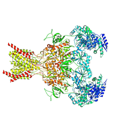
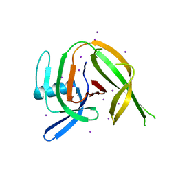
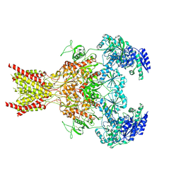 | | Phencyclidine-bound GluN1a-GluN2B NMDA receptors | | 分子名称: | 1-(PHENYL-1-CYCLOHEXYL)PIPERIDINE, 2-acetamido-2-deoxy-beta-D-glucopyranose, 2-acetamido-2-deoxy-beta-D-glucopyranose-(1-4)-2-acetamido-2-deoxy-beta-D-glucopyranose, ... | | 著者 | Chou, T.-H, Furukawa, H. | | 登録日 | 2021-09-22 | | 公開日 | 2022-07-20 | | 実験手法 | ELECTRON MICROSCOPY (4.3 Å) | | 主引用文献 | Structural insights into binding of therapeutic channel blockers in NMDA receptors.
Nat.Struct.Mol.Biol., 29, 2022
|
|
7SAD
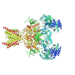 | | Memantine-bound GluN1a-GluN2B NMDA receptors | | 分子名称: | 2-acetamido-2-deoxy-beta-D-glucopyranose, 2-acetamido-2-deoxy-beta-D-glucopyranose-(1-4)-2-acetamido-2-deoxy-beta-D-glucopyranose, Glutamate receptor ionotropic, ... | | 著者 | Chou, T.-H, Furukawa, H. | | 登録日 | 2021-09-22 | | 公開日 | 2022-07-20 | | 実験手法 | ELECTRON MICROSCOPY (3.96 Å) | | 主引用文献 | Structural insights into binding of therapeutic channel blockers in NMDA receptors.
Nat.Struct.Mol.Biol., 29, 2022
|
|
7SAA
 | |
7SAC
 | |
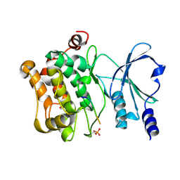
6YZE
 | | Zinc metalloprotease ProA from native source | | 分子名称: | ZINC ION, Zinc metalloproteinase | | 著者 | Schmelz, S, Blankenfeldt, W. | | 登録日 | 2020-05-06 | | 公開日 | 2021-02-03 | | 最終更新日 | 2024-01-24 | | 実験手法 | X-RAY DIFFRACTION (2.18 Å) | | 主引用文献 | Zinc metalloprotease ProA of Legionella pneumophila increases alveolar septal thickness in human lung tissue explants by collagen IV degradation.
Cell.Microbiol., 23, 2021
|
|
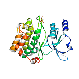
5N74
 | | Microtubule end binding protein complex | | 分子名称: | Karyogamy protein KAR9, Microtubule-associated protein RP/EB family member 1 | | 著者 | Kumar, A, Steinmetz, M. | | 登録日 | 2017-02-18 | | 公開日 | 2017-06-14 | | 最終更新日 | 2024-01-17 | | 実験手法 | X-RAY DIFFRACTION (2.3 Å) | | 主引用文献 | Short Linear Sequence Motif LxxPTPh Targets Diverse Proteins to Growing Microtubule Ends.
Structure, 25, 2017
|
|
2Z9I
 | |
8CPO
 | | Crystal structure of the PolB16_OarG intein variant S1A, N183A, C111A, C165A | | 分子名称: | PolB16 Intein Cys-less | | 著者 | Kattelmann, S, Pasch, T, Mootz, H.D, Kuemmel, D. | | 登録日 | 2023-03-03 | | 公開日 | 2023-05-17 | | 最終更新日 | 2024-06-19 | | 実験手法 | X-RAY DIFFRACTION (2.6 Å) | | 主引用文献 | Structural and biochemical analysis of a novel atypically split intein reveals a conserved histidine specific to cysteine-less inteins.
Chem Sci, 14, 2023
|
|
8CPN
 | | Crystal structure of the PolB16_OarG intein variant S1A, N183A | | 分子名称: | IODIDE ION, PolB16 intein | | 著者 | Kattelmann, S, Pasch, T, Mootz, H.D, Kummel, D. | | 登録日 | 2023-03-03 | | 公開日 | 2023-05-17 | | 最終更新日 | 2024-06-19 | | 実験手法 | X-RAY DIFFRACTION (1.85 Å) | | 主引用文献 | Structural and biochemical analysis of a novel atypically split intein reveals a conserved histidine specific to cysteine-less inteins.
Chem Sci, 14, 2023
|
|
3D3M
 | |
3IAD
 | |
2P3I
 | | Crystal structure of Rhesus Rotavirus VP8* at 295K | | 分子名称: | 2-O-methyl-5-N-acetyl-alpha-D-neuraminic acid, SULFATE ION, VP4 | | 著者 | Blanchard, H. | | 登録日 | 2007-03-09 | | 公開日 | 2008-03-11 | | 最終更新日 | 2023-10-25 | | 実験手法 | X-RAY DIFFRACTION (1.75 Å) | | 主引用文献 | Effects on sialic acid recognition of amino acid mutations in the carbohydrate-binding cleft of the rotavirus spike protein
Glycobiology, 19, 2009
|
|
2P3K
 | | Crystal structure of Rhesus rotavirus VP8* at 100K | | 分子名称: | 2-O-methyl-5-N-acetyl-alpha-D-neuraminic acid, GLYCEROL, SULFATE ION, ... | | 著者 | Blanchard, H. | | 登録日 | 2007-03-09 | | 公開日 | 2008-03-11 | | 最終更新日 | 2024-03-13 | | 実験手法 | X-RAY DIFFRACTION (1.56 Å) | | 主引用文献 | Effects on sialic acid recognition of amino acid mutations in the carbohydrate-binding cleft of the rotavirus spike protein
Glycobiology, 19, 2009
|
|
2P3J
 | |
6HV6
 | | Crystal structure of PatoxP, a cysteine protease-like domain of Photorhabdus asymbiotica toxin PaTox | | 分子名称: | 1,2-ETHANEDIOL, 4-(2-HYDROXYETHYL)-1-PIPERAZINE ETHANESULFONIC ACID, Toxin PAU_02230 | | 著者 | Bogdanovic, X, Wirth, C, Hunte, C. | | 登録日 | 2018-10-10 | | 公開日 | 2018-12-05 | | 最終更新日 | 2024-06-19 | | 実験手法 | X-RAY DIFFRACTION (2.001 Å) | | 主引用文献 | A cysteine protease-like domain enhances the cytotoxic effects of thePhotorhabdus asymbioticatoxin PaTox.
J. Biol. Chem., 294, 2019
|
|
4JDI
 | |
4JDJ
 | |
4JDH
 | |
3O9K
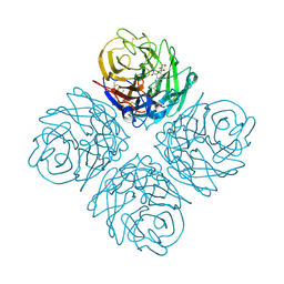 | | Influenza NA in complex with compound 6 | | 分子名称: | 5-acetamido-2,6-anhydro-3,5-dideoxy-3-[(2E)-3-(4-methylphenyl)prop-2-en-1-yl]-D-glycero-D-galacto-non-2-enonic acid, Neuraminidase | | 著者 | Russell, R.J, Kerry, P.S. | | 登録日 | 2010-08-04 | | 公開日 | 2010-12-15 | | 最終更新日 | 2020-07-29 | | 実験手法 | X-RAY DIFFRACTION (2.4945 Å) | | 主引用文献 | Novel sialic acid derivatives lock open the 150-loop of an influenza A virus group-1 sialidase.
Nat Commun, 1, 2010
|
|
3O9J
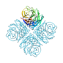 | | Influenza NA in complex with compound 5 | | 分子名称: | 2-acetamido-2-deoxy-alpha-D-glucopyranose, 5-acetamido-2,6-anhydro-3,5-dideoxy-3-prop-2-en-1-yl-D-glycero-D-galacto-non-2-enonic acid, CALCIUM ION, ... | | 著者 | Russell, R.J, Kerry, P.S. | | 登録日 | 2010-08-04 | | 公開日 | 2010-12-15 | | 最終更新日 | 2020-07-29 | | 実験手法 | X-RAY DIFFRACTION (2.0002 Å) | | 主引用文献 | Novel sialic acid derivatives lock open the 150-loop of an influenza A virus group-1 sialidase.
Nat Commun, 1, 2010
|
|
4JDK
 | |
1AVO
 | |
4D8S
 | | Influenza NA in complex with antiviral compound | | 分子名称: | CALCIUM ION, Neuraminidase, pentan-3-yl 2-acetamido-2,4-dideoxy-alpha-L-threo-hex-4-enopyranosiduronic acid | | 著者 | Kerry, P.S, Russell, R.J.M.R. | | 登録日 | 2012-01-11 | | 公開日 | 2013-02-13 | | 最終更新日 | 2020-07-29 | | 実験手法 | X-RAY DIFFRACTION (2.398 Å) | | 主引用文献 | Exploring the interactions of unsaturated glucuronides with influenza virus sialidase.
J.Med.Chem., 55, 2012
|
|
1EJE
 | | CRYSTAL STRUCTURE OF AN FMN-BINDING PROTEIN | | 分子名称: | FLAVIN MONONUCLEOTIDE, FMN-BINDING PROTEIN, NICKEL (II) ION, ... | | 著者 | Christendat, D, Saridakis, V, Bochkarev, A, Arrowsmith, C, Edwards, A.M, Northeast Structural Genomics Consortium (NESG) | | 登録日 | 2000-03-02 | | 公開日 | 2000-10-11 | | 最終更新日 | 2024-02-07 | | 実験手法 | X-RAY DIFFRACTION (2.2 Å) | | 主引用文献 | Structural proteomics of an archaeon.
Nat.Struct.Biol., 7, 2000
|
|


