7QXU
 
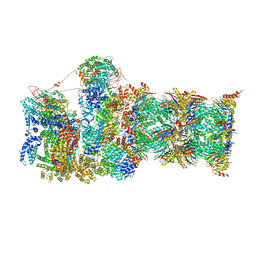
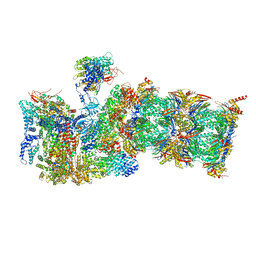 | | Proteasome-ZFAND5 Complex Z+C state | | 分子名称: | 26S protease regulatory subunit 6A, 26S protease regulatory subunit 6B, 26S protease regulatory subunit 7, ... | | 著者 | Zhu, Y, Lu, Y. | | 登録日 | 2022-01-27 | | 公開日 | 2023-02-08 | | 最終更新日 | 2024-09-04 | | 実験手法 | ELECTRON MICROSCOPY (4.3 Å) | | 主引用文献 | Molecular mechanism for activation of the 26S proteasome by ZFAND5.
Mol.Cell, 83, 2023
|
|
7QXW
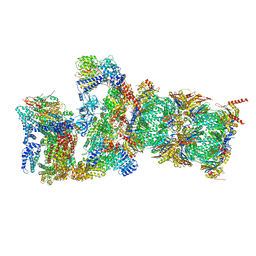 | | Proteasome-ZFAND5 Complex Z+D state | | 分子名称: | 26S protease regulatory subunit 6A, 26S protease regulatory subunit 6B, 26S protease regulatory subunit 7, ... | | 著者 | Zhu, Y, Lu, Y. | | 登録日 | 2022-01-27 | | 公開日 | 2023-02-08 | | 最終更新日 | 2024-10-16 | | 実験手法 | ELECTRON MICROSCOPY (4.1 Å) | | 主引用文献 | Molecular mechanism for activation of the 26S proteasome by ZFAND5.
Mol.Cell, 83, 2023
|
|
7QYA
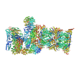 | | Proteasome-ZFAND5 Complex Z-B state | | 分子名称: | 26S protease regulatory subunit 4, 26S protease regulatory subunit 6A, 26S protease regulatory subunit 6B, ... | | 著者 | Zhu, Y, Lu, Y. | | 登録日 | 2022-01-27 | | 公開日 | 2023-02-08 | | 最終更新日 | 2024-09-04 | | 実験手法 | ELECTRON MICROSCOPY (4.8 Å) | | 主引用文献 | Molecular mechanism for activation of the 26S proteasome by ZFAND5.
Mol.Cell, 83, 2023
|
|
7QXN
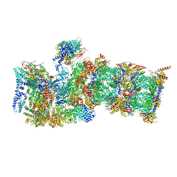 | | Proteasome-ZFAND5 Complex Z+A state | | 分子名称: | 26S protease regulatory subunit 4, 26S protease regulatory subunit 6A, 26S protease regulatory subunit 6B, ... | | 著者 | Zhu, Y, Lu, Y. | | 登録日 | 2022-01-26 | | 公開日 | 2023-02-08 | | 最終更新日 | 2024-09-04 | | 実験手法 | ELECTRON MICROSCOPY (3.7 Å) | | 主引用文献 | Molecular mechanism for activation of the 26S proteasome by ZFAND5.
Mol.Cell, 83, 2023
|
|
7QYB
 | | Proteasome-ZFAND5 Complex Z-C state | | 分子名称: | 26S protease regulatory subunit 4, 26S protease regulatory subunit 6A, 26S protease regulatory subunit 6B, ... | | 著者 | Zhu, Y, Lu, Y. | | 登録日 | 2022-01-27 | | 公開日 | 2023-02-08 | | 最終更新日 | 2024-09-04 | | 実験手法 | ELECTRON MICROSCOPY (4.1 Å) | | 主引用文献 | Molecular mechanism for activation of the 26S proteasome by ZFAND5.
Mol.Cell, 83, 2023
|
|
6B41
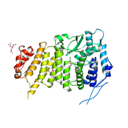 | | Menin bound to M-525 | | 分子名称: | Menin, methyl {(1S,2R)-2-[(S)-cyano[1-({1-[4-({1-[4-(dimethylamino)butanoyl]azetidin-3-yl}sulfonyl)phenyl]azetidin-3-yl}methyl)piperidin-4-yl](3-fluorophenyl)methyl]cyclopentyl}carbamate, praseodymium triacetate | | 著者 | Stuckey, J.A. | | 登録日 | 2017-09-25 | | 公開日 | 2018-01-24 | | 最終更新日 | 2023-10-04 | | 実験手法 | X-RAY DIFFRACTION (2.61 Å) | | 主引用文献 | Design of the First-in-Class, Highly Potent Irreversible Inhibitor Targeting the Menin-MLL Protein-Protein Interaction.
Angew. Chem. Int. Ed. Engl., 57, 2018
|
|
1Y9X
 | |
6NBT
 | |
6OQ5
 | | Structure of the full-length Clostridium difficile toxin B in complex with 3 VHHs | | 分子名称: | 5D, 7F, E3, ... | | 著者 | Chen, P, Lam, K, Jin, R. | | 登録日 | 2019-04-25 | | 公開日 | 2019-07-10 | | 最終更新日 | 2023-10-11 | | 実験手法 | X-RAY DIFFRACTION (3.87 Å) | | 主引用文献 | Structure of the full-length Clostridium difficile toxin B.
Nat.Struct.Mol.Biol., 26, 2019
|
|
6OQ7
 | | Structure of the GTD domain of Clostridium difficile toxin B in complex with VHH E3 | | 分子名称: | E3, MAGNESIUM ION, MANGANESE (II) ION, ... | | 著者 | Chen, P, Lam, K, Jin, R. | | 登録日 | 2019-04-25 | | 公開日 | 2019-07-10 | | 最終更新日 | 2023-10-11 | | 実験手法 | X-RAY DIFFRACTION (2.39 Å) | | 主引用文献 | Structure of the full-length Clostridium difficile toxin B.
Nat.Struct.Mol.Biol., 26, 2019
|
|
7ML7
 | |
8U6Y
 | | Preholo-Proteasome from Beta 3 D205 deletion | | 分子名称: | Proteasome assembly chaperone 2, Proteasome chaperone 1, Proteasome maturation factor UMP1, ... | | 著者 | Walsh Jr, R.M, Rawson, S, Velez, B, Blickling, M, Razi, A, Hanna, J. | | 登録日 | 2023-09-14 | | 公開日 | 2024-04-17 | | 最終更新日 | 2024-10-16 | | 実験手法 | ELECTRON MICROSCOPY (2.8 Å) | | 主引用文献 | Mechanism of autocatalytic activation during proteasome assembly.
Nat.Struct.Mol.Biol., 31, 2024
|
|
8U7U
 | | Proteasome 20S Core Particle from Beta 3 D205 deletion | | 分子名称: | Proteasome subunit alpha type-1, Proteasome subunit alpha type-2, Proteasome subunit alpha type-3, ... | | 著者 | Walsh Jr, R.M, Rawson, S, Velez, B, Blickling, M, Razi, A, Hanna, J. | | 登録日 | 2023-09-15 | | 公開日 | 2024-04-17 | | 最終更新日 | 2024-09-04 | | 実験手法 | ELECTRON MICROSCOPY (2.16 Å) | | 主引用文献 | Mechanism of autocatalytic activation during proteasome assembly.
Nat.Struct.Mol.Biol., 31, 2024
|
|
6OQ8
 | |
6OQ6
 | |
3ON9
 | |
3ONA
 | |
3WBM
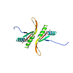
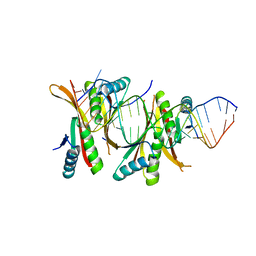 | | Crystal Structure of protein-RNA complex | | 分子名称: | DNA/RNA-binding protein Alba 1, RNA (25-MER) | | 著者 | Ding, J, Wang, D.C. | | 登録日 | 2013-05-20 | | 公開日 | 2013-12-11 | | 最終更新日 | 2023-11-08 | | 実験手法 | X-RAY DIFFRACTION (2 Å) | | 主引用文献 | Biochemical and structural insights into RNA binding by Ssh10b, a member of the highly conserved Sac10b protein family in Archaea.
J.Biol.Chem., 289, 2014
|
|
8HUG
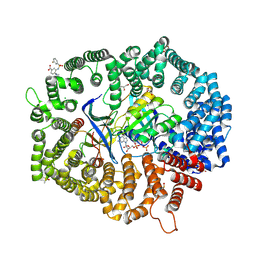 | | F1 in complex with CRM1-Ran-RanBP1 | | 分子名称: | 4-[4-(3-chlorophenyl)piperazin-1-yl]-3-[(3-fluorophenyl)sulfonylamino]benzoic acid, CHLORIDE ION, CRM1 isoform 1, ... | | 著者 | Sun, Q, Lei, Y. | | 登録日 | 2022-12-23 | | 公開日 | 2023-12-27 | | 最終更新日 | 2024-07-10 | | 実験手法 | X-RAY DIFFRACTION (2.15 Å) | | 主引用文献 | Discovery of a Hidden Pocket beneath the NES Groove by Novel Noncovalent CRM1 Inhibitors.
J.Med.Chem., 66, 2023
|
|
8HUF
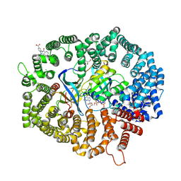 | | B28 in complex with CRM1-Ran-RanBP1 | | 分子名称: | 3-[(4-chlorophenyl)carbonylamino]-4-[4-(2,5-dimethylphenyl)piperazin-1-yl]benzoic acid, BROMIDE ION, CHLORIDE ION, ... | | 著者 | Sun, Q, Lei, Y. | | 登録日 | 2022-12-23 | | 公開日 | 2023-12-27 | | 最終更新日 | 2024-07-10 | | 実験手法 | X-RAY DIFFRACTION (2.29 Å) | | 主引用文献 | Discovery of a Hidden Pocket beneath the NES Groove by Novel Noncovalent CRM1 Inhibitors.
J.Med.Chem., 66, 2023
|
|
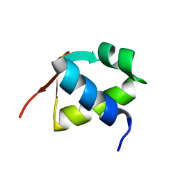
4GMB
 | | Crystal structure of human WD repeat domain 5 with compound MM-402 | | 分子名称: | MM-402, WD repeat-containing protein 5 | | 著者 | Karatas, H, Townsend, E.C, Chen, Y, Bernard, D, Cao, F, Liu, L, Lei, M, Dou, Y, Wang, S. | | 登録日 | 2012-08-15 | | 公開日 | 2014-02-19 | | 最終更新日 | 2023-11-15 | | 実験手法 | X-RAY DIFFRACTION (2.781 Å) | | 主引用文献 | Discovery of a Highly Potent, Cell-Permeable Macrocyclic Peptidomimetic (MM-589) Targeting the WD Repeat Domain 5 Protein (WDR5)-Mixed Lineage Leukemia (MLL) Protein-Protein Interaction.
J.Med.Chem., 60, 2017
|
|
7BZH
 | |
3F57
 | |
3DIY
 | |
3DIQ
 | |


