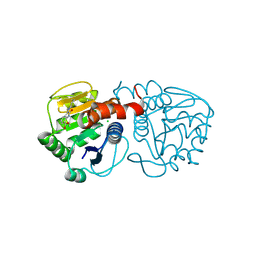
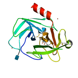
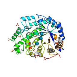
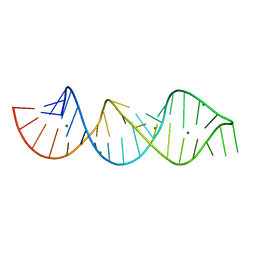
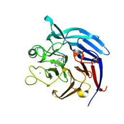
6AF7
 
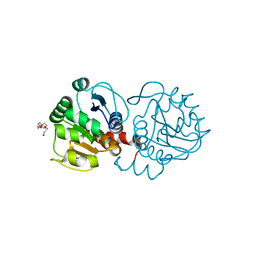 | | DJ-1 C106S unbound | | 分子名称: | CHLORIDE ION, PENTAETHYLENE GLYCOL, Protein/nucleic acid deglycase DJ-1 | | 著者 | Caaveiro, J.M.M, Tashiro, S, Tsumoto, K. | | 登録日 | 2018-08-08 | | 公開日 | 2018-08-29 | | 最終更新日 | 2023-11-22 | | 実験手法 | X-RAY DIFFRACTION (1.3 Å) | | 主引用文献 | Discovery and Optimization of Inhibitors of the Parkinson's Disease Associated Protein DJ-1.
ACS Chem. Biol., 13, 2018
|
|
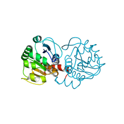
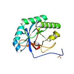
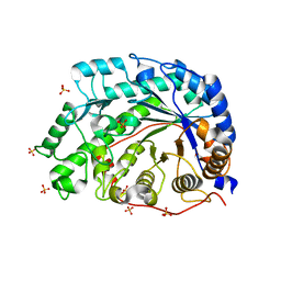
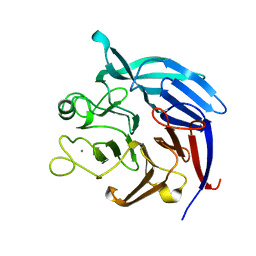
6AFI
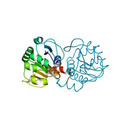 | | DJ-1 with compound 11 | | 分子名称: | 1-ethylindole-2,3-dione, CHLORIDE ION, Protein/nucleic acid deglycase DJ-1 | | 著者 | Caaveiro, J.M.M, Tashiro, S, Tsumoto, K. | | 登録日 | 2018-08-08 | | 公開日 | 2018-08-29 | | 最終更新日 | 2023-11-22 | | 実験手法 | X-RAY DIFFRACTION (1.65 Å) | | 主引用文献 | Discovery and Optimization of Inhibitors of the Parkinson's Disease Associated Protein DJ-1.
ACS Chem. Biol., 13, 2018
|
|
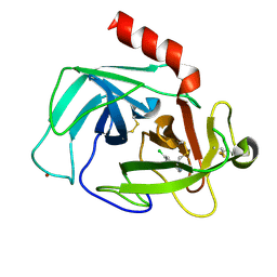
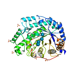
6AFE
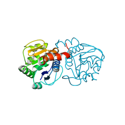 | | DJ-1 with compound 7 | | 分子名称: | 7-(trifluoromethyl)-1~{H}-indole-2,3-dione, CHLORIDE ION, Protein/nucleic acid deglycase DJ-1 | | 著者 | Caaveiro, J.M.M, Tashiro, S, Tsumoto, K. | | 登録日 | 2018-08-08 | | 公開日 | 2018-08-29 | | 最終更新日 | 2023-11-22 | | 実験手法 | X-RAY DIFFRACTION (1.5 Å) | | 主引用文献 | Discovery and Optimization of Inhibitors of the Parkinson's Disease Associated Protein DJ-1.
ACS Chem. Biol., 13, 2018
|
|
6AF9
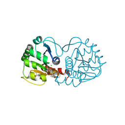 | |
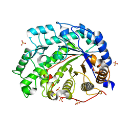
6AFF
 | | DJ-1 with compound 8 | | 分子名称: | CHLORIDE ION, Protein/nucleic acid deglycase DJ-1, methyl 2,3-bis(oxidanylidene)-1~{H}-indole-7-carboxylate | | 著者 | Caaveiro, J.M.M, Tashiro, S, Tsumoto, K. | | 登録日 | 2018-08-08 | | 公開日 | 2018-08-29 | | 最終更新日 | 2023-11-22 | | 実験手法 | X-RAY DIFFRACTION (1.6 Å) | | 主引用文献 | Discovery and Optimization of Inhibitors of the Parkinson's Disease Associated Protein DJ-1.
ACS Chem. Biol., 13, 2018
|
|
6AFJ
 | | DJ-1 with compound 13 | | 分子名称: | CHLORIDE ION, Protein/nucleic acid deglycase DJ-1, butyl 2-[2,3-bis(oxidanylidene)indol-1-yl]ethanoate | | 著者 | Caaveiro, J.M.M, Tashiro, S, Tsumoto, K. | | 登録日 | 2018-08-08 | | 公開日 | 2018-08-29 | | 最終更新日 | 2023-11-22 | | 実験手法 | X-RAY DIFFRACTION (1.48 Å) | | 主引用文献 | Discovery and Optimization of Inhibitors of the Parkinson's Disease Associated Protein DJ-1.
ACS Chem. Biol., 13, 2018
|
|
4KRT
 | |
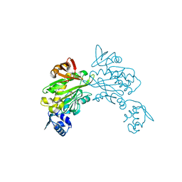
4K60
 | | Crystal Structure of Human Chymase in Complex with Fragment 6-bromo-1,3-dihydro-2H-indol-2-one | | 分子名称: | 2-acetamido-2-deoxy-beta-D-glucopyranose, 6-bromo-1,3-dihydro-2H-indol-2-one, Chymase, ... | | 著者 | Collins, B.K, Padyana, A.K. | | 登録日 | 2013-04-15 | | 公開日 | 2013-05-29 | | 最終更新日 | 2020-07-29 | | 実験手法 | X-RAY DIFFRACTION (1.5 Å) | | 主引用文献 | Discovery of Potent, Selective Chymase Inhibitors via Fragment Linking Strategies.
J.Med.Chem., 56, 2013
|
|
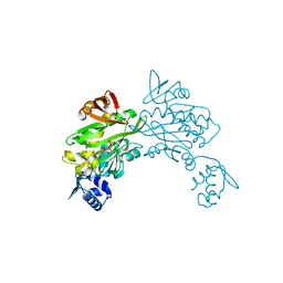
4K2Y
 | | Crystal Structure of Human Chymase in Complex with Fragment Inhibitor 6-chloro-1,3-dihydro-2H-indol-2-one | | 分子名称: | 2-acetamido-2-deoxy-beta-D-glucopyranose, 6-chloro-1,3-dihydro-2H-indol-2-one, Chymase, ... | | 著者 | Collins, B.K, Padyana, A.K. | | 登録日 | 2013-04-09 | | 公開日 | 2013-05-29 | | 最終更新日 | 2020-07-29 | | 実験手法 | X-RAY DIFFRACTION (2.3 Å) | | 主引用文献 | Discovery of Potent, Selective Chymase Inhibitors via Fragment Linking Strategies.
J.Med.Chem., 56, 2013
|
|
4K69
 | | Crystal Structure of Human Chymase in Complex with Fragment Linked Benzimidazolone Inhibitor: (3S)-3-{3-[(6-bromo-2-oxo-2,3-dihydro-1H-indol-4-yl)methyl]-2-oxo-2,3-dihydro-1H-benzimidazol-1-yl}hexanoic acid | | 分子名称: | (3S)-3-{3-[(6-bromo-2-oxo-2,3-dihydro-1H-indol-4-yl)methyl]-2-oxo-2,3-dihydro-1H-benzimidazol-1-yl}hexanoic acid, 2-acetamido-2-deoxy-beta-D-glucopyranose, Chymase, ... | | 著者 | Collins, B.K, Padyana, A.K. | | 登録日 | 2013-04-15 | | 公開日 | 2013-05-29 | | 最終更新日 | 2020-07-29 | | 実験手法 | X-RAY DIFFRACTION (1.5 Å) | | 主引用文献 | Discovery of Potent, Selective Chymase Inhibitors via Fragment Linking Strategies.
J.Med.Chem., 56, 2013
|
|
4KRU
 | |
4K5Z
 | | Crystal Structure of Human Chymase in Complex with Fragment Inhibitor 6-chloro-2,3-dihydro-1H-isoindol-1-one | | 分子名称: | 2-acetamido-2-deoxy-beta-D-glucopyranose, 6-chloro-2,3-dihydro-1H-isoindol-1-one, Chymase, ... | | 著者 | Collins, B.K, Padyana, A.K. | | 登録日 | 2013-04-15 | | 公開日 | 2013-05-29 | | 最終更新日 | 2020-07-29 | | 実験手法 | X-RAY DIFFRACTION (1.8 Å) | | 主引用文献 | Discovery of Potent, Selective Chymase Inhibitors via Fragment Linking Strategies.
J.Med.Chem., 56, 2013
|
|
1BXE
 | | RIBOSOMAL PROTEIN L22 FROM THERMUS THERMOPHILUS | | 分子名称: | CHLORIDE ION, PROTEIN (RIBOSOMAL PROTEIN L22) | | 著者 | Unge, J, Aberg, A, Al-Karadaghi, S, Nikulin, A, Nikonov, S, Davydova, N, Nevskaya, N, Garber, M, Liljas, A. | | 登録日 | 1998-10-02 | | 公開日 | 1998-10-07 | | 最終更新日 | 2018-03-14 | | 実験手法 | X-RAY DIFFRACTION (1.9 Å) | | 主引用文献 | The crystal structure of ribosomal protein L22 from Thermus thermophilus: insights into the mechanism of erythromycin resistance.
Structure, 6, 1998
|
|
1WDS
 | | The role of an inner loop in the catalytic mechanism of soybean beta-amylase | | 分子名称: | Beta-amylase, SULFATE ION, alpha-D-glucopyranose, ... | | 著者 | Kang, Y.N, Adachi, M, Utsumi, S, Mikami, B. | | 登録日 | 2004-05-17 | | 公開日 | 2005-04-05 | | 最終更新日 | 2024-05-29 | | 実験手法 | X-RAY DIFFRACTION (1.64 Å) | | 主引用文献 | Structural analysis of threonine 342 mutants of soybean beta-amylase: role of a conformational change of the inner loop in the catalytic mechanism.
Biochemistry, 44, 2005
|
|
1WDP
 | | The role of an inner loop in the catalytic mechanism of soybean beta-amylase | | 分子名称: | Beta-amylase, SULFATE ION | | 著者 | Kang, Y.N, Adachi, M, Utsumi, S, Mikami, B. | | 登録日 | 2004-05-17 | | 公開日 | 2005-04-05 | | 最終更新日 | 2024-03-13 | | 実験手法 | X-RAY DIFFRACTION (1.27 Å) | | 主引用文献 | Structural analysis of threonine 342 mutants of soybean beta-amylase: role of a conformational change of the inner loop in the catalytic mechanism.
Biochemistry, 44, 2005
|
|
1WDR
 | | The role of an inner loop in the catalytic mechanism of soybean beta-amylase | | 分子名称: | Beta-amylase, SULFATE ION, alpha-D-glucopyranose, ... | | 著者 | Kang, Y.N, Adachi, M, Utsumi, S, Mikami, B. | | 登録日 | 2004-05-17 | | 公開日 | 2005-04-05 | | 最終更新日 | 2021-11-10 | | 実験手法 | X-RAY DIFFRACTION (1.35 Å) | | 主引用文献 | Structural analysis of threonine 342 mutants of soybean beta-amylase: role of a conformational change of the inner loop in the catalytic mechanism.
Biochemistry, 44, 2005
|
|
1WDQ
 | | The role of an inner loop in the catalytic mechanism of soybean beta-amylase | | 分子名称: | Beta-amylase, SULFATE ION, alpha-D-glucopyranose-(1-4)-alpha-D-glucopyranose | | 著者 | Kang, Y.N, Adachi, M, Utsumi, S, Mikami, B. | | 登録日 | 2004-05-17 | | 公開日 | 2005-04-05 | | 最終更新日 | 2024-05-29 | | 実験手法 | X-RAY DIFFRACTION (1.28 Å) | | 主引用文献 | Structural analysis of threonine 342 mutants of soybean beta-amylase: role of a conformational change of the inner loop in the catalytic mechanism.
Biochemistry, 44, 2005
|
|
4PCJ
 | |
1MRR
 | |
4OL9
 | |
7WKI
 | |
3A9H
 | | Crystal Structure of PQQ-dependent sugar dehydrogenase holo-form | | 分子名称: | CALCIUM ION, PYRROLOQUINOLINE QUINONE, Putative uncharacterized protein, ... | | 著者 | Sakuraba, H, Yokono, K, Yoneda, K, Ohshima, T. | | 登録日 | 2009-10-26 | | 公開日 | 2010-09-08 | | 最終更新日 | 2023-11-01 | | 実験手法 | X-RAY DIFFRACTION (2.5 Å) | | 主引用文献 | Catalytic properties and crystal structure of quinoprotein aldose sugar dehydrogenase from hyperthermophilic archaeon Pyrobaculum aerophilum
Arch.Biochem.Biophys., 502, 2010
|
|
3A9G
 | | Crystal Structure of PQQ-dependent sugar dehydrogenase apo-form | | 分子名称: | CALCIUM ION, Putative uncharacterized protein, alpha-D-glucopyranose-(1-1)-alpha-D-glucopyranose | | 著者 | Sakuraba, H, Yokono, K, Yoneda, K, Ohshima, T. | | 登録日 | 2009-10-26 | | 公開日 | 2010-09-08 | | 最終更新日 | 2023-11-01 | | 実験手法 | X-RAY DIFFRACTION (2.39 Å) | | 主引用文献 | Catalytic properties and crystal structure of quinoprotein aldose sugar dehydrogenase from hyperthermophilic archaeon Pyrobaculum aerophilum
Arch.Biochem.Biophys., 502, 2010
|
|
3RKY
 | |
3RKW
 | |


